R E S E A R C H
Open Access
Wilms
’
tumor 1-associating protein plays
an aggressive role in diffuse large B-cell
lymphoma and forms a complex with
BCL6 via Hsp90
Yue Kuai
1,2, Xin Gong
1, Liya Ding
1, Fang Li
1, Lizhen Lei
1, Yuqi Gong
1, Qingmeng Liu
3, Huajiao Tan
1, Xinxia Zhang
4,
Dongyu Liu
5, Guoping Ren
6, Hongyang Pan
4, Yaoyao Shi
7, Friederike Berberich-Siebelt
8, Zhengrong Mao
1*and Ren Zhou
1*Abstract
Background:Wilms’tumor 1-associating protein (WTAP) is a nuclear protein, which is ubiquitously expressed in many tissues. Furthermore, in various types of malignancies WTAP is overexpressed and plays a role as an oncogene. The function of WTAP in diffuse large B-cell lymphoma (DLBCL), however, remains unclear.
Methods:Immunohistochemistry was applied to evaluate the levels of WTAP expression in DLBCL tissues and
normal lymphoid tissues. Overexpression and knock-down ofWTAPin DLBCL cell lines, verified on mRNA and protein level served to analyze cell proliferation and apoptosis in DLBCL cell lines by flow cytometry. Finally, co-immunoprecipitation (Co-IP), IP, and GST-pull down assessed the interaction of WTAP with Heat shock protein 90 (Hsp90) and B-cell lymphoma 6 (BCL6) as well as determined the extend of its ubiquitinylation.
Results:WTAP protein levels were consistently upregulated in DLBCL tissues. WTAP promoted DLBCL cell
proliferation and improved the ability to confront apoptosis, while knockdown ofWTAPin DLBCL cell lines allowed a significant higher apoptosis rate after treatment with Etoposide, an anti-tumor drug. The stable expression of WTAP was depended on Hsp90. In line, we demonstrated that WTAP could form a complex with BCL6 via Hsp90 in vivo and in vitro.
Conclusion:WTAP is highly expressed in DLBCL, promoting growth and anti-apoptosis in DLBCL cell lines. WTAP is a client protein of Hsp90 and can appear in a complex with BCL6 and Hsp90 in DLBCL. Down-regulation of WTAP could improve the chemotherapeutic treatments in DLBCL.
Keywords:WTAP, BCL6, Hsp90, Complex, DLBCL
Background
Wilms’ tumor 1-associating protein (WTAP) is a nuclear protein first identified as a binding protein of Wilms’ tumor 1 (WT1) both in vitro and in vivo [1]. The import-ance of WTAP has been verified by knockout experiments, as WTAP-null mice die at embryonic day 6.5 [2] and WTAP heterozygous mice die at embryonic day 10.5 [3]. The biological function of WTAP in normal cells has been
partly explored. Dynamic expression of WTAP and WT1 in smooth muscle cells (SMCs) can regulate cell prolifera-tion [4]. WTAP expression can be enhanced by insulin-like growth factor-1 (IGF-1) conferring anti-apoptotic proper-ties on smooth muscle cells [5]. Furthermore, in human umbilical vein endothelial cells, WTAP facilitates cell cycle progression by stabilizing cyclin A2 mRNA through an ef-fect on its 3’UTR [2]. Meanwhile, accumulating evidences have shown that WTAP may play an aggressive role in some malignant tumors. For instance, WTAP is overex-pressed in glioblastoma and regulates motility of glioblast-oma cells by controlling the activity of EGFR [6]. In * Correspondence:[email protected];[email protected]
1Department of Pathology and Pathophysiology, Institute of Pathology and
Forensic Medicine, Zhejiang University School of Medicine, Hangzhou, China Full list of author information is available at the end of the article
cholangiocarcinoma cells, WTAP regulates migration and invasion, where microarray studies showed that WTAP could induce the expression of MMP7, MMP28, cathepsin H and Muc1 [7]. Moreover, WTAP is a novel oncogene in acute myelogenous leukemia (AML) and a client protein of Heat shock protein 90 (Hsp90) [8]. Client proteins are drawn into a multi-protein complex by Hsp90, a molecular chaperone assisting in folding and stabilization of proteins [9]. Without the company of Hsp90, client proteins may be misfolded, subsequently ubiquitylated and proteasomally degraded [10]. Hence, Hsp90 is a rational therapeutic tar-get for cancer [11].
B-cell lymphoma 6 (BCL6) is known client protein of Hsp90, i.e. BCL6 might function as a stress response gene that forms part of a larger, post-transcriptionally regulated program governed by Hsp90 [12]. In the pathogenesis of Diffuse large B-cell lymphoma (DLBCL), BCL6 transcriptional repressor is the most frequently in-volved oncoprotein, which is required to sustain prolifer-ation and survival of DLBCL cells through regulprolifer-ation of specific targets such as PRDM1, c-Myc or PAX-5 [13]. However, relatively little is known about the contribu-tion of WTAP–one further client protein of Hsp90.
DLBCLs are the most common B-cell non-Hodgkin lymphoma (NHL) in the world, comprising about 30–35% of all NHLs [14], which exhibit a heterogeneity in morph-ology, immunophenotype, genetics, and biological behav-ior [15]. Activated B-cell (ABC) and germinal-center B-cell (GCB) subgrous of DLBCL have been defined by gene-expression profiling, leaving approximately 10 to 20% of cases “unclassified” [16, 17]. Up to one third of DLBCL cases have abnormalities of BCL6 and ~ 20% of cases have translocations ofBCL2[18]. Although there are some patients, who can be cured of DLBCL, a substantial fraction of them (∼40%) die of this disease [19], pointing to the growing need to explore more specific drugs.
There are some in vitro studies, which provide evi-dence for the interaction of Hsp90 with WTAP as well as Hsp90 with BCL6. Since in a recent study Hsp90 was found to be frequently expressed in DLBCLs [20], we hypothesized that WTAP expression in DLBCL could be regulated by Hsp90 activity. In such a case, Hsp90 inhib-ition would affect the maintenance of WTAP and the protein’s function contributed by WTAP. Moreover, we speculated that WTAP might form a complex with BCL6 via Hsp90. Indeed, we could demonstrate that WTAP is not only highly expressed in DLBCLs and de-tectable in a complex with Hsp90 and BCL6, but medi-ates proliferation, while counteracting apoptosis.
Methods Cell culture
HEK293T cell line was obtained from the Type Culture
Collection of the Chinese Academy of Sciences
(Shanghai, China); Dr. Xin Jiang (China) kindly provided DLBCL cell lines OCI- Ly10, OCI-Ly19, SU-DHL2 and SU-DHL4. The HEK293T cells were maintained in DMEM supplemented with 10% FBS (Gibco). The DLBCL cell lines were maintained in IMDM with 10% FBS (Gibco). Cultures were maintained in a 5% CO2 hu-midified atmosphere at 37 °C.
Construction of vector
The WTAP gene was PCR-amplified from HEK293T
cDNA and ligated into the pLVX-Puro vector (Clontech Laboratories) and pcDNA3.1-his-myc-B vector (Invitro-gen), named WTAP-pLVX-Puro and pcDNA 3.1-WTAP, respectively. The WTAP-encoding sequence was also inserted into the pGEX-4 T-3 (Amersham biosciences) plasmid, called GST-WTAP thereafter. The BCL6 gene was PCR-amplified from HEK293T cDNA and ligated into the pcDNA3.1-his-myc-B (Invitrogen), named BCL6-His.
WTAP gain and loss of function experiments
WTAP-overexpressed lentivirus was packaged by different
recombinant plasmids along with helper plasmids
(psPAX2 and pMD2.G) in HEK293T cells, and virus super-natants were collected at 48 h and 72 h post-transfection.
After concentration, recombinant WTAP-pLVX-Puro
virus or control (pLVX-Puro) virus were infected into OCI-Ly19 cells. Additionally, WTAP-knock-down lenti-viral infectious supernatant was obtained from Ibsbio
company (China). WTAP target sequence was “GGGC
AACACAACCGAAGAT”, the control sequence was
“TTCTCCGAACGTGTCACGT”. WTAP-specific
lenti-virus was infected into OCI-Ly10 cells for knock-down, and control cells were generated using a non-target scram-ble. After infection, stable clones were selected with puro-mycin (Invitrogen) at a final concentration of 2μg/ml.
Immunohistochemistry
A total of 30 DLBCLs paraffin-embedded samples and 30 normal lymphoid tissues were obtained from the Second hospital of Shaoxing and the First Affiliated Hospital of Zhejiang University, respectively, after the necessary in-formed consent of patients. In each case, diagnosis of DLBCL was made according to the World Health Organization Classification of Tumors of Hematopoietic and Lymphoid Tissues [21]. This study was approved by the Ethics Committee of Zhejiang University (Hangzhou, China). IHC staining was performed using the EnVision system (Maixin Biotech) with antibodies for WTAP (1:1200, Abcam, ab155655). Data were calculated as previ-ously described [22].
RNA isolation and quantitative real time PCR (qRT-PCR)
reverse-transcribed using a PrimeScript RT reagent kit (Takara). Real-time PCR was performed three times in triplicate with SYBR Premix Ex TaqTM (Takara), using the 480II Real-Time PCR System (Roche). WTAP-F: TGTGCTGTGTAAGGGCATTCGTACTCATGC, WTA P-R: ACTGGGCAAACTTGGCAGTCATAAACCCAC; GAPDH-F: ACCACAGTCCATGCCATCAC; GAPDH -R: TCCACCACCCTGTTGCTGTA. The reactions were incubated at 95 °C for 5 min, followed by 40 cycles of 95 °C for 30 s, 60 °C for 30 s and 72 °C for 30 s. The relative amount of WTAP mRNA was normalized to that of GAPDH. Relative expression levels were normal-ized to GAPDH and calculated with the 2-ΔΔct method. Each experiment was performed independently at least three times.
Protein isolation and western blotting
Nuclear and cytoplasmic proteins were obtained according to the manufacturer’s instruction (Beibokit, China). The total protein samples were harvested with 1 × SDS loading buffer or RIPA and then resolved by SDS- polyacrylamide gel electrophoresis (PAGE), electrophoretically transferred to PVDF membranes (Millipore) and blocked in nonfat milk in TBS/Tween-20. Antibodies were specific for BCL6 (N3) (Santa cruz, sc-858), WTAP (Abcam, ab195380), anti-Hsp90 (Abcam, ab203126). The anti-GAPDH (Cell Signaling Technology, 5174), anti-HistoneH2A (Cell
Sig-naling Technology, 7631), anti-LaminA/C (Abcam,
ab108595), anti-ubiquitin, Lys48-specific Clone Apu2 (Millipore, 2145090), anti-His and anti-GST antibodies were purchased from CWBiotech. The experiments for the immunoblotting were performed at least three times.
Proliferation assay
Cells (1 × 10⁴/well) were seeded into 96-well plates in triplicates, and were cultured in 100μL IMDM contain-ing 10% FBS for 3 days. The Cell Countcontain-ing Kit-8 (CCK8) was used to evaluate cell proliferation. Briefly, 10 μL CCK8 solution was added to each plate, and cells were incubated for 4 h at 37 °C. Cell viability was measured at 450 nm by SpectraMax M5.
Apoptosis assay
OCI-Ly19 cells were infected with control or WTAP-pLVX-Puro lentivirus and then cultured for 48 h; an optimum concentration of puromycin was used until the cells in a blank group died. We cultured cells with hydrogen peroxide (300 mM) combined with serum deprivation for 16 h. 2 × 10⁵ cells were harvested with ice-cold phosphate buffered saline (PBS). SU-DHL2 and SU-DHL4 were cultured in PU-H71 (1.5 μM) for 48 h, and then added Etoposide (10 µM) for another 24 h. Cells were also collected and stained with Annexin V-PE along with 7-AAD to detect apoptosis by flow cytometry.
WTAP-knocked down OCI-Ly10 cells or the control cells were cultured in the presence of Etoposide or DMSO for 24 h. The final concentration of Etoposide was 10 μM. Cells were also collected and stained with Annexin V-PE along with 7-AAD. The quantitative ana-lysis of the percentage of positive cells was reported from three independent experiments.
Immunofluorescence staining
After cultures with PU-H71 or DMSO for 48 h, OCI-Ly10 cells were washed twice in PBS, spread onto slides, air dried and fixed in acetone at −20 °C for 15 min. Immunofluorescence staining was performed as described previously [13]. After treatment of 0.3% Triton X-100 (KeyGen Biotech) for 10 min, the slides were blocked with Goat serum (Boster Biological Technology) for 1 h at room temperature. The cells were then incu-bated with the primary antibody against WTAP (diluted 1:50, Santa Cruz, sc-374280) and BCL6 (diluted 1:100, Abcam, ab33901) overnight at 4 °C. Signals were de-tected with Alexa Fluor 488 and Alexa Fluor 594, re-spectively. Nuclei in live cells were stained with 4', 6-diamidino-2-phenylindole (DAPI), and visualized with laser confocal microscopy (NikonA1R).
Co-immunoprecipitation (Co-IP)
HEK293T cells were co-transfected with BCL6-His and pcDNA3.1-WTAP vectors by LipofectamineTM 3000 (Invitrogen). 48 h after transfection, collected cells were lysed on ice for 1 h using RIPA buffer (Beyotime) with 1 mM Phenylmethanesulfonyl fluoride. Co-IP was per-formed as described previously [13]. Briefly, 1 μl anti-WTAP antibody (Abcam, ab195380) was added to the whole-cell lysate. The immune complexes were incu-bated at 4 °C with rotation and collected using 20 μl protein G Plus-Agarose Immuno-precipitation Reagent (Santa Cruz). After that, the collected agarose beads were boiled in SDS sample buffer and analyzed by west-ern blotting. LaminA/C is a nuclear protein and served as a negative control. All the experiments were per-formed in triplicates.
Immunoprecipitation
OCI-Ly19 were harvested from a six-well plate, the cells were lysed on ice for 1 h using RIPA buffer (Beyotime) with 1 mM Phenylmethanesulfonyl fluoride. 1 μl anti-WTAP
antibody (Abcam, ab195380), 5 μl anti-BCL6 (N3)
by western blotting. All the experiments were per-formed in triplicates.
GST fusion proteins and his-tag-purified protein pull-down assay
A total of 100μl GST fusion proteins of GST-WTAP or
GST vector was purified and immobilized on
GST-immunomagnetic beads (Beaverbio, China) to pull down the GST-bound form of His-tag fusion protein at 4 °C for 4 h. BCL6-His fusion protein was extracted from HEK293T cells transfected with BCL6-His and purified by His-immunomagnetic beads according to the manufacturer’s instructions (Beaverbio, China). at 4 °C for 5 min × 3 with a buffer containing 1 ml PBS and 1% TritonX-100, the bound proteins were eluted by boiling in 2 × loading buffer followed by SDS-PAGE and detected by western blotting analysis with GST anti-bodies (CWBiotech, China) and anti-Hsp90 antianti-bodies (Abcam, ab203126).
GST fusion proteins and cell lysates pull-down assay
A total of 100 μl of GST fusion proteins of
GST-WTAP or GST was purified and immobilized on GST-immunomagnetic beads (Beaverbio, China)
to pull down the GST-bound form of proteins ex-tracted from the HEK293T cells transfected with BCL6-His.The following progress was the same as above described.
Statistical analysis
Differences between the expression score of immunohis-tochemistry in lymphoid tissues and DLBCLs were assessed using the Student’s t-test. For in vitro and in vivo assays, data were analyzed using Student’s t-test or one-way ANOVA to compare the results among experi-mental groups and control groups. Statistical analysis was carried out using Graph Pad Prism version 5. Sig-nificance level was set atP< 0.05.
Results
WTAP was overexpressed in DLBCLs
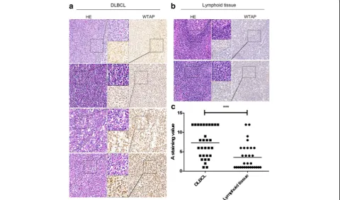
To determine the expression status of WTAP in DLBCLs, we performed immunohistochemistry staining using DLBCL patients’tissues (n= 30) (Additional file1: Table S1) and normal lymphoid tissues (n= 30). Indeed, stainings revealed nuclear expression of WTAP (Fig.1a). Importantly, WTAP protein was widely overexpressed in the cancerous areas of DLBCL tissues and, compared
with the control group (Fig.1b), a higher level of WTAP was observed in DLBCLs tissues (Fig.1c). Many, but not all DLBCL tissues overexpressed it compared with nor-mal lymphoid tissues. The score of each sample was cal-culated as previously described [22].
Roles of WTAP in proliferation and apoptosis of DLBCL cells
To examine roles of WTAP in DLBCL, we overex-pressed or knocked-down WTAP using lentivirus. We checked the efficiency of infection by western blotting and real-time PCR (Fig. 2a). Two days after infection with overexpressing or knocking-down lentivirus, an optimum concentration of puromycin was used until cells in a blank group died. Proliferation assays were car-ried out with the selected cells. WTAP positively influ-enced cell proliferation, as the proliferation of OCI-Ly19 cells, which were infected with WTAP overexpressed lentivirus, was higher than the control group (Fig.2b). On the reverse, when WTAP had been knocked-down, the proliferation of OCI-Ly10 cells was inhibited (Fig.2c). To determine the role of WTAP during the process of DLBCL cell apoptosis, we cultured WTAP overexpressing cells with hydrogen peroxide combined with serum deprivation for 16 h and then we checked the apoptosis level with flow cytometry. A significant lower apoptosis rate was revealed in the group overexpressing exogenous WTAP (Fig. 2d). In knocked-down cells, there was no obvious difference between the two groups (data not shown). However, when we treated WTAP-knock-down cells and control cells with Etoposide or DMSO, respectively, a distinctive higher apoptosis rate was evident in the WTAP-reduced group upon Etoposide treatment (Fig. 2e, f). Together, WTAP supports proliferation and counteracts Etopside-mediated apoptosis induction. Hence, we speculate that a DLBCL patient, who has a higher level of WTAP expression, may experience a poor therapeutic efficiency of chemotherapy.
Hsp90 can stabilize WTAP on the protein level
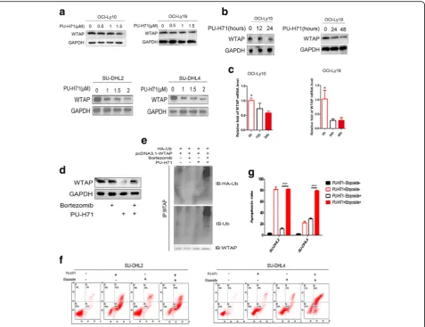
In a recent paper, H Bansal et al. have proven that in vitro Hsp90 could interact with WTAP, whereas in AML cell lines the expression of WTAP could be affected by inhibitors of Hsp90 [8]. Here, we were wondering about the relationship between WTAP and Hsp90 in DLBCL cell lines. Firstly, we chose the cell lines OCI-Ly10 and SU-DHL2, resembling the ABC types as well as OCI-Ly19 and SU-DHL4 which are GCB types as ex-perimental subjects. After being cultured in the presence of PU-H71, an Hsp90 inhibitor, the level of expression of WTAP was changing with concentration and time (Fig. 3a, b). Meanwhile, WTAP is described to associate with the METTL3/14 heterodimer and to influence RNA processing including RNA stability [23]. Interest-ingly, we show here that reduced WTAP protein levels provoke less WTAP RNA (Fig. 3c). Since 3 AU-rich
elements (ARE) could be detected with the help of an ARE database (data not shown), it is tempting to specu-late that WTAP stabilizes its own RNA via these RNA-destabilizing motifs.
Lack of Hsp90 interaction might expose WTAP to ubi-quination and proteasomal degradation [8]. Hence, we pretreated cells with Bortezomib, a 26 s proteasome in-hibitor or DMSO for 2 h, and subsequently cultured them with PU-H71 for 6 h. We found that Bortezomib could prevent the actions of PU-H71 (Fig. 3d). In line, higher levels of ubiquitinated WTAP were observed in the presence of the combination of Bortezomib and PU-H71 compared with either agent alone (Fig. 3e), re-vealing that PU-H71-mediated degradation was depend on ubiquitin-proteasome pathway. Then we wondered if PU-H71, i.e. Hsp90 inhibition, impacts on apoptosis as well. We pretreated SU-DHL2 and SU-DHL4 cells with PU-H71 or DMSO for 24 h and then cultured them with Etoposide for another 24 h. Indeed, groups pretreated with PU-H71 demonstrated significantly higher levels of apoptosis (Fig.3f). In sum, similar to other Hsp90 clients, WTAP expression levels seem to be primarily regulated via protein stability.
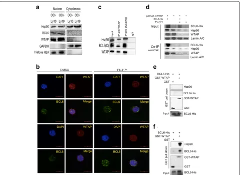
WTAP forms a complex with BCL6 via Hsp90 in vivo and in vitro
WTAP and BCL6 could form a complex in cells, without either protein being overexpressed, immunoprecipitation experiments were carried out using OCI-Ly19 cell lysates. The DLBCL cell line OCI-Ly19 is known to express BCL6 and additionally, as shown by western blotting, endogen-ous WTAP and Hsp90 (Fig. 4a). The immunoprecipita-tions were carried out and after subsequent western blotting analysis with anti-WTAP antibody, WTAP and Hsp90 could be detected not only in the Co-IPs carried out with the anti-WTAP antibody, but could also be discovered in the anti-BCL6 Co-IPs, thus indicating that WTAP, Hsp90 and BCL6 formed a complex in vivo
(Fig. 4c). Similarly, we performed Co-IPs after
transfection of BCL6-his and pcDNA3.1-WTAP express-ing plasmids into HEK293T cells. In parallel, cell were additionally treated with PU-H71. Firstly, WTAP inter-acts not only with endogenous Hsp90, but also with ex-ogenous BCL6 if provided (Fig. 4d). Secondly, PU-H71 treatment, i.e. Hsp90 inhibition, obstructed the complex formation (Fig. 4d). Consequently, WTAP must be as-sumed to interact with BCL6 directly or indirectly via the chaperone Hsp90. Now GST pull-down assay revealed the presence of proteins in vitro, but affinity-purified GST-WTAP and the His-tagged fusion protein BCL6-His, did not bind to each other directly (Fig.4e). In contrast, when whole cell lysates–presumably containing Hsp90–
Fig. 2WTAP supports proliferation and counteracts apoptosis of DLBCL cells.aOCI-Ly19 and OCI-Ly10 cells, 48 h post-infection. Expression of WTAP was checked on the level of protein and mRNA by using western blotting and qPCR. Mean ± s.d. of three technical replicates were plotted. **P< 0.01, ***P< 0.001.b,cWe infected OCI-Ly19 and OCI-Ly10 by WTAP-overexpressing lentivirus or WTAP-knocked down lentivirus. Then cells were cultured in 96 cell panels for 0, 24, 48, and 72 h. Cell viability was measured by CCK8 assay.*,P< 0.05, **,P< 0.01 and ***,P< 0.001 compared with control group.
were provided during GST-WTAP pull-down assay,
BCL6-His and Hsp90 could be pulled down by
GST-WTAP, but not GST (Fig. 4f). From this, we con-clude that WTAP forms a complex with BCL6 via Hsp90 in vivo and in vitro.
Discussion
WTAP is a nuclear protein which was first regarded as a housekeeping protein because of its ubiquitous expression pattern [1]. However, with an increase in the number of studies, WTAP has predominantly been described in the context of proliferation, apoptosis, migration and invasion of tumor cells [6, 7]. Accordingly, in mammalian cells, WTAP forms a nuclear complex with METTL3 and
METTL14, which methylates N6-adenosines of RNA in-volved in cell cycle regulation, post-transcriptional mRNA metabolism and splicing [26–29]. WTAP, as the name in-dicates, partners with the Wilms’tumor 1 (WT1), which has an oncogenic potential in leukemogenesis [30]. In AML, WTAP itself was identified as an oncogene [8]. In the present study, we show that WTAP is overexpressed in DLBCL and that it supports proliferation and inhibits apoptosis of DLBCL cells.
In particular, overexpression of WTAP could
up-regulate the proliferation in DLBCL cell lines and knocked-down of WTAP would suppress cell prolifera-tion. Apoptosis assays showed that overexpressed WTAP reduces the apoptosis rate of cells. Per se, knockdown of
WTAP had no significant influence on apoptosis. How-ever, after treated with Etoposide, an anti-tumor drug, the apoptosis rate in this experimental group was obvi-ously higher than in the untreated control group. These results strongly suggest that WTAP acts as an aggressor in DLBCL and that the patients who exhibit a higher level of WTAP expression might have a poor curative ef-fect on chemotherapy. This prompted us to address the underlying mechanism of WTAP stable and enhanced expression in DLBCLs.
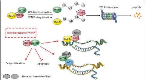
DLBCL cells seem to be in a stressed state, because of the presence of mutant proteins and rapid proliferation, which puts additional pressure on controlling proteosta-sis [10]. In this stage, molecular chaperones are essential, as they ensure that changes that affect proteins are
“buffered” to guarantee proteostasis and thus cellular homeostasis (Fig. 5). Thus, Hsp90 plays an important part in stabilization of client proteins. As we know, chaperone Hsp90 maintains the stability of many tumor-promoting oncoproteins [31]. Considering the elucidated oncogenic function of WTAP in DLBCLs, the complex with Hsp90 may indicate a target for DLBCL treatment. In case one is able to separate the client pro-teins from Hsp90, they will be degraded by the ubiquitin-proteasome pathway [31] (Fig. 5). Accordingly, we show in the present study that Hsp90 stabilizes WTAP as a protein. In addition, we determined that WTAP-Hsp90-BCL6 could form a protein complex in vivo and in vitro. Therefore, addressing Hsp90 by an in-hibitor would leave its many client proteins– including
BCL6 and WTAP – unprotected and prone to degradation.
Strikingly, WTAP mRNA was additionally reduced
upon Hsp90 inhibition. Since WTAP – via its associ-ation with the METTL3/14 heterodimer – impacts on RNA stability [23], we hypothesize that in malignant
DLBCL cells abundant WTAP protects WTAP mRNA
from degradation, while this positive feedback loop can be interrupted by Hsp90-directed drugs and successive WTAP ubiquitination and proteasomal degradation. Interestingly, a similar scenario has been documented for BCL6, also bound to Hsp90 [12], degraded upon Hsp90 inhibition and subsequent less abundant RNA as well. One might speculate the here reported trimer of WTAP-Hsp90-BCL6 to stabilize all RNAs, which possess ARE elements recognized by WTAP-METTL3/14.
BCL6 was initially discovered as a transcriptional re-pressor in B-cell lymphomas, in which it drives the ma-lignant phenotype by binding to hundreds of target genes, and then repressing these genes by recruiting sev-eral different chromatin modifying corepressor com-plexes to repress DNA damage checkpoints and block B-cell terminal differentiation [24]. Hsp90 could bind with BCL6 to perform as a transcriptional complex re-pressor, which has been verified by ChIP (Chromosome
immunoprecipitation) in previous studies. ATR, TP53 and CD69 are target genes of BCL6. Their messenger RNA abundance can be derepressed by PU-H71 in a time-dependent manner. In other words, Hsp90 may thus function as a corepressor for BCL6 by maintaining it in a stable conformation directly within the gene repressing complexes [12] (Fig. 5). Indeed, as a tran-scriptional repressor, BCL6 inhibits numerous target genes by recruiting co-repressors (most potently in a ternary complex with BCOR and SMRT) and stabilized by Hsp90 at regulatory gene elements [12,32,33]. BCL6 also regulates B-cell responses to chemokines and cyto-kines [32, 34], cell cycle control, either directly or indir-ectly, by repressing target genes or interacting with Miz1 to represses transcription of CDKN1 [35, 36]. BCL6 could form a complex or directly function as a transcrip-tion factor or an oncology protein.
Whether the here described complex of WTAP-Hsp90-BCL-6 is involved in transcriptional activity and/ or elicits other functions is still not known. Previous studies have shown WTAP could bind to Wilms’ tumor 1 (WT1), a transcription factor, forming a complex which affects transcriptional targets in smooth muscle cells (SMCs) [4]. Those targets have long been known to act as tumor suppressors in Wilms’ tumor in other
neoplasms, including lung, kidney, and breast carcinoma, as well as in leukemia like acute myeloid leukemia (AML) and myelodysplastic syndrome [37, 38]. How-ever, WT1 seems to play an opposite role to an onco-gene. This may be due to a two-sided function of WT1 promoting differentiation of cells in the genitourinary tract, while maintaining an immature mesenchymal state in other tissues [39]. In line, the complex of WTAP-WT1 is involved in Wnt signaling in colon cancer development [40]. In DLBCL, WT1-positive patients showed significantly worse overall and disease-free sur-vival under the same treatment protocols [41]. WTAP conforms complexes with–at least–two transcriptional factors, BCL6 and WT1, respectively. Whether Hsp90 is involved in both scenarios is unknown (Fig. 5). Maybe WTAP is primarily stabilized in the complex with Hsp90 and from this has the possibility to from alternate
com-plexes with WT1 and METTL3/14 to exert its
pro-tumorigenic potential. However, although the number of DLBCLs tissues examined in this study was not enough for generalization and more thorough studies in the future will clarify if high WTAP expression is a general feature of DLBCLs or describes a more aggressive subtype, we pro-claim in any case to consider Hsp90 inhibition to be in-cluded during chemotherapy if WTAP expression is high.
Additional file
Additional file 1:Table S1.The information of DLBCL samples. (DOC 30 kb)
Abbreviations
ABC:Activated B cell; BCL6: B-cell Lymphoma 6; DLBCL: Diffuse large B-cell lymphomas; GAPDH: Glyceraldehyde-3-phosphate dehydrogenase; GCB: Germinal Center B cell; Hsp90: Heat shock protein 90; WTAP: Wilms’ tumor 1-associating protein
Acknowledgements
This work was supported by the grant from the Nature Science Foundation of Zhejiang Province (Nos. Y17H160070), National Natural Science Foundation of China (No.81071937), Health and Family Planning Commission of Zhejiang Province (No.2018KY362). Prof. Chenfang Dong kindly provided pCMV-HA-UbiquitinC vector. We thank Prof. Andreas Jung from Ludwig-Maximilians-University Munic for his critical review of the manuscript.
Authors’contributions
YK and XG carried out the molecular genetic studies, participated in the sequence alignment. YK and RZ designed the study. YK, ZM, RZ drafted the manuscript and ZM, RZ also provide the research funds. FBS participated in writting. LD and FL carried out the immunoassays and statistics. LL, YG, QL and GR collected patients’samples. GR and QL read the slides and gave the diagnosis. DL and HT participated in the immunoassays. XZ, HP and YS made their comments for some experiment results. All authors read and approved the final manuscript.
Competing interests
The authors declare that they have no competing interests.
Publisher’s Note
Springer Nature remains neutral with regard to jurisdictional claims in published maps and institutional affiliations.
Author details
1Department of Pathology and Pathophysiology, Institute of Pathology and
Forensic Medicine, Zhejiang University School of Medicine, Hangzhou, China.
2
Department of Medical Oncology, Institute of Clinical Science, Sir Run Run Shaw Hospital, Zhejiang University School of Medicine, Hangzhou, China.
3Department of Pathology, the Second Hospital of Shaoxing, Shaoxing,
China.4Epitomics (Hangzhou) Inc, Hangzhou, China.5Department of
Orthopedics, the Second Affiliated Hospital, Zhejiang University School of Medicine, Hangzhou, China.6Department of Pathology, the First Affiliated
Hospital, Zhejiang University School of Medicine, Hangzhou, China.
7Department of Pathology, Sir Run Run Show Affiliated Hospital, Zhejiang
University School of Medicine, Hangzhou, China.8Institute of Pathology, University of Würzburg, 97080 Würzburg, Germany.
Received: 27 February 2018 Accepted: 7 August 2018
References
1. Little NA, Hastie ND, Davies RC. Identification of WTAP, a novel Wilms’ tumour 1-associating protein. Hum Mol Genet. 2000;9(15):2231–9. PubMed PMID: WOS:000089664400004
2. Horiuchi K, Umetani M, Minami T, Okayama H, Takada S, Yamamoto M, et al. Wilms’tumor 1-associating protein regulates G2/M transition through stabilization of cyclin A2 mRNA. Proc Natl Acad Sci U S A. 2006;103(46): 17278–83.https://doi.org/10.1073/pnas.0608357103. PubMed PMID: 17088532; PubMed Central PMCID: PMC1634838
3. Naruse C, Fukusumi Y, Kakiuchi D, Asano M. A novel gene trapping for identifying genes expressed under the control of specific transcription factors. Biochem Biophys Res Commun. 2007;361(1):109–15.https://doi.org/ 10.1016/j.bbrc.2007.06.161. PubMed PMID: 17644066
4. Small TW, Penalva LO, Pickering JG. Vascular biology and the sex of flies: regulation of vascular smooth muscle cell proliferation by wilms’tumor 1-associating protein. Trends Cardiovasc Med. 2007;17(7):230–4.https://doi. org/10.1016/j.tcm.2007.08.002. PubMed PMID: 17936204
5. Small TW, Pickering JG. Nuclear degradation of Wilms tumor 1-associating protein and survivin splice variant switching underlie IGF-1-mediated survival. J Biol Chem. 2009;284(37):24684–95.https://doi.org/10.1074/jbc. M109.034629. PubMed PMID: 19605357; PubMed Central PMCID: PMC2757172
6. Jin DI, Lee SW, Han ME, Kim HJ, Seo SA, Hur GY, et al. Expression and roles of Wilms’tumor 1-associating protein in glioblastoma. Cancer Sci. 2012; 103(12):2102–9.https://doi.org/10.1111/cas.12022. PubMed PMID: 22957919 7. Jo HJ, Shim HE, Han ME, Kim HJ, Kim KS, Baek S, et al. WTAP regulates
migration and invasion of cholangiocarcinoma cells. J Gastroenterol. 2013; 48(11):1271–82.https://doi.org/10.1007/s00535-013-0748-7. PubMed PMID: 23354623
8. Bansal H, Yihua Q, Iyer SP, Ganapathy S, Proia D, Penalva LO, et al. WTAP is a novel oncogenic protein in acute myeloid leukemia. Leukemia. 2014;28(5): 1171–4.https://doi.org/10.1038/leu.2014.16. PubMed PMID: WOS: 000336661500031
9. Isaacs JS, Xu WP, Neckers L. Heat shock protein 90 as a molecular target for cancer therapeutics. Cancer Cell. 2003;3(3):213–7.https://doi.org/10.1016/ S1535-6108(03)00029-1. PubMed PMID: WOS:000181951400005
10. Wandinger SK, Richter K, Buchner J. The Hsp90 chaperone machinery. J Biol Chem. 2008;283(27):18473–7.https://doi.org/10.1074/jbc. R800007200. PubMed PMID: WOS:000257165600001
11. Abramson JS, Chen W, Takahashi H, Juszczynski P, Kutok JL, Shipp MA. Heat shock protein 90 (HSP90) is a rational therapeutic target in diffuse large B-cell lymphoma. Blood. 2006;108(11):249a. PubMed PMID: WOS:
000242440001089
12. Cerchietti LC, Lopes EC, Yang SN, Hatzi K, Bunting KL, Tsikitas LA, et al. A purine scaffold Hsp90 inhibitor destabilizes BCL-6 and has specific antitumor activity in BCL-6-dependent B cell lymphomas. Nat Med. 2009; 15(12):1369–U3.https://doi.org/10.1038/nm.2059. PubMed PMID: WOS: 000272407800016
13. Shi YY, Kuai Y, Lei LZ, Weng YY, Berberich-Siebelt F, Zhang XX, et al. The feedback loop of LITAF and BCL6 is involved in regulating apoptosis in B cell non-Hodgkin’s-lymphoma. Oncotarget. 2016;7(47):77444–56.https://doi. org/10.18632/oncotarget.12680. PubMed PMID: WOS:000389633400090 14. Siegel R, Naishadham D, Jemal A. Cancer statistics, 2013. Ca-Cancer J Clin.
15. Wang WG, Cui WL, Wang L, Zhu F, Wan XC, Ping B, et al. Loss of B-cell Receptor Expression Defines a Subset of Diffuse Large B-cell Lymphoma Characterized by Silent BCR/PI3K/AKT Signaling and a Germinal Center Phenotype Displaying Low-risk Clinicopathologic Features. Am J Surg Pathol. 2015;39(7):902–11.https://doi.org/10.1097/Pas.0000000000000396. PubMed PMID: WOS:000356944900003
16. Alizadeh AA, Eisen MB, Davis RE, Ma C, Lossos IS, Rosenwald A, et al. Distinct types of diffuse large B-cell lymphoma identified by gene expression profiling. Nature. 2000;403(6769):503–11.https://doi.org/10.1038/35000501. PubMed PMID: 10676951
17. Rosenwald A, Wright G, Chan WC, Connors JM, Campo E, Fisher RI, et al. The use of molecular profiling to predict survival after chemotherapy for diffuse large-B-cell lymphoma. N Engl J Med. 2002;346(25):1937–47.https:// doi.org/10.1056/NEJMoa012914. PubMed PMID: 12075054
18. Shaffer AL, Rosenwald A, Staudt LM. Lymphoid malignancies: the dark side of B-cell differentiation. Nat Rev Immunol. 2002;2(12):920–32.https://doi.org/ 10.1038/nri953. PubMed PMID: 12461565
19. Lenz G, Staudt LM. Aggressive lymphomas. N Engl J Med. 2010;362(15): 1417–29.https://doi.org/10.1056/NEJMra0807082. PubMed PMID: 20393178 20. Valbuena JR, Rassidakis GZ, Lin P, Atwell C, Georgakis GV, Younes A, et al.
Expression of heat-shock protein-90 in non-Hodgkin’s lymphomas. Modern Pathol. 2005;18(10):1343–9.https://doi.org/10.1038/modpathol.3800459. PubMed PMID: WOS:000232145600009
21. Vardiman JW. The World Health Organization (WHO) classification of tumors of the hematopoietic and lymphoid tissues: An overview with emphasis on the myeloid neoplasms. Chem-Biol Interact. 2010;184(1–2):16–20.https://doi. org/10.1016/j.cbi.2009.10.009. PubMed PMID: WOS:000276877800004 22. Yu F, Xie D, Ng SS, Lum CT, Cai MY, Cheung WK, et al. IFITM1 promotes the
metastasis of human colorectal cancer via CAV-1. Cancer Lett. 2015;368(1): 135–43.https://doi.org/10.1016/j.canlet.2015.07.034. PubMed PMID: WOS: 000361265500017
23. Scholler E, Weichmann F, Treiber T, Ringle S, Treiber N, Flatley A, et al. Interactions, localization, and phosphorylation of the m (6) a generating METTL3-METTL14-WTAP complex. RNA. 2018;24(4):499–512.https://doi.org/ 10.1261/rna.064063.117. PubMed PMID: 29348140; PubMed Central PMCID: PMC5855951
24. Cardenas MG, Oswald E, Yu W, Xue F, MacKerell AD Jr, Melnick AM. The expanding role of the BCL6 Oncoprotein as a Cancer therapeutic target. Clin Cancer Res. 2017;23(4):885–93. https://doi.org/10.1158/1078-0432.CCR-16-2071. PubMed PMID: 27881582; PubMed Central PMCID: PMC5315622 25. Ci WM, Polo JM, Melnick A. B-cell lymphoma 6 and the molecular
pathogenesis of diffuse large B-cell lymphoma. Curr Opin Hematol. 2008; 15(4):381–90.https://doi.org/10.1097/Moh.0b013e328302c7df. PubMed PMID: WOS:000257379600016
26. Horiuchi K, Kawamura T, Iwanari H, Ohashi R, Naito M, Kodama T, et al. Identification of Wilms’tumor 1-associating protein complex and its role in alternative splicing and the cell cycle. J Biol Chem. 2013;288(46):33292–302.
https://doi.org/10.1074/jbc. M113.500397. PubMed PMID: 24100041; PubMed Central PMCID: PMC3829175
27. Liu J, Yue Y, Han D, Wang X, Fu Y, Zhang L, et al. A METTL3-METTL14 complex mediates mammalian nuclear RNA N6-adenosine methylation. Nat Chem Biol. 2014;10(2):93–5.https://doi.org/10.1038/nchembio.1432. PubMed PMID: 24316715; PubMed Central PMCID: PMC3911877
28. Ping XL, Sun BF, Wang L, Xiao W, Yang X, Wang WJ, et al. Mammalian WTAP is a regulatory subunit of the RNA N6-methyladenosine methyltransferase. Cell Res. 2014;24(2):177–89.https://doi.org/10.1038/cr. 2014.3. PubMed PMID: 24407421; PubMed Central PMCID: PMC3915904 29. Schwartz S, Mumbach MR, Jovanovic M, Wang T, Maciag K, Bushkin GG, et
al. Perturbation of m6A writers reveals two distinct classes of mRNA methylation at internal and 5’sites. Cell Rep. 2014;8(1):284–96.https://doi. org/10.1016/j.celrep.2014.05.048. PubMed PMID: 24981863; PubMed Central PMCID: PMC4142486
30. Sugiyama H. WT1 (Wilms’tumor gene 1): biology and cancer immunotherapy. Jpn J Clin Oncol. 2010;40(5):377–87.https://doi.org/10. 1093/jjco/hyp194. PubMed PMID: 20395243
31. Whitesell L, Lindquist SL. HSP90 and the chaperoning of cancer. Nat Rev Cancer. 2005;5(10):761–72.https://doi.org/10.1038/nrc1716. PubMed PMID: 16175177
32. Basso K, Saito M, Sumazin P, Margolin AA, Wang K, Lim WK, et al. Integrated biochemical and computational approach identifies BCL6 direct target genes controlling multiple pathways in normal germinal center B cells.
Blood. 2010;115(5):975–84.https://doi.org/10.1182/blood-2009-06-227017. PubMed PMID: 19965633; PubMed Central PMCID: PMC2817639 33. Hatzi K, Jiang Y, Huang C, Garrett-Bakelman F, Gearhart MD, Giannopoulou
EG, et al. A hybrid mechanism of action for BCL6 in B cells defined by formation of functionally distinct complexes at enhancers and promoters. Cell Rep. 2013;4(3):578–88.https://doi.org/10.1016/j.celrep.2013.06.016. PubMed PMID: 23911289; PubMed Central PMCID: PMC3854650 34. Basso K, Dalla-Favera R. Roles of BCL6 in normal and transformed germinal
center B cells. Immunol Rev. 2012;247(1):172–83.https://doi.org/10.1111/j. 1600-065X.2012.01112.x. PubMed PMID: 22500840
35. Ci WM, Polo JM, Cerchietti L, Shaknovich R, Wang L, Yang SN, et al. The BCL6 transcriptional program features repression of multiple oncogenes in primary B cells and is deregulated in DLBCL. Blood. 2009;113(22):5536–48.
https://doi.org/10.1182/blood-2008-12-193037. PubMed PMID: WOS: 000266634700045
36. Phan RT, Saito M, Basso K, Niu H, Dalla-Favera R. BCL6 interacts with the transcription factor Miz-1 to suppress the cyclin-dependent kinase inhibitor p21 and cell cycle arrest in germinal center B cells. Nat Immunol. 2005;6(10): 1054–60.https://doi.org/10.1038/ni1245. PubMed PMID: 16142238 37. Call KM, Glaser T, Ito CY, Buckler AJ, Pelletier J, Haber DA, et al. Isolation And
Characterization Of a Zinc Finger Polypeptide Gene at the Human Chromosome-11 Wilms Tumor Locus. Cell. 1990;60(3):509–20.https://doi. org/10.1016/0092-8674(90)90601-A. PubMed PMID: WOS:A1990CP23700017 38. Miwa H, Beran M, Saunders GF. Expression Of the Wilms-Tumor Gene (Wt1)
In Human Leukemias. Leukemia. 1992;6(5):405–9. PubMed PMID: WOS: A1992JB14100009
39. Hohenstein P, Hastie ND. The many facets of the Wilms’tumour gene, WT1. Hum Mol Genet. 2006;15(Suppl_2):R196–201.https://doi.org/10.1093/hmg/ ddl196. PubMed PMID: 16987884
40. Zhang JW, Tsoi H, Li XX, Wang H, Gao J, Wang KN, et al. Carbonic anhydrase IV inhibits colon cancer development by inhibiting the Wnt signalling pathway through targeting the WTAP-WT1-TBL1 axis. Gut. 2016; 65(9):1482–93.https://doi.org/10.1136/gutjnl-2014-308614. PubMed PMID: WOS:000381274700013