© 2019 by the Serbian Biological Society 711
Different levels of epidermal growth factor signaling modifies the differentiation of
specific cell types in mouse postnatal retina
Sanja Ivković1,*, Irena Jovanović Macura1, Tijana Antonijević1, Selma Kanazir1 and Domingos Henrique2
1Department for Neurobiology, Institute for Biological Research “Siniša Stanković” - National Institute of Republic of Serbia,
University of Belgrade, Bulevar despota Stefana 142, 11060 Belgrade, Serbia
2Instituto de Medicina Molecular, Faculdade de Medicina da Universidade de Lisboa, Avenida Professor Egas Moniz,
1649-028 Lisbon, Portugal
*Corresponding author: [email protected]
Received: June 17, 2019; Revised: August 22, 2019; Accepted: September 4, 2019; Published online: September 5, 2019
Abstract: Epidermal growth factor (EGF) signaling has been implicated in the regulation of the differentiation and pro-liferation of retinal progenitors. We assessed how different levels of EGF signaling, achieved either by increasing receptor expression or via addition of the exogenous ligand, or an increase in both, can affect the differentiation of progenitors in the first week of postnatal retinal development in the model system of retinal explants (REs). Proliferating progenitor cells in REs were infected with either the control CLV3/ESR-related peptide family (CLE)-green fluorescent protein (GFP)- or with EGF receptor (EGFR)-GFP-expressing retrovirus, and grown in the control medium or in the presence of exogenous EGF (10 ng/mL). The differentiation of infected cells into Müller glia (Sox9+), rod photoreceptors (rhodopsin+) and
hori-zontal cells (calbindin+) was analyzed. In all the examined conditions, infected cells differentiated into Müller glia and
rod photoreceptors that normally develop postnatally. Horizontal cells finished their development during the embryonic stages and progenitors infected with control-GFP virus did not differentiate into GFP+/calbindin- in either control or
EGF-supplemented medium, however, cells infected with EGFR-GFP differentiated into horizontal cells (GFP+/calbindin+) in
both culture conditions. These results imply that altering the levels of EGFR and/or the amount of the EGF ligand can overcome progenitor competence restriction.
Keywords: retina; EGFR; progenitors; differentiation; postnatal development
How to cite this article: Ivković S, Jovanović-Macura I, Antonijević T, Kanazir S, Henrique D. Different levels of epidermal growth factor signaling modifies the differentiation of specific cell types in mouse postnatal retina. Arch Biol Sci. 2019;71(4):711-9.
INTRODUCTION
The vertebrate neural retina is comprised of six types of neurons: rod and cone photoreceptors, amacrine cells, retinal ganglion cells (RGCs), horizontal cells, bipolar cells and one type of glia, Müller glia. These cell types are all derived from one population of prolif-erating multipotent progenitors [1] during embryonic and postnatal development through sequential and tightly regulated differentiation steps involving both cell intrinsic and extrinsic factors [2]. The seemingly homogenous populations of progenitor cells differ in their ability to respond to gradients of multiple extra-cellular signals present in the developing tissue.
Epidermal growth factor (EGF) signaling is one of the signaling pathways that regulate retinal devel-opment. Different cellular response to EGF can be
achieved either by a different concentration of ligand or with graded activation of receptors, accomplished by regulating ligand levels, as in the case of Drosophila
neuronal differentiation [9]. EGF in postnatal retinal development has thus far been closely investigated as a proliferation and differentiation factor for Müller glia [8,10]. Nevertheless, several lines of investigation have revealed that EGFR signaling has a nonmitotic function and is involved in cell fate decisions [10-12]. Studies on retinal development showed that the intro-duction of additional EGFR into progenitor cells in retinal explant cultures did not enhance proliferation but induced an increase in the proportion of clones that contain Müller glial cells [10]. These results sug-gest that receptor level is usually the limiting factor.
Different manipulations of the level of receptor expression in vivo in the brain and retina showed that if the critical level of active receptor is present on the cell surface it can allow sustained activation of intracellular signal transduction and change the properties and potential of such a progenitor cell [10,13-15]. These findings demonstrate that respon-siveness to extracellular signals during development can be modulated by the introduction of additional receptors and/or ligands and suggest that the level of expression of receptors for these signals contributes to the regulation of cell fate.
It is unclear how restriction is the specific devel-opmental stage that affects the competence potential of progenitor cells and whether competence can be altered by modifications of the levels of EGF signaling. Higher levels of EGFR-mediated signaling alone do not specify glial fate, indicating that the competence to generate glia is temporally regulated by additional mechanisms [10]. In the present study, we analyzed how altering the levels of EGF
signal-ing with the addition of extra EGFR via retroviral infection or by addition of exogenous EGF ligand or both, affects cell fate choice of P0 postna-tal retinal progenitors. By reducing the discrepancy in EGFR expression among early and late progenitor cells, additional changes in progenitor cells that regulate their competence to generate glia were revealed [14]. We showed that altered levels of EGF signaling enabled postnatal proliferat-ing progenitors to develop into neu-ronal cell types whose differentiation
is completed during embryonic development, and that they do not normally differentiate postnatally. These data indicate that modifying the levels of EGF signal-ing can change the competence of postnatal retinal progenitors.
MATERIALS AND METHODS
Ethics statement
Animal experiments were approved by the Animal Ethics Committee of Instituto de Medicina Molecular (AEC_027_2010_DH_Rdt_general_IMM) (Lisbon, Portugal) and according to National Regulations. Retrovirus production
EGFR-GFP and CLE-GFP plasmids were generated by Sun et al. [16] from the original pCLE retroviral vector [17] and were a kind gift from Dr. Sally Temple (University at Albany, State University of New York, Albany, NY). Replication-deficient viruses with vsv-G coats were generated from these constructs as de-scribed previously [18]. Briefly, viral titers were deter-mined in colony-forming units (CFUs) by incubating C6 glioma cells with serial dilutions of retrovirus in 10 steps. At 48 h post-infection, the number of GFP+
cell clusters was counted. The CFUs were calculated by multiplying the number of GFP+ cell clusters by
the dilution factor. The titer of both the CLE-GFP and EGFR-GFP viruses was 106/µL. Viruses were added to
explants immediately after culturing.
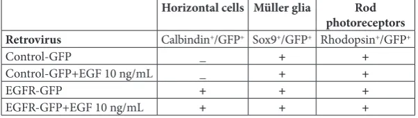
Table 1. Schematic presentation of the differentiation potential of retinal progeni-tors in 7-DIV REs in different experimental paradigms. P0 progeniprogeni-tors infected with control-GFP and EGFR-GFP retrovirus differentiate into Müller glia and rod photore-ceptors in either control or EGF-supplemented medium. P0 progenitors infected with EGFR-GFP and grown in either control or EGF-supplemented medium differentiate into horizontal cells, while the progenitors infected with control-GFP do not.
Horizontal cells Müller glia Rod
photoreceptors
Retrovirus Calbindin+/GFP+ Sox9+/GFP+ Rhodopsin+/GFP+
Control-GFP _ + +
Control-GFP+EGF 10 ng/mL _ + +
EGFR-GFP + + +
EGFR-GFP+EGF 10 ng/mL + + +
Mouse strain and sample collection
C57BL/6 mice were fed ad libitum and housed in spe-cific-pathogen-free (SPF) facilities. For the production of P0 pups, three female mice were housed with one male mouse and the date of the vaginal plug formation was established in order to ensure the precise timing of the birth. At birth (P0), the eyes were enucleated (the eye balls were removed with curved forceps and the optic nerve was severed) from P0 animals (n=18) after decapitation and processed for culturing. Eyes used for the same experimental condition were never taken from the same animal.
In vitro culture of retinal explants (REs)
The enucleated eyes were transferred to a Petri dish containing Dulbecco’s modified Eagle’s medium (DMEM) (Sigma, St. Louis, USA) supplemented with 50 IU (μg)/mL penicillin-streptomycin (Invitrogen, Paisley, UK).Retinas were removed and cultured as described [19]. Briefly, the retinas were placed on membrane culture inserts (Millicell CM, Millipore, Bedford, MA, USA; pore size 0.4 μm) in 6-well plates (vitreous side down), and cultured in a culture me-dium modified from [19], in 50% minimal essential medium (MEM)-HEPES (Invitrogen, San Diego, CA, USA), 25% Hank’s balanced salt solution (Invitrogen), 5.75 mg/mL glucose. 25 U/mL penicillin, 25 mg/mL streptomycin, 200 mM L-glutamine, 1x Gibco B27 (Thermo Fischer Scientific, USA) and 1x Gibco N2 (Thermo Fischer Scientific)).
Treatments of retinal explants
EGF-treated explants were cultured for 7 days in vitro
(DIV) in medium containing 10 ng/mL EGF (Sigma), and the medium was changed every other day. Control explants were cultured in normal culture medium. At 7 DIV, the REs were fixed in 4% paraformaldehyde (PFA) and processed for immunofluorescence (IF). Retroviral infection
After the retinas were placed in a culture dish, 5 µL of either the control CLE-GFP or EGFR-GFP-expressing retrovirus was added in drops on the upper surface of the explants. The explants were left on 37oC and
the media was changed as described. Two-4 different retroviral infections were performed for each experi-mental paradigm analyzed.
Immunofluorescence
Eyes and retinal explants were fixed in 4% paraform-aldehyde at 4°C overnight, cryoprotected in 30% sucrose and embedded in 7.5% gelatin; 15% sucrose and 12 µm sections were used in the analysis. For IF, sections were degelatinized at 37°C for 15 min in 1x phosphate buffer solution (PBS) and permeabilized using Triton X-100 (0.5%) for 15 min. This was fol-lowed by blocking (10% normal goat serum, 0.1% TritonX-100) for 1 h at room temperature (RT) The primary antibodies used in this study were as follows: mouse anti-transcription factor Sox9 (1:500, DSHB), mouse anti-calbindin (1:500, Sigma), mouse anti-rho-dopsin (1:500, DSHB) and chicken anti-GFP (1:1000, Abcam). The sections were incubated in the primary antibodies overnight at 4°C, washed and incubated with appropriate Alexa Fluor (488 or 594) conjugated secondary antibodies (1:400, Molecular Probes) for 1 h at RT.
Imaging and analysis
Images were obtained with a Leica DM5000B mi-croscope (Leica, Wetzlar, Germany). The findings reported as microscopic images were representative of observations performed in two-to-four REs.
RESULTS
Retroviral infection and culturing of retinal explants
divid-ing progenitors via the EGFR-GFP retrovirus (Sup-plementary Fig. S1A, B) immediately after culturing. To understand how different levels of EGF signaling affect the development of specific cell types in postna-tal retina, we cultured P0 REs under four different con-ditions: (i) progenitors infected with control-GFP and grown in the control media; (ii) progenitors infected with control-GFP and grown in the presence of the exogenous EGF (10 ng/mL); (iii) progenitors infected with EGFR-GFP and grown in the control medium; (iv) progenitors infected with EGFR-GFP and grown in the presence of exogenous EGF (10 ng/mL) (the schematic overview of the experimental paradigm is presented in Supplementary Fig. S1A). The continuous presence of the receptor in conditions with and without added EGF provided graded levels of EGF signaling and established the experimental paradigm in which we could compare the outcomes of the increasing levels of EGF signaling. Retroviral infection allowed us to follow the destiny of the infected progenitors (GFP+
cells) at 7 DIV (Supplementary Fig. S1C).
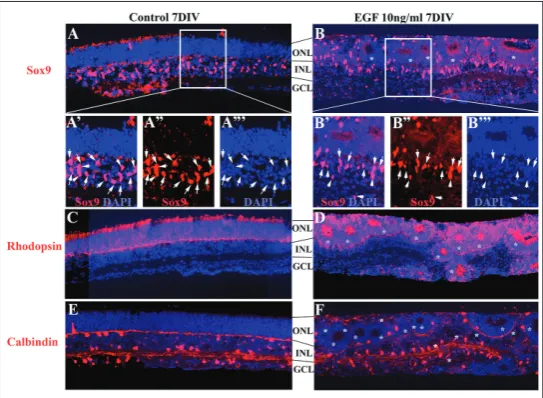
The addition of the exogenous EGF did not alter the development of Müller glia, rod photoreceptors and horizontal cells
REs were isolated from P0 eyes and cultured either in the control medium (Fig. 1A, A’, A’’, C, E), or in the presence of exogenous EGF (10 ng/mL) (Fig. 1B, B’ B’’, D, F). After 7 days, the REs were analyzed for the expression of cell type specific markers such as Sox9, the marker for Müller glia, calbindin, the marker for horizontal cells and rhodopsin, the marker for rod photoreceptors. Horizontal cells appear during em-bryonic development, and rods and Müller glia are the cell types that appear postnatally [20]. The pres-ence of EGF led to the formation of rosettes in the outer nuclear layer (ONL) (Fig. 1B, D, E, asterisk) but the general pattern of expression was preserved for all the markers analyzed. In control explant nuclei of Sox9+, the cells were distributed in the inner nuclear
layer (INL), forming a ribbon bordering the basal part of the outer nuclear layer (ONL) (Fig. 1A, A’, A’’). A similar pattern was observed in the EGF-treated REs where the Sox9+ cell distribution followed
the rosette organization marking the basal border of the ONL (Fig. 1B, B’, B’’). In the control REs, rhodopsin staining labeled the rod photoreceptors in the ONL (Fig. 1C). Similarly, in EGF-treated REs, rhodopsin staining delineated the ONL organized in rosettes (Fig. 1D). calbindin+ cells were
dis-tributed throughout the INL, with nuclei located in the apical and basal border of the INL in control REs (Fig. 1E). In the EGF-treated Res, a similar pattern of calbindin+
cells assuming a rosette organization was present as well (Fig. 1F).
P0 progenitors differentiate into Müller glia in the presence of different levels of EGF signaling
Müller glia are the only glial cell type in the retina and develop primarily during the first postnatal week. It has been shown that premature Müller cell differentiation is normally achieved by increased expres-sion of EGFR and/or exposure to high local concentrations of ligand [10]. We analyzed
Fig. 1. The addition of exogenous EGF did not alter the development of
Müller glia, rod photoreceptors and horizontal cells. Micrographs of 7DIV REs showing the organization and general pattern of expression of Müller glia (Sox9+, red) (A, B), rod photoreceptors (rhodopsin+, red) (C, D) and
horizontal cells (calbindin+, red) (E, F) in control and EGF-treated REs. The
if the virus-infected cells differentiate into Müller glia in all the conditions examined. Double-labeling with anti-Sox9 and anti-GFP antibodies revealed double-labeled cells (Fig. 2, arrows) in all conditions. Pro-genitors infected with the control virus developed into Sox9+ cells (Müller glia) in the absence or presence of
exogenous EGF (Fig. 2A, B, arrows). Similarly, pro-genitors infected with the EGFR-GFP virus differenti-ated into Müller glia in both control media and in the presence of exogenous EGF (Fig. 2C, D). However, in REs infected with the control virus, some infected cells (GFP+) were Sox9 negative (-), indicating that
infected progenitors were capable of differentiating into other cell types in the presence or absence of EGF (Fig. 2A, B, arrowheads). Similarly, some progenitors infected with the EGFR-GFP virus and grown with or without EGF were Sox9-, indicating that these cells
can adopt fates other than Müller glia even with con-tinuous EGFR expression (Fig. 2C, D, arrowheads). Thus, different levels of EGF signaling did not inhibit control and EGFR-infected progenitor cells from
dif-ferentiating into Müller glia during the first postnatal week in retinal development, but did not exclusively drive progenitors into the glial fate.
P0 progenitors differentiate into rod photoreceptors in the presence of different levels of EGF signaling
The other cell types whose differentiation peaks during the first postnatal week are rod photoreceptors. The normal fate of P0 pro-genitor cells in rat retina in vivo is to devel-op predominantly into rod photoreceptors [21]. We examined whether P0 progenitor differentiation in rods (rhodopsin+) is
af-fected by different levels of EGF signaling. Double-labeling with anti-rhodopsin and anti-GFP antibodies revealed the presence of double-labeled cells (Fig. 3, arrows) in all of the conditions. Progenitors infected with the control virus developed into rhodopsin+
cells (rod photoreceptors) in the absence and in the presence of exogenous EGF (Fig. 3A, B, arrows). Similarly, progenitors infected with the EGFR-GFP virus differentiated into rhodopsin+/GFP+ cells in both control media
and in the presence of exogenous EGF (Fig. 3C, D). In all the conditions examined, the bulk of progenitors, infected with either control or EGFR-GFP virus, developed into rhodopsin- cells (arrowheads).
Thus, different levels of EGF signaling did not inhibit the expression of rhodopsin in the infected, GFP+ cells
during the first postnatal week of retinal development. Increased levels of EGF signaling permit
postnatal differentiation of progenitors into horizontal cells
Horizontal cells are retinal interneurons that appear early on, from embryonic day 11 (E11), and they fin-ish their differentiation by E18. We examined whether different levels of EGF signaling affected the develop-ment of cell types whose differentiation process was finalized before birth, during embryonic development. We sought to determine how different experimental paradigms affect the differentiation of infected pro-genitors into horizontal cells (calbindin+) [22] in all
analyzed experimental paradigms. Double-labeling with anti-calbindin and anti-GFP antibodies revealed no double-labeled cells in REs infected with control vi-rus, with or without the addition of the exogenous EGF (Fig. 4A, B), as was expected. However, a proportion of P0 progenitors infected with EGFR-GFP virus were calbindin+/GFP+ in REs grown in control medium (Fig.
4C’, C’’, C”’, arrows). Similarly, REs infected with EGFR-GFP and grown with the addition of the exogenous EGF resulted in the presence of double-labeled, cal-bindin+/GFP+, cells (Fig. 4D’’, arrows). Therefore, the
continuous expression of EGFR in progenitor cells, either in control media or in the presence of exogenous EGF, permitted for the differentiation of horizontal cells in postnatal REs.
DISCUSSION
The major signaling pathways and their molecular constituents are evolutionarily highly conserved.
The morphogen, any diffusible signaling molecule, can, upon reaching the receiving cell, induce multiple different cell fates de-pending on its levels of expression. On the other hand, a number of receptors in the receiving cell can regulate different signal-ing thresholds and consequently, different differentiation outcomes. Therefore, the quantitative nature of signaling has raised a variety of questions [23-27]. Similarly, the quantitative nature of EGF signaling in development has been addressed in several studies and in different experimental mod-els [28-29]. The introduction of extra EGFR
in vivo increased the proportion of clones that contained Müller glial cells suggesting that receptor levels are normally limited [10]. The introduction of extra EGFR into ventricular zone (VZ) cells in the brain via retroviral infection resulted in premature expression of features characteristic of late SVZ progenitors [13]. Similarly, the intro-duction of EGFR via retroviral infection into oligodendrocyte progenitors hindered their final differentiation and extended their migratory behavior [15]. Activation of EGFR in retinal precursors can regulate proliferation and differentiation in vitro and
in vivo [7,8,10,14,30,31]. These studies indi-cate that the level of EGFRs expressed by progenitor cells in the brain cortex and retina can contribute to the timing of their maturation and choice of response to pleiotropic environmental signals.
Retinal progenitor proliferation peaks around the day of birth and declines until about the end of the first postnatal week [20,32]. Cell division in postnatal retina ceases by P5-P6 days in the center of the retina, and by P11 in the periphery. Among cells produced postnatally, 73% differentiate as rods, 20% as bipolar cells, 6% as Müller cells and 1% as amacrine and gan-glion cells [20]. In our study, P0 progenitors were in-fected with control and EGFR virus and grown either in control media or media supplemented with exogenous EGF (10 ng/mL), forming experimental groups with different levels of EGF signaling. This concentration was chosen in order to avoid overstimulation of pro-genitor proliferation and to allow for biased differentia-tion, as reported previously [10,13]. The competence
Fig. 3. P0 progenitors differentiate into rod photoreceptors in the presence of different levels of EGF signaling.Micrographs of7DIV REs infected with control-GFP (A, B) or EGFR-GFP (C, D) retrovirus were analyzed for the expression of GFP (green) in rod photoreceptors (Rhodopsin+, red) in dif-ferent experimental conditions. A – REs infected with control-GFP virus and grown in control media. B – REs infected with control-GFP and grown in the presence of EGF. C – REs infected with EGFR-GFP and grown in control media. D – REs infected with EGFR-GFP and grown in the presence of EGF. Arrows indicate double-labeled GFP+/rhodopsin+ cells. Arrowheads indicate
GFP+/rhodopsin- cells. The results are presented as lower magnification (20x)
of P0 progenitor cells to differentiate into Müller glia (Sox9+) and rod photoreceptors (rhodopsin+) cells was
not affected by different levels of EGF signaling. It is possible that the number of Sox9+ and rhodopsin+ cells
was different between different conditions as a result of the effect that different levels of EGF signaling can have on proliferation. However, our study was focused on the influence of different levels of EGF signaling on progenitor competence, and consequently on their final differentiation outcome. Surprisingly, P0 progenitors infected with EGFR virus developed into horizontal cells (calbindin+) with or without the presence of
exog-enous EGF, while progenitors infected with the control virus did not. This suggested that the addition of extra receptor permitted the infected progenitors to differen-tiate into earlier fates that normally differendifferen-tiate during the embryonic period, such as horizontal cells.
Horizontal cells, together with amacrine cells, lie within the INL and modulate signaling between
pho-toreceptors and bipolar cells. They exhibit a variety of unique biological properties such as unusual migratory behavior, unique mor-phological plasticity and the ability to divide at a relatively late stage during development [33]. Finally, data indicating that fully dif-ferentiated horizontal cells can give rise to metastatic retinoblastoma challenge the assumption that tumors are derived solely from progenitor/stem-like cells [34]. It is possible that P0 progenitors with forced expression of EGFR regain the competence to differentiate into horizontal cells in the time frame generally not permissive for this cell fate choice. EGFR+/calbindin+ cells
pre-sent in REs grown without exogenous EGF indicated that the receptor is the limiting factor and that there is enough endogenous ligand in the explant to sustain the signal-ing, although it is possible that the addition of extra EGF affected the proliferation of EGFR+/calbindin+ cells. As the horizontal
cells have the ability to divide in a fully differentiated state [34], it is possible that under specific conditions of RE culturing, these cells entered the cycle and were thus capable of being infected with the retro-virus. However, this seems unlikely as we could not detect any GFP+/calbindin+ cells
in REs infected with the control virus in the presence of exogenous EGF. A more likely scenario is that some of the EGFR+ P0 progenitors recapitulated
the embryonic developmental program, resulting in the expression of the horizontal cell-specific marker.
The presence of continuous expression of EGFR in infected cells did not hinder the expression of the markers of final cell fates – Sox9, rhodopsin and calbindin. Moreover, the addition of the exogenous EGF also allowed for the expression of these markers, indicating that the increased level of EGF signaling is not sufficient to maintain these progenitors in the undifferentiated state. Several studies have suggested that progenitor cell competence to respond to specific environmental signals can either be lost or acquired during development. For example, competence to di-vide in response to TGFα was acquired between E15 and E18 [7], as was competence to respond to signals that promote rod development [35]. Thus, altering the
Fig. 4. The increased levels of EGF signaling permit postnatal progenitor differ-entiation into horizontal cells. Micrographs of7DIV REs infected with control-GFP (A, B) or EGFR-GFP (C, D) retrovirus were analyzed for theexpression of GFP (green) in horizontal cells (calbindin+, red) in different experimental
conditions. A – REs infected with control-GFP virus and grown in control media. B – REs infected with control-GFP and grown in the presence of EGF. C
– REs infected with EGFR-GFP and grown in control media. D – REs infected with EGFR-GFP and grown in the presence of EGF. Arrows indicate double-labeled GFP+/calbindin+ cells. The results are presented as lower magnification
levels of EGF signaling cannot extend the time frame of progenitor competence indefinitely.
Horizontal cells form an extensive network that allows for the communication between photoreceptors and bipolar cells. The ablation of horizontal cells led to rod photoreceptor degeneration and induced ex-tensive retinal network remodeling [36]. It is thus pos-sible that in the case of photoreceptor degeneration, the ability to induce horizontal cell differentiation de novo and manipulate the levels of EGF signaling can facilitate the repair of retinal circuits or even enable photoreceptor regeneration. This is of particular im-portance because one of the main problems related to photoreceptor transplantation and regeneration is their inability to become incorporated into the exist-ing retinal network and become fully functional.
The determination and differentiation of hetero-geneous cell types within the context of complex tis-sues is the culmination of the expression of many gene products and their subsequent intra- and intercellular signaling events. Fully understanding the mechanisms underlying these processes is fundamental to many ar-eas of biology. This knowledge will have widespread ap-plication in the treatments of developmental disorders and diseases, such as cancer, and will be critical for the successful bioengineering and transplantation of tissue types to replace damaged or degenerated structures.
Funding: This work was funded by Marie Curie Welcome II
fel-lowship (2011-2015) and Fundação para a Ciência e Tecnologia (www.fct.mctes.pt). The funders had no role in study design, data collection and analysis, decision to publish or preparation of the manuscript.
Author contributions: SI conducted the experimental research and presented the obtained results. She supervised all phases of the research and reviewed the manuscript. IJM and TA performed the literature search, prepared all the images and critically re-viewed the manuscript. SK and DH supervised the research and the preparation of the manuscript. All authors read and approved the final manuscript.
Conflict of interest disclosure: The authors have no conflicts of interest to declare,
REFERENCES
1. Cepko CL, Austin CP, Yang X, Alexiades M, Ezzedine D. Cell fate determination in the vertebrate retina. Proc Natl Acad Sci U S A. 1996;93:589-95.
2. Livesey R, Cepko C. Vertebrate neural cell-fate determination: lessons from the retina. Nat. Rev. Neurosci. 2001;2:109-18. 3. Raz E, Shilo B-Z. Establishment of ventral cell fates in the
Drosophila embryonic ectoderm requires DER, the EGF receptor homolog. Genes Dev. 1993;7:1937-48.
4. Diaz-Benjumea FJ, Hafen E. The sevenless signaling cassette mediates Drosophila EGF receptor function during epider-mal development. Development. 1994;120:569-78.
5. Schweitzer R, Shaharabany M, Seger R, Shilo B-Z. Secreted Spitz triggers the DER signaling pathway and is a limiting component in embryonic ventral ectoderm determination. Genes Dev. 1995;9:1518-29.
6. Katz WS, Hill RJ, Clandinin TR, Sternberg PW. Different levels of the C. elegans growth factor LIN-3 promote distinct vulval precursor fates. Cell. 1995;82:297-307.
7. Lillien L, Cepko C. Control of proliferation in the retina: temporal changes in responsiveness to FGF and TGF alpha. Development. 1992;115:253-66.
8. Close JL, Liu J, Gumuscu B, Reh TA. Epidermal growth fac-tor recepfac-tor expression regulates proliferation in the post-natal rat retina. Glia. 2006;54:94-104.
9. Traverse S, Seedorf K, Paterson H, Marshall CJ, Cohen P, Ullrich A. EGF triggers neuronal differentiation of PC12 cells that overexpresses the EGF receptor. Curr Biol. 1994;4:694-701.
10. Lillien L. Changes in retinal cell fate induced by overexpres-sion of EGF receptor. Nature. 1995;377:158-62.
11. Neophytou C, Vernallis AB, Smith A, Raff MC. Müller-cell-derived leukaemia inhibitory factor arrests rod photorecep-tor differentiation at a postmitotic pre-rod stage of develop-ment. Developdevelop-ment. 1997;124:2345-54.
12. Rhee KD, Goureau O, Chen S, Yang X-J. Cytokine-induced activation of signal transducer and activator of transcription in photoreceptor precursors regulates rod differentiation in the developing mouse retina. J Neurosci. 2004;24:9779 -88. 13. Burrows RC, Wancio D, Levitt P, Lillien L. Response diver-sity and the timing of progenitor cell maturation are regu-lated by developmental changes in EGF-R expression in the cortex. Neuron. 1997;19:251-67.
14. Lillien L, Wancio D. Changes in epidermal growth factor receptor expression and competence to generate glia regulate timing and choice of differentiation in the retina. Mol Cell Neurosci. 1998;10:296-308.
15. Ivkovic S, Canoll P, Goldman JE. Constitutive EGFR signal-ing in oligodendrocyte progenitors leads to diffuse hyper-plasia in postnatal white matter. J Neurosci. 2008;28:914-22. 16. Sun Y, Goderie SK, Temple S. Asymmetric distribution of EGFR receptor during mitosis generates diverse CNS pro-genitor cells. Neuron. 2005;45:873-86.
17. Gaiano N, Kohtz JD, Turnbull DH, Fishell G. A method for rapid gain-of-function studies in the mouse embryonic ner-vous system. Nature. 1999;2:812-9.
18. Kakita A, Goldman JE. Patterns and dynamics of SVZ cell migrationin the postnatal forebrain: monitoring living pro-genitors in slice preparations. Neuron. 1999;23:461-72. 19. Hatakeyama J, Kageyama R. Retrovirus-mediated gene
transfer to retinal explants. Methods. 2002;28:387-95. 20. Young RW. Cell proliferation during postnatal development
21. Turner DL, Cepko CL. A common progenitor for neurons and glia persists in rat retina late in development. Nature. 1987;328:131-6.
22. Haverkamp S, Wassle H. Immunocytochemical analysis of the mouse retina. J Comp Neurol. 2000;424:1-23.
23. McAvoy JW, Chamberlin CG. Fibroblast growth factor induces different responses in lens epithelial cells depending on its concentration. Development. 1989;107:221-8. 24. Green JBA, Smith JC. Graded changes in dose of a Xenopus
activin A homologue elicit stepwise transitions in embryonic cell fate. Nature. 1990;347:391-4.
25. Roelink H, Porter JA, Chiang C, Tanabe Y, Chang TJ, Beachy PA, Jessell TM. Floor plate and motor neuron induction by different concentrations of the amino-terminal cleav-age product of sonic hedgehog autoproteolysis. Cell. 1995;81:445-55.
26. Johnson RL, Tabin C. The long and short of hedgehog sig-naling. Cell. 1995;81:313-6.
27. Hynes M, Porter JA, Chiang C, Chang D, Tessier-Lavigne M, Beachy PA, Rosenthal A. Induction of midbrain dopami-nergic neurons by sonic hedgehog. Neuron. 1995;15:35-44. 28. Goentoro LA, Reeves GT, Kowal CP, Martinelli L, Schup-bach T, Shvartsman SY. Quantifying the Gurken morphogen gradient in Drosophila oogenesis. Dev Cell. 2006;11:263-72. 29. Bergmann S, Sandler O, Sberro H, Shnider S, Schejter E,
Shilo BZ, Barkai N. Pre-steady-state decoding of the Bicoid morphogen gradient. PLoS Biol.2007;5:e46.
30. Anchan RM, Reh TA, Angello J, Balliet A, Walker M. EGF and TGF-alpha stimulate retinal neuroepithelial cell prolif-eration in vitro. Neuron. 1991;6:923-36.
31. Anchan RM, Reh TA. Transforming growth factor-beta-3 is mitogenic for rat retinal progenitor cells in vitro. J Neuro-biol. 1995;28:133-45.
32. Sidman RL, 1960. The structure of the eye. In: Smelser GK, editor. Seventh International Congress of Anatomists. New York: Academic Press; 1960. p. 487-505.
33. Poche RA, Resse BE. Retinal horizontal cells: challenging paradigms of neural development and cancer biology. Devel-opment. 2009;136:2141-51.
34. Ajioka I, Martins RA, Bayazitov IT, Donovan S, Johnson DA, Frase S, Cicero SA, Boyd K, Zakharenko SS, Dyer MA. Dif-ferentiated horizontal interneurons clonally expand to form metastatic retinoblastoma in mice. Cell. 2007;131:378-90. 35. Watanabe T, Raff MC. Rod photoreceptor development in
vitro: intrinsic properties of proliferating neuroepithelial cells change as development proceeds in the rat retina. Neu-ron. 1990;4:461-7.
36. Sonntag S, Dedek K, Dorgau B, Schultz K, Schmidt K-F, Cimiotti K, Weiler R, Lowel S, Willecke K, Janssen-Bienhold U. Ablation of retinal horizontal cells from adult mice leads to rod degeneration and remodeling in the outer retina. J Neurosci. 2012; 32:10713-24.
Supplementary Material