© 2019 by the Serbian Biological Society How to cite this article: Dunjić S, Cumbo M, Gvozdenov M, Tomić B, Pruner I, 49 Radojković D, Đorđević V. Prothrombin expression in cancer-derived cell lines. Arch Biol Sci. 2019;71(1):49-54.
Prothrombin expression in cancer-derived cell lines
Sofija Dunjić1,*, Marija Cumbo1, Maja Gvozdenov1, Branko Tomić1, Iva Pruner1, Dragica Radojković1 and
Valentina Đorđević1
1 Institute of Molecular Genetics and Genetic Engineering, University of Belgrade, Vojvode Stepe 444a, 11000 Belgrade, Serbia
*Corresponding author: [email protected]
Received: August 29, 2018; Revised: September 27, 2018; Accepted: October 3, 2018; Published online: October 10, 2018
Abstract: The link between thrombotic disorders and cancer has been known for over 150 years, although the precise mechanism of this relationship has not yet been resolved. Current data show that thrombin has a significant role in can-cer metabolism, invasiveness, adhesion and survival. However, data regarding the expression of the thrombin precursor prothrombin in various cancer cell lines are scarce. Therefore, it was our objective to determine whether common cancer-derived cell lines (Caco-2, MCF-7, SK-BR-3, U-87 and U-251) express prothrombin. The prothrombin RNA expression level was assessed by qPCR, and the presence of prothrombin was analyzed by Western blot analysis. Our results show that Caco-2 cells originating from colorectal adenocarcinoma express prothrombin, whereas other analyzed cell lines do not. Our results provide a background for further research into the role of (pro)thrombin in cancer etiopathology.
Keywords: prothrombin; prothrombin expression; cancer cells; Caco-2; colorectal carcinoma
INTRODUCTION
Cancer is a major health issue worldwide and one of the leading causes of death in the modern world, ac-counting for more than 8 million deaths annually [1]. Cancer patients are prone to developing prothrombotic disorders with higher frequencies than the general population, which contribute significantly to the mor-bidity and mortality of the primary disease [2]. This was first recognized by the French clinician Armand Trousseau, who noticed that gastric cancer patients have a higher risk of developing venous thrombosis [3]. It was later reported that idiopathic thrombotic events could be a sign of an underlying cancer [4]. Risk of venous thromboembolism (VTE) is 5 times higher in cancer patients than in the general population [5] and occurring VTE correlates with a poorer prognosis [6].
Prothrombin (72 kDa) is a precursor of thrombin that is predominantly synthesized in the liver and secreted into the bloodstream. Thrombin is a serine protease with a central role in hemostasis that involves both pro- and anticoagulant activities [7]. Apart from its main role, there is a significant amount of evidence regarding the effect of thrombin on cancer patho-physiology. Thrombin influences cancer growth and
invasion through protease-activated receptor (PAR) activation [8]. These G protein-coupled receptors are present on membranes of both healthy and cancer cells [9,10]. Activation of PAR receptors triggers increased transcription of growth factors and the different pro-teins that regulate cell proliferation and death and whose overexpression leads to cancer invasion and metastasis [8,10]. Thrombin is also a potent activator of angiogenesis, which occurs through the activation of metalloproteinases and upregulation of expression of vascular endothelial growth factor (VEGF) and its receptors [11,12]. In addition, thrombin is involved in cancer progression through coagulation-dependent mechanisms by generation of fibrin clots. Therefore, it creates a microenvironment that protects cancer from the immune system, and it acts as a scaffold for various growth and proangiogenic factors [13,14].
MATERIALS AND METHODS
Cell culture
Permanent cancer cell lines used in this study were: Caco-2 (ATCC® HTB-37™), MCF-7 (HTB-22™), SK-BR-3 (ATCC® HTB-30™), U-87 (ATCC® HTB-14™) and U-251 (ECACC 09063001). Cells were cultured in Dulbecco’s Modified Eagle’s medium (DMEM, high glucose; Invitrogen, USA) supplemented with 10% fetal bovine serum (FBS; Gibco, USA) and an antibiotic/an-timycotic mix that consisted of penicillin, streptomycin and amphotericin B at a final concentration of 100 μg/μL (PAA Laboratories, Austria). All media were enriched with Vitamin K (Konakion M, Roche, Switzerland) at a final concentration of 5 μg/μL. The cells were incubated at 37°C in a humidified atmosphere of 5% CO2.
Prothrombin expression analysis at the RNA level
Total RNA was isolated from cultured cells using TRI reagent solution (Invitrogen, USA), according to the manufacturer’s instructions. Healthy human liver RNA, which was kindly provided by Dr. Snežana Kojić, Insti-tute of Molecular Genetics and Genetic Engineering, University of Belgrade, Serbia, served as the control. Reverse transcription was completed using a High Capacity cDNA Reverse Transcription Kit (Applied Biosystems, USA) containing 1 μg of total RNA.
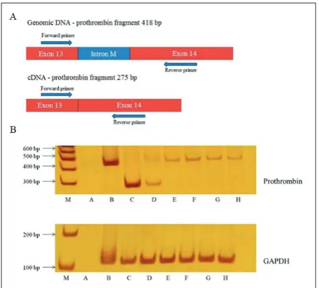
Prothrombin expression was determined by PCR using cDNA from cell lines as a template. The designed primers (Supplementary Table S1) were complementary to the region of the prothrombin gene consisting of the last intron and surrounding exon end sequence (Fig. 1A). This enabled differentiation of the desired PCR products from possible genomic DNA contamination. The prothrombin fragment amplified from cDNA (275 bp) is shorter in length than the prothrombin fragment amplified from genomic DNA (418 bp) (Fig. 1A). Human genomic DNA isolated from the peripheral blood sample (200 ng) served as a PCR control. As an internal control, a fragment of the gene encoding glyceraldehyde-3-phosphate dehydrogenase (GAPDH, 112 bp) was used (Supplementary Table S1). PCR fragments were analyzed by 10% non-denaturing polyacrylamide gel electrophoresis (PAGE) and visual-ized by silver staining.
Real-Time PCR (qPCR) for relative quantification of prothrombin mRNA expression in Caco-2 and healthy human liver samples was performed using TaqMan Gene Expression Assays (Applied Biosystems, USA) on the Applied Biosystems 7500 Real-Time PCR. Each reaction was 10 μL in volume and performed in trip-licates that consisted of 1x Universal PCR MasterMix, 1x TaqMan Gene Expression Assays and 1.5 μL of cDNA. Relative gene expression levels were analyzed using GAPDH as a reference gene in ABI Prism 7500 software (Applied Biosystems, USA).
Prothrombin expression analysis at the protein level
Cultured cells were collected, washed in PBS buffer (137 mM NaCl, 3.4 mM KCl, 1.8 mM KH2PO4, 10 mM Na2HPO4, pH 7.2) and lysed using EBC buffer (50 mM Tris-HCl pH 7.5, 120 mM NaCl, 0.5% NP40, protease inhibitors mix (Roche Diagnostics GmbH, Germany)). Lysates were concentrated using Amicon Ultra 30K centrifugal filters (Millipore, USA) and final protein concentrations were measured by the Bradford method [15]. Prothrombin expression was deter-mined by Western blotting. Two hundred µg of total cell lysates were separated by sodium dodecyl sulfate
(SDS)-PAGE and transferred to an Immobilon P mem-brane (Millipore, USA). To achieve better detection, 600 µg of Caco-2 lysate were loaded to SDS-PAGE. Healthy human liver protein lysate (200 µg), kindly provided by Dr. Snežana Kojić, Institute of Molecular Genet-ics and Genetic Engineering, University of Belgrade, Serbia, and standard human plasma (0.5 µL) (Siemens, Germany) were used as positive controls, and HemosIL Factor II Deficient Plasma (0.5 µL) (Instrumentation Laboratory, USA) was used as a negative control. Ac-tin was used as a loading control. Prothrombin was detected by incubation of the membrane with goat polyclonal Thrombin K-20 (Santa Cruz Biotechnol-ogy, USA) as a primary antibody (dilution 1:1000) at 4°C overnight. Actin was detected by incubation with non-commercially synthesized mouse polyclonal primary antibody, diluted 1:3000 (a generous gift from Dr. Snežana Kojić, Institute of Molecular Genetics and Genetic Engineering, University of Belgrade, Serbia) at 4°C overnight. After short successive washes in TBST buffer (1 M Tris-HCl, pH 7.5, 5 M NaCl), the membranes were incubated with the corresponding rabbit anti-goat and rabbit anti-mouse IgG horserad-ish peroxidase conjugate (dilution 1:80000) for 1 h at 4°C. Signal detection was performed by enhanced chemiluminescence (Western Lightning Plus-ECL, Perkin Elmer, USA)[16].
RESULTS
Analysis of prothrombin gene expression
To determine prothrombin expression, we performed PCR using cDNA from all 5 cell lines as a template. Results are shown on Fig. 1B. The prothrombin frag-ment (275 bp) was detected in healthy liver tissue (control) and Caco-2 cell line samples. However, none of the other cell lines showed any prothrombin gene expression. Insignificant genomic DNA contamination (a 418 bp fragment) was detected. The GAPDH band (212 bp) was present in all cell line samples, proving satisfactory cDNA quality (Fig. 1B).
The relative level of prothrombin gene expression was assessed by qPCR. The obtained relative quantifica-tion values (RQ) of the prothrombin mRNA level are shown in Table 1. The Caco-2 cell line showed ~100
times lower prothrombin expression in comparison to the healthy liver tissue sample.
Analysis of prothrombin protein expression
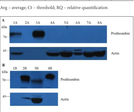
Western blotting was used to determine the presence of prothrombin in cancer cell lysates. The prothrombin band was detected in the standard human plasma and healthy liver samples, whereas it was undetectable in the cancer cell lines (200 µg lysates) (Fig. 2A). However, prothrombin was detected in the Caco-2 lysate when a higher amount of lysate (600 µg) was separated by SDS-PAGE (Fig. 2B).
DISCUSSION
Prothrombin and its active form, thrombin, are key regulators of hemostasis, whose imbalance can lead to hemostatic disorders, with VTE being one of the most common. Apart from its main role in
hemo-Table 1. Relative quantification of prothrombin gene expression achieved by qPCR.
Sample Avg Ct Avg dCt RQ RQ min RQ max
Liver 23.206 -1.492 1 0.856 1.168
Caco-2 25.256 5.202 0.010 0.008 0.011
Avg – average; Ct – threshold; RQ – relative quantification
stasis, thrombin has multiple roles in carcinogenesis [10,13,17]. Previous studies have shown that thrombin has a potent mitogenic effect and is also involved in nu-merous cell functions [13]. Therefore, it was important to investigate whether various cancer cell lines express prothrombin. We opted for MCF-7 and SK-BR-3 cell lines originating from breast cancer, Caco-2, which is derived from colorectal adenocarcinoma, and U-87 and U-251 from glioblastoma. These cell lines are derived from some of the most common cancers associated with frequent thrombotic disorders.
Breast cancer is one of the most common human malignancies, accounting for 25% of all cancers in females worldwide [18]. Compared to subjects with-out cancer, the risk of VTE in patients with cancer is increased three- to four-fold [19]. Colorectal adeno-carcinoma is the third most common type of cancer. Patients with colorectal cancer have a higher occur-rence of venous thromboembolism, with as many as 15.2% of patients associated with VTE [20-22]. Gliomas represent some of the most frequent and most lethal neoplasms of the central nervous system. Glioblastoma multiforme (GBM) is known for being one of the most life-threatening and aggressive types of cancer, accounting for 16% of all primary CNS malignancies [23]. The rate of VTE in patients with GBM is very high, at almost 30% [24]. Because of the increased levels of VEGF, GBM presents one of the most vascularized types of cancer [25].
In our study, of all the analyzed cell lines, prothrom-bin expression was detected only in Caco-2 cells. According to our results, the Caco-2 cell line showed notably lower prothrombin expression in comparison to the liver sample. It should be mentioned that RNA was isolated from the liver tissue (not only hepatocytes), while Caco-2 RNA was isolated from a cultured cell line.
To detect prothrombin expression at the protein level, cell lysates were used as a sample for Western blotting. The prothrombin band was initially present in positive controls (healthy liver lysate and standard human plasma), but it was not detected in the cell-line samples. Considering that prothrombin expression in Caco-2 cells was confirmed at the RNA level at a significantly lower quantity than in the healthy liver sample, three-fold higher quantities of Caco-2 cell lysate were loaded on SDS-PAGE when the prothrombin
band was detected in Caco-2 cells. It is known that Caco-2 cells are highly heterogeneous and tend to differentiate into subpopulations under certain condi-tions [26,27]. There is the possibility that only a certain group of Caco-2 cells could synthesize prothrombin, which is in correlation with our findings of low and inconsistent prothrombin expression. However, based on our results we cannot claim which subpopulation is expressing prothrombin. Also, (pro)thrombin, via activation of PAR receptors, could additionally influ-ence cell differentiation, as has been shown by Martin et al. [28] in smooth muscle precursor cells. Further research focused on establishing the prothrombin expression profiles of specific cell subpopulations is needed. This should pave the way for more answers to existing questions, such as which mechanism lies in the background of prothrombin expression in specific cancer cell subpopulations and how does this affect cancer development and progression.
Adams et al. [29] showed that Caco-2 cells express functional PAR-1 receptors and that thrombin influ-ences the motility and growth of Caco-2 cells. Con-sidering that Caco-2 cells express prothrombin and functional PAR receptors, it is likely that (pro)thrombin impacts colon cancer etiopathology. Furthermore, our results might provide first insights into prothrombin origin in this type of cancer, since all the analyses were performed using cell lines, thus eliminating the possibility of prothrombin uptake from the colon-cancer microenvironment. On the other hand, due to the heterogeneity of colon cancer, the mechanism of prothrombin activation and its effects on cancer metabolism need further clarification.
origin of prothrombin expression. The authors have also shown that thrombin, through PAR receptors, is involved in VEGF secretion in U-87 and U-251 cell lines in a dose-dependent manner. Zucker et al. [32] have demonstrated that prothrombin could be acti-vated on both endothelial and cancer cells through the stimulatory effect of VEGF on the coagulation cascade. However, the source of prothrombin and the mechanism of its further activation remain unclear. Even though the cell lines used in this study, with the exception of Caco-2, did not show any prothrombin expression, there is the possibility that glioblastoma and breast cancer cells uptake exogenous thrombin or activate prothrombin from the surrounding blood vessels, which supports tumor progression by way of activation of PAR receptors.
Further research involving thrombin and its role in cancer metabolism could examine thrombin as a cancer treatment. Studies have shown that anticoagula-tion therapies have inhibited tumor progression and metastasis, as was shown for breast and prostate cancer [33-36]. Furthermore, dabigatran, a direct thrombin inhibitor, enhanced the antitumor effects of cisplatin in vivo [37]. A plethora of studies indicate that throm-bin could be a potentially powerful target for treating various tumors, which is why it is of great importance to further research and identify the mechanisms by which (pro)thrombin is expressed and acts in cancer.
CONCLUSIONS
Expression of the prothrombin gene was determined for Caco-2 cells, while for the other cell lines (MCF-7, SK-BR-2, U-87 and U-251) prothrombin expression was not detected. Our results point to the potential role of prothrombin expression in certain cancers, such as colorectal adenocarcinoma. However, further research is needed to elucidate cancer-induced prothrombin expression and cancer-specific mechanisms in the acti-vation of prothrombin expression, as well as the role of prothrombin in cancer development and progression.
Funding: This work was funded by the Ministry of Education, Science and Technological Development of the Republic of Serbia, OI 173008.
Acknowledgments: The authors are very grateful to Dr. Snežana Kojić from the Institute of Molecular Genetics and Genetic
En-gineering, University of Belgrade, Serbia, for providing the liver tissue lysate and RNA, as well as actin antibody. Also, the authors would like to thank Dr. Marija Svirltih, Dr. Isidora Petrović and Jelena Marjanović Vićentić from the Institute of Molecular Genetics and Genetic Engineering, University of Belgrade, Serbia for the assistance with breast and glioblastoma cancer cell lines. Author contributions: SD wrote the manuscript. MC, MG and SD contributed to the cultivation of cell lines, Western blotting and qPCR methods. BT contributed to the evaluation and inter-pretation of the results. IP and VD designed the research. IP, DR and VD were responsible for supervision of the manuscript. All authors have revised and approved the final manuscript. Conflict of interest disclosure: None of the authors has a conflict of interest to disclose.
REFERENCES
1. Torre LA, Bray F, Siegel RL, Ferlay J, Lortet-Tieulent J, Jemal A. Global Cancer Statistics, 2012. CA Cancer J Clin. 2015;65(2):87-108.
2. Falanga A, Marchetti M, Russo L. The Mechanisms of Cancer-associated Thrombosis. Thromb Res. 2015;135:S8-S11. 3. Trousseau A. Clinique médicale de l’Hôtel-Dieu de Paris.
Paris: Baillière; 1865. French.
4. Eichinger S. Cancer Associated Thrombosis: Risk Factors and Outcomes. Thromb Res. 2016;140:S12-S7.
5. Falanga A, Russo L, Verzeroli C. Mechanisms of Thrombosis in Cancer. Thromb Res. 2013;131:S59-S62.
6. Khorana AA, Francis CW. Cancer-Associated Thrombosis: New Findings in Translational Science, Prevention and Treat-ment. New York: CRC Press; 2007. 279 p.
7. Chung DW XW, Davie EW. The Blood Coagulation Factors and Inhibitors: Their Primary Structure, Complementary DNAs, Genes, and Expression. In: Marder VJ, Aird WC, Bennett JS, Schulman S, White GC, editors. Hemostasis and Thrombosis: Basic Principles and Clinical Practice. Philadel-phia: Lippincott Williams & Wilkins; 2012. p. 110-45. 8. Wojtukiewicz MZ, Hempel D, Sierko E, Tucker SC, Honn
KV. Protease-Activated Receptors (PARs)—Biology and Role in Cancer Invasion and Metastasis. Cancer Metastasis Rev. 2015;34(4):775-96.
9. Coughlin SR. Thrombin Signaling and Protease-Activated Receptors. Nature. 2000;407(6801):258.
10. Ebrahimi S, Rahmani F, Behnam-Rassouli R, Hoseinkhani F, Parizadeh MR, Keramati MR, Khazaie M, Avan, Hassanian SM. Proinflammatory Signaling Functions of Thrombin in Cancer. J Cell Physiol. 2017;232(9):2323-9.
11. Tsopanoglou NE, Maragoudakis ME. Role of Thrombin in Angiogenesis and Tumor Progression. Semin Thromb Hemost. 2004;30(1):63-9.
13. Rickles FR, Patierno S, Fernandez PM. Tissue factor, Throm-bin and Cancer. Chest. 2003;124(3 Suppl):58s-68s.
14. Danckwardt S, Hentze MW, Kulozik AE. Pathologies at the Nexus of Blood Coagulation and Inflammation: Thrombin in Hemostasis, Cancer and Beyond. J Mol Med. 2013;91(11):1257-71.
15. Bradford MM. A Rapid and Sensitive Method for the Quan-titation of Microgram Quantities of Protein Utilizing the Principle of Protein-dye Binding. Anal Biochem. 1976;72(1-2):248-54.
16. Burnette WN. “Western blotting”: Electrophoretic Transfer of Proteins from Sodium Dodecyl Sulfate-Polyacrylamide Gels to Unmodified Nitrocellulose and Radiographic Detection with Antibody and Radioiodinated Protein A. Anal Biochem. 1981;112(2):195-203.
17. Wojtukicwicz MZ, Tang DG, Nelson KK, Walz DA, Diglio CA, Honn KV. Thrombin Enhances Tumor Cell Adhesive and Metastatic Properties via Increased αIIbβ3 Expression on the Cell Surface. Thromb Res. 1992;68(3):233-45.
18. Makki J. Diversity of Breast Carcinoma: Histological Subtypes and Clinical Relevance. Clin Med Insights Pathol. 2015;8:23. 19. Khan UT, Walker AJ, Baig S, Card TR, Kirwan CC, Grainge
MJ. Venous Thromboembolism and Mortality in Breast Can-cer: Cohort Study with Systematic Review and Meta-analysis. BMC Cancer. 2017;17(1):747.
20. Levitan N, Dowlati A, Remick SC, Tahsildar HI, Sivinski LD, Beyth R, Rimm AA. Rates of Initial and Recurrent Throm-boembolic Disease Among Patients with Malignancy Versus Those Without Malignancy: Risk Analysis Using Medicare Claims Data. Medicine. 1999;78(5):285-91.
21. Rickles FR, Edwards RL. Activation of Blood Coagula-tion in Cancer: Trousseau’s Syndrome Revisited. Blood. 1983;62(1):14-31.
22. Fleming M, Ravula S, Tatishchev SF, Wang HL. Colorec-tal Carcinoma: Pathologic Aspects. J Gastrointest Oncol. 2012;3(3):153-73.
23. Thakkar JP, Dolecek TA, Horbinski C, Ostrom QT, Light-ner DD, Barnholtz-Sloan JS, Villano JL. Epidemiologic and Molecular Prognostic Review of Glioblastoma. Cancer Epi-demiol Biomarkers Prev. 2014;23(10):1985-96.
24. Preusser M, de Ribaupierre S, Wöhrer A, Erridge SC, Hegi M, Weller M, Stupp R. Current Concepts and Management of Glioblastoma. Ann Neurol. 2011;70(1):9-21.
25. Dutra-Oliveira A, Monteiro RQ, Mariano-Oliveira A. Prote-ase-Activated Receptor-2 (PAR2) Mediates VEGF Production Through the ERK1/2 Pathway in Human Glioblastoma Cell Lines. Biochem Biophys Res Commun. 2012;421(2):221-7. 26. Sambuy Y, De Angelis I, Ranaldi G, Scarino M, Stammati
A, Zucco F. The Caco-2 Cell Line as a Model of the Intes-tinal Barrier: Influence of Cell and Culture-related Factors on Caco-2 Cell Functional Characteristics. Cell Biol Toxicol. 2005;21(1):1-26.
27. Vachon PH, Beaulieu J-F. Transient Mosaic Patterns of Mor-phological and Functional Differentiation in the Caco-2 Cell Line. Gastroenterology. 1992;103(2):414-23.
28. Martin K, Weiss S, Metharom P, Schmeckpeper J, Hynes B, O’Sullivan J, Caplice N. Thrombin Stimulates Smooth Mus-cle Cell Differentiation from Peripheral Blood MononuMus-clear Cells via Protease-Activated Receptor-1, RhoA, and Myocar-din. Circ Res. 2009;105(3):214-8.
29. Adams GN, Rosenfeldt L, Frederick M, Miller W, Waltz D, Kombrinck K, McElhinney KE, Flick MJ, Monia BP, Revenko AS, Palumbo JS. Colon Cancer Growth and Dissemination Relies Upon Thrombin, Stromal PAR-1 and Fibrinogen. Can-cer Res. 2015;75(19):4235-43.
30. Yamahata H, Takeshima H, Kuratsu J-I, Sarker KP, Tanioka K, Wakimaru N, Nakata M, Kitajima I, Maruyama I. The Role of Thrombin in the Neo-vascularization of Malignant Gliomas: An Intrinsic Modulator for the Up-regulation of Vascular Endothelial Growth Factor. Int J Oncol. 2002;20(5):921-8. 31. Elste AP, Petersen I. Expression of Proteinase-Activated
Receptor 1-4 (PAR 1-4) in Human Cancer. J Mol Histol. 2010;41(2-3):89-99.
32. Zucker S, Mirza H, Conner CE, Lorenz AF, Drews MH, Bahou WF, Jesty J. Vascular Endothelial Growth Factor Induces Tissue Factor and Matrix Metalloproteinase Pro-duction in Endothelial Cells: Conversion of Prothrombin to Thrombin Results in Progelatininase A Activation and Cell Proliferation. Int J Cancer. 1998;75(5):780-6.
33. DeFeo K, Hayes C, Chernick M, Van Ryn J, Gilmour SK. Use of Dabigatran Etexilate to Reduce Breast Cancer Progression. Cancer Biol Ther. 2010;10(10):1001-8.
34. Wojtukiewicz MZ, Hempel D, Sierko E, Tucker SC, Honn KV. Thrombin-Unique Coagulation System Protein with Multi-faceted Impacts on Cancer and Metastasis. Cancer Metastasis Rev. 2016;35(2):213-33.
35. Afratis NA, Karamanou K, Piperigkou Z, Vynios DH, Theo-charis AD. The Role of Heparins and Nano-Heparins as Ther-apeutic Tool in Breast Cancer. Glycoconj J. 2017;34(3):299-307.
36. Nieman MT, LaRusch GA, Fang C, Zhou Y, Schmaier AH. Oral Thrombostatin FM19 Inhibits Prostate Cancer. Thromb Haemost. 2010;104(5):1044.
37. Alexander ET, Minton AR, Peters MC, Van Ryn J, Gilmour SK. Thrombin Inhibition and Cisplatin Block Tumor Progres-sion in Ovarian Cancer by Alleviating the Immunosuppres-sive Microenvironment. Oncotarget. 2016;7(51):85291.
Supplementary Data
Supplementary Table S1.