SYMPLEKIN AND TRANSFORMING ACIDIC COILED-COIL CONTAINING
PROTEIN 3 SUPPORT THE CANCER CELL MITOTIC SPINDLE
Kathryn M. Cappell
A dissertation submitted to the faculty of the University of North Carolina at Chapel Hill in partial fulfillment of the requirements for the degree of Doctorate of Philosophy in the Department of Pharmacology, School of Medicine.
Chapel Hill 2011
Approved by:
ABSTRACT
KATHRYN CAPPELL: Symplekin and Transforming Acidic Coiled-Coil Containing Protein 3 Support the Cancer Cell Mitotic Spindle
(Under the direction of Dr. Angelique Whitehurst)
An increased rate of proliferation in cancer cells, combined with abnormalities in spindle architecture, places tumors under increased mitotic stress. Previously, our laboratory performed a genome-wide paclitaxel chemosensitizer screen to identify genes whose depletion sensitizes non-small cell lung cancer (NSCLC) cells to mitotic stress induced by paclitaxel treatment. This screen uncovered a cohort of genes that are required for viability only in the presence of paclitaxel. Two genes uncovered in this screen were the polyadenylation scaffold symplekin and the gametogenic protein transforming acidic coiled-coil containing protein 3 (TACC3).
Herein, we examine the impact of polyadenylation and gametogenesis on the tumor cell mitotic spindle. First, we demonstrate that depletion of SYMPK and other polyadenylation components sensitizes many NSCLC cells, but not normal immortalized lines, to paclitaxel by inducing mitotic errors and leading to abnormal mitotic progression. Second, we demonstrate that multiple gametogenic genes are required for normal microtubule dynamics and mitotic spindle formation in the presence of paclitaxel. Additionally, we show that the gametogenic protein TACC3 is uniquely required for mitosis only in transformed cell lines but not normal immortalized cell lines and that this unique dependency can be targeted in vitro with a small molecule.
ACKNOWLEDGEMENTS
First and foremost, I would like to thank my mentor. Angelique Whitehurst has proven to be a fantastic mentor who has guided me on the path towards becoming a scientist. Angelique is always full of energy and excitement to share her many ideas and this has made it a great experience to be her graduate student. She has challenged me where I needed it but at the same time introduced me to the joys of scientific research. I will always be grateful for the time I spent in her laboratory and look forward to watching as her career develops in the future.
I would particularly like to thank our laboratory managers Brittany Larson and Moriah Scarbrough who have provided much assistance to my projects and Charlene Ross in the UNC-Animal Core who assisted with mouse studies. I am also thankful to the Department of Pharmacology, the Cancer Cell Biology Training Program and the UNC MD/PhD program. During rough patches of my PhD, these programs have really stood by me and for that I will always be grateful. Dr. Eugene Orringer and Dr. David Siderovski deserve special credit for all the support they have given me personally and for making UNC such a wonderful place to be an MD/PhD student. I would also like to thank my committee, including Dr. Pilar Blancafort, Dr. David Siderovski, Dr. Mohanish Deshmukh and Dr. Channing Der for their insights.
TABLE OF CONTENTS
LIST OF TABLES ...vi
LIST OF FIGURES...vii
LIST OF ABBREVIATIONS AND SYMBOLS...viii
INTRODUCTION...1
Hallmarks of cancer and the stress phenotype of cancer cells...2
Mitotic stress in cancer ...4
Paclitaxel and genome-wide screen to identify modulators of chemosensitivity ...9
Role of symplekin in polyadenylation and tumorigenesis...17
The cancer-testis antigens ...24
Transforming acidic coiled-coil containing protein 3...27
Thesis Summary...29
SYMPLEKIN IS REQUIRED FOR APPROPRIATE MICROTUBULE FUNCTION AND MITOSIS...31
Summary ...32
Introduction...33
Results...34
Discussion ...48
Experimental Procedures ...51
TACC3 AND MULTIPLE GAMETOGENIC GENES SUPPORT THE CANCER CELL MITOTIC SPINDLE ...55
Summary ...56
Results...58
Discussion ...75
Experimental Procedures ...76
CLINICAL RELEVANCE AND FUTURE DIRECTIONS...79
Summary ...80
Future directions ...81
Clinical implications ...89
Conclusions...94
LIST OF TABLES
LIST OF FIGURES
Figure 1.1 The spindle assembly checkpoint ...5
Figure 1.2 Variation in response of mitotic cells to anti-mitotic therapies ...13
Figure 1.3 Protein factors involved in cleavage and polyadenylation and impact of their depletion in two genome-wide screens ...20
Figure 2.1 SYMPK is required for spindle integrity after exposure to paclitaxel...35
Figure 2.2 SYMPK is required for normal mitotic progression in tumor cells ...37
Figure 2.3 SYMPK is necessary for mitosis in multiple tumor cell lines ...39
Figure 2.4 Depletion of SYMPK impairs tumor growth in vivo...41
Figure 2.5 Depletion of SYMPK reduces microtubule stability ...43
Figure 2.6 SYMPK depletion leads to loss of CKAP5 ...45
Figure 2.7 SYMPK alters CKAP5 levels post-transcriptionally ...47
Figure 2.8 Polyadenylation is required for CKAP5 expression and mitosis...49
Figure 3.1 Multiple gametogenic genes sensitize H1155 NSCLC cells to paclitaxel...62
Figure 3.2 Loss of gametogenic genes impairs formation of the bipolar mitotic spindle ...64
Figure 3.3 Oncogenic changes alter mitotic properties ...68
Figure 3.4 Oncogenic changes drive a dependency on TACC3...71
Figure 3.5 TACC3 dependency is targetable with KHS101 ...74
Figure 4.1 Model of adaptation to mitotic stress in tumor cells...82
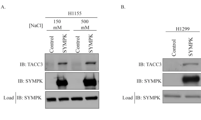
Figure 4.2 Interaction of SYMPK and TACC3...88
LIST OF ABBREVIATIONS AND SYMBOLS
3’ UTR 3’ untranslated regionACRBP Acrosin binding protein
APC Anaphase promoting complex
ARNT Aryl hydrocarbon receptor nuclear translocator
AURKA Aurora kinase A
BLI Bioluminescent imaging
BRDT Bromodomain testis specific
Bub3 Budding uninhibited by benzimidazoles 3 BubR1 Budding uninhibited by benzimidazoles R1 cdc20 Cell division cycle 20
cDNA Complementary DNA
CKAP5 Cytoskeleton associated protein 5
CNTRL Control
CPE Cytoplasmic polyadenylation element
CPEB Cytoplasmic polyadenylation element binding protein CPSF1-6 Cleavage and polyadenylation specific factor 1-6 CSTF1-3 Cleavage stimulation factor 1-3
Ct Cycle threshold
CT-antigen Cancer-testis antigen DAC 5-aza-2’-deoxycytidine
DAPI 4’-6-Diamidino-2-phenylindole
DNA Deoxyribonucleic acid
DSE Downstream element
EGTA Ethylene glycol tetraacetic acid FATE1 Fetal and adult testis expressed 1 FDA Food and Drug Administration
FMR1NB Fragile X mental retardation neighbor 1 GAPDH Glyceraldehyde-3-phosphate dehydrogenase GFP Green fluorescent protein
GFP-H2B Green fluorescent protein conjugated to histone 2B HBEC Human bronchial epithelial cell
HEPES 4-(2-hydroxyethyl)-1-piperazineethanesulfonic acid HIF Hypoxia inducible factor
hTERT Human telomerase reverse transcriptase MCAK Kinesin family member 2C
MPS1 TTK protein kinase
NA Nuclear aperature
NSCLC Non-small cell lung cancer
NuMa Nuclear mitotic apparatus protein 1 NXF2 Nuclear RNA export factor 2 PABP Poly(A) binding protein
Pac Paclitaxel
PAP Poly(A) polymerase
PARP Poly(ADP-ribose) polymerase 1 PAS Polyadenylation signal
PCR Polymerase chain reaction PLK4 Polo-like kinase 4
RNA Ribonucleic acid
RT-PCR Real-time polymerase chain reaction SAC Spindle assembly checkpoint
SD Standard deviation
SDS Sodium dodecyl sulfate SEM Standard error of the mean shRNA Short hairpin RNA
siRNA Small interfering RNA
STARD6 StAR-related lipid transfer (START) domain containing 6
SYMPK Symplekin
CHAPTER I
Hallmarks of cancer and the stress phenotype of cancer cells
An increased proliferation rate in cancer cells, coupled with dysregulation of normal controls on cellular growth, drives several hallmarks of tumors. These hallmarks were originally elegantly proposed by Hanahan and Weinberg and include self-sufficiency in growth signals, insensitivity to anti-growth signals, evasion of apoptosis, sustained angiogenesis, limitless replicative potential, and metastasis (1). Over the past several decades, scientists have unraveled the signaling networks driving the hallmarks of cancer and found many hallmarks derive from activation of oncogenes or loss of tumor suppressors. Knowledge of the signaling pathways driving the hallmarks of cancer has led to significant improvements in cancer treatment using drugs that target oncogenic signaling networks. However, researchers have also realized new complexities in cancer including a tremendous degree of genetic heterogeneity between tumors, the existence of subpopulations within a single tumor that may respond differently to drugs, the propensity for tumors to develop resistance to targeted therapeutics, and a strong influence of stress pathways on the tumor cell phenotype. These complexities demand new directions for scientific inquiry but also represent potential inroads for the development of novel therapeutics.
Stress phenotypes in cancer cells – One recent development in cancer biology has been
However, BRCA mutant breast cancers have markedly increased sensitivity to PARP inhibitors that target DNA damage repair (3). Thus, a second perturbation of the DNA damage repair pathway in BRCA mutant breast cancers can greatly increase tumor cell killing. The stress phenotypes therefore represent a pressure point in the cancer cell that can be therapeutically exploited.
While the idea that these stress phenotypes represent additional hallmarks of cancer cells is new, stress pathways have in actuality been indirectly targeted in cancer treatment for decades. Two notable examples of this are cytotoxic agents that induce DNA damage and mitotic stress in tumor cells. DNA damage stress is commonly targeted in cancers using platinum compounds or radiation, both of which damage the DNA. Mitotic stress is targeted with agents such as the Vinca alkaloids and paclitaxel, which damage the tumor cell mitotic spindle. These cytotoxic therapies represent the mainstream of cancer treatment today. Although cytotoxic therapies activate stress pathways in tumor cells, potent effects on normal tissues limit the usefulness of cytotoxic therapies. These off-target effects are likely due to the fact that cytotoxic drugs target components that are present in both tumor and normal cells, in contrast to agents such as PARP inhibitors that preferentially affect BRCA mutant cancer cells. A better understanding of how cytotoxic drugs activate stress pathways in cancer may lead to more directed targeting of stress phenotypes unique to cancer cells, leading to improved efficacy and less effects on normal tissues.
Mitotic stress in cancer
Mitotic stress is widely observed in cancer cells and likely accounts for the clinical success of numerous anti-mitotic therapies including paclitaxel and the Vinca alkaloids. Mitotic stress arises from an increased rate of proliferation in combination with structural changes in the cancer cell including aneuploidy, supernumerary centrosomes, and altered microtubule stability. These three structural alterations are interdependent since development of any one of these alterations can drive development of the others. Importantly, these structural alterations in the tumor cell mitotic spindle represent unique vulnerabilities that could provide a method to more specifically target mitotic stress in tumor cells.
Aneuploidy and the spindle assembly checkpoint - Aneuploidy describes cells that have an uneven number of chromosomes that is either more or less then the normal diploid chromosomal complement. Greater then 90% of human solid tumors are aneuploid (5). The widespread occurrence of aneuploidy in solid tumors is somewhat of a paradox because aneuploidy consistently reduces proliferative capacity both at the organismal (6, 7) and the cellular level (8) yet cancers grow at an extremely high rate. This paradox can be partially explained by aneuploidy leading to increased expression of oncogenes or loss of tumor suppressors (9). For example, changes in chromosomal composition can lead to amplification of the oncogene ERBB2 (10) or loss of the tumor suppressor PTEN (11). These alterations in expression of oncogenic genes could provide a proliferative advantage to the tumor cell and account for the widespread occurrence of aneuploidy in tumors.
Figure 1.1
Figure 1.1 The spindle assembly checkpoint
anaphase onset (12). Activation of this checkpoint protects the cell from uneven segregation of chromosomes during mitosis and therefore acts as a fail-safe to avoid the development of aneuploidy. Thus, for a cell to develop aneuploidy, it must bypass the SAC.
There are several possible mechanisms by which aneuploid cells may avoid detection by the SAC including mutations in checkpoint genes, altered expression of checkpoint proteins, aberrant spindle attachments, or the generation of multi-polar spindles (13). Originally, it was thought that tumor cells must exhibit widespread mutation or loss of checkpoint genes to allow for bypass of the SAC. However, mutations in checkpoint proteins were later shown to be relatively rare (14-16), which is inconsistent with the widespread incidence of aneuploidy in tumor cells (13). In fact, checkpoint proteins are often over-expressed in tumor cells (17, 18) and overexpression of checkpoint proteins has been shown to drive the development of aneuploidy (18, 19). The mechanism by which overexpression of checkpoint proteins drives aneuploidy may involve an increased mitotic delay leading to nondisjunction of sister chromatids and tetraploidy (18). In addition to alterations in the SAC components themselves, tumor cells may also become aneuploid by developing spindle attachments that are not detected by the SAC. Two examples of these types of defects are changes in chromatid cohesion or merotelic attachments. Altered chromatid cohesion occurs when the two sister chromatids adhere to each other abnormally during mitosis, allowing for improper segregation of an additional chromatid to one daughter cell without activation of the SAC (13, 20, 21). Merotelic attachments, which occur when a single kinetechore attaches to both spindle poles, are also not detected by the SAC and could lead to aneuploidy (22, 23). Finally, the ability of some cancer cells to undergo multipolar mitoses, as discussed below, could also account for aneuploidy development (13). It is currently unclear which of these mechanisms is the major driver of aneuploidy in tumor cells.
that specifically target aneuploid cells. As the vast majority of normal cells in the human body are euploid, aneuploidy-specific compounds could have greatly reduced off-target effects on normal tissues. A recent study has identified compounds that specifically kill aneuploid as compared to euploid cells in culture (25). Future studies to examine the impact of these drugs in vivo may uncover a more specific method for targeting mitotic stress in tumors.
Supernumerary centrosomes and multipolar mitoses - An additional structural barrier
to mitosis in tumor cells is abnormal expression of greater then two centrosomes in a single mitotic cell. The presence of these supernumerary centrosomes in tumor cells is common and may correlate with tumor aggressiveness (26-29). Supernumerary centrosomes can arise through a number of mechanisms including fusion of two cells, failed cytokinesis, over-duplication, and following activation of oncogenic signaling pathways (27, 30-32). The presence of additional centrosomes in a single mitotic cell can lead to nucleation of more then two spindle poles and therefore places the cell at risk for a multipolar mitosis (33). Multipolar mitoses result in abnormal segregation of chromosomes into three or more daughter cells and can foster the development of aneuploidy (33, 34). However, it is unclear whether this is a significant source for aneuploidy in tumors since the daughter cells from a multipolar division are often inviable (33, 35, 36). Regardless of whether additional centrosomes drive the development of aneuploidy, it is clear that supernumerary centrosomes represent a source of mitotic stress in the cancer cell.
presence of additional centrosomes. Centrosome clustering may represent either a unique adaptation of tumor cells to the extra centrosome or an additional dependence of tumors on centrosomal clustering pathways that exist in all cells (27). Since normal human cells rarely express redundant centrosomes, the pathways guiding centrosome clustering may be uniquely required in tumor cells and represent tumor-specific drug targets. The requirement for clustering in cancer cells has spurred efforts to identify genes specifically required for this process, including a recent genome-wide screen (41). Importantly, both CKAP5 (41, 42) and TACC3 (42), discussed in Chapters 2 and 3 of this work, have been implicated in centrosomal clustering.
Changes in microtubule stability and composition - A final mitotic stress phenotype
often observed in tumor cells is profound changes in the stability and composition of the microtubule network. Microtubules are structures composed of heterodimers of alpha and beta tubulin. There are six isotypes of alpha and 7 isotypes of beta tubulin (43). Microtubules are dynamic structures that continually cycle between a state of growth and shrinkage. This cycling is called microtubule dynamicity and is vital for rapid formation of the mitotic spindle. In tumor cells, this equilibrium is shifted such that tumor cell microtubules are more stable then those in normal immortalized cells (44-46). Increased microtubule stability in tumor cells has been postulated to drive the development of aneuploidy because kinetechore-microtubule attachments that are excessively stable increase the likelihood of improper segregation of the chromosomes between the daughter cells (44-46). The enhanced microtubule stability in tumor cells may partially explain the success of chemotherapeutic drugs targeting the microtubules.
composition could likewise influence stability of microtubules in tumor lines. Third, changes in microtubule stability could develop in tumor cells due to altered expression of microtubule stabilizing proteins. For example, the microtubule stabilizing proteins HEC1 (53, 54), TACC3 (55) and CKAP5 (56, 57) are all highly over-expressed in cancer cells. This overexpression may drive increased microtubule stability in tumor cells. An important future direction of study is determining the factors uniquely supporting microtubule stability in tumor cells and understanding how these influence the development of aneuploidy.
Overall, tumor cells face profound mitotic stress due to aneuploidy, the presence of supernumerary centrosomes, and altered microtubule stability. Identifying the molecular mechanisms by which these stresses interface with each other and with tumorigenic phenotypes is an important future direction of study. In particular, these stresses may have a strong influence on the response of tumor cells to anti-mitotic therapies.
Paclitaxel and genome-wide screen to identify modulators of chemosensitivity
Discovery of paclitaxel - Paclitaxel is an anti-mitotic chemotherapy that targets
microtubules in dividing cells (43). Paclitaxel was originally isolated from the bark of the Pacific Yew tree and later shown to have potent efficacy in killing tumor cells (58). The earliest clinical trials of paclitaxel were hugely successful with efficacy seen in some cancers, such as ovarian carcinoma, which previously had few treatments available. Early trials in ovarian carcinoma demonstrated a 30% overall response rate and some patients exhibited complete remissions (59). At the time, this level of response rate in ovarian carcinoma was outstanding. Since then, paclitaxel has become widely utilized to treat a range of cancers including breast, ovarian, lung, head and neck cancers, and Kaposi sarcoma, and is now an important part of the clinical armamentarium (43).
Pacific Yew. Researchers attempted to isolate paclitaxel from almost every part of the Yew tree but were unable to derive a sufficient quantity of drug to satisfy the appetites of researchers, clinicians, and patients (60). Large quantities of trees had to be cut down to obtain enough paclitaxel to meet demand. There was so much demand, however, that there were fears the Pacific Yew would be driven to extinction (61). Fortunately, a synthetic method to make paclitaxel was developed, compromising one of the most difficult chemical syntheses to date (62). Second, paclitaxel had poor solubility, which made it difficult to administer the drug to patients. This almost led researchers to abandon paclitaxel but was somewhat solved by the use of the agent Cremophor, a derivative of castor oil, to resuspend paclitaxel (60). Third, there was an initial concern that paclitaxel had too much toxicity on normal cells. However, further research indicated there was a sufficient therapeutic window to allow paclitaxel to become a clinically useful drug. The fact that paclitaxel, now one of the most widely utilized chemotherapies, had to overcome many significant hurdles demonstrates the challenges faced in cancer drug discovery.
Problems of paclitaxel treatment – The widespread clinical usage of paclitaxel
or nab-paclitaxel. Nab-paclitaxel (Abraxane or ABI-007) is a Cremophor-free formulation that uses albumin-stabilized nanoparticles to deliver paclitaxel and was recently approved by the FDA for the treatment of breast cancer (65) and has shown efficacy in NSCLC (66). Many of the other side effects seen with paclitaxel are observed with other cytotoxic chemotherapeutic agents and stem from the ability of paclitaxel to kill rapidly dividing cells of the gut, bone marrow, and hair follicle. New formulations of paclitaxel that allow for increased concentration of paclitaxel within tumors may reduce effects on normal tissues by allowing for decreased drug dosage (67, 68). Alternatively, it may be possible to combine paclitaxel treatment with other agents to avoid several of the dose-related side effects of paclitaxel (4). Future efforts to address the problems of drug resistance and side effects related to paclitaxel treatment could have important impacts on patient care.
Biological mechanism of paclitaxel - Shortly after the discovery of the efficacy of
state of shrinkage or catastrophe, a process called microtubule dynamicity (75). Since paclitaxel has effects on microtubule dynamicity at a much lower dose then that needed to induce outright polymerization, the efficacy of paclitaxel likely derives from its ability to disrupt microtubule dynamicity (43). The disruption of microtubule dynamics in paclitaxel-treated cells impairs the ability of the mitotic spindle to make proper kinetechore-microtubule attachments. These improper attachments lead to activation of the spindle assembly checkpoint (SAC), mitotic delay, and triggering of cell death pathways (63). In summary, paclitaxel functions by binding to the beta tubulin subunit and inducing alterations in microtubule dynamics, which causes impaired mitotic progression and cell death.
Heterogeneity in response to paclitaxel – Ideally paclitaxel treatment causes death of
the cancer cell in mitosis as described above. Unfortunately, there is much more complexity in the ways in which both patients and tissue culture cells respond to paclitaxel. In particular, while some patients demonstrate a marked response to paclitaxel, others show no response at all (59). This heterogeneity of response is paralleled in tissue culture cells where there is great variation in cell fate after paclitaxel treatment both between and within tumor lines (35, 76-80). In tissue culture cells, paclitaxel treatment can lead to a range of responses (Figure 1.2). These responses include dying directly in mitosis, exiting from mitosis, returning to interphase, or exiting as a micronucleated cell that may either die or continue to cycle. This heterogeneity of response in vitro may explain the heterogeneity of response to paclitaxel treatment in patients.
Figure 1.2
Figure 1.2 Variation in response of mitotic cells to anti-mitotic therapies
mitosis, paclitaxel treatment is not effective until the next round of replication. Therefore, researchers have attempted to understand what guides the decision of a cell to die in mitosis after treatment with paclitaxel. The most effective studies have used single-cell time-lapse imaging to follow cells treated with paclitaxel and map the fate of individual treated cells (35, 76-80). The prevailing theory suggests that whether a cell dies in mitosis after treatment with paclitaxel relates to a balance between two competing networks: the rate of degradation of cyclin B1 and the activation rate of apoptotic pathways (63). In this model, cyclin B1 is slowly degraded in the SAC-arrested cell by a proteasome-dependent mechanism (35, 76). Concurrently, activation of the SAC triggers apoptotic pathways that are designed to detect mitotic cells that cannot form the proper kinetechore-microtubule attachments. When the rate of activation of apoptotic pathways exceeds the rate of cyclin B1 destruction, the cell dies in mitosis. In contrast, if the rate of cyclin B1 destruction exceeds the rate of apoptotic pathway activation, the cell slips through the checkpoint. The heterogeneity observed in response of different tumor lines to paclitaxel may therefore derive from variation in apoptotic factors between individual cells and cell lines (35, 79, 80, 82). Therefore, agents that allow for prolonged activation of the SAC may have increased efficacy in killing cancer cells during mitosis (82).
death following a micronucleated exit is independent of the degree of mitotic damage. Therefore, heterogeneity in propensity for micronucleated cell death may depend on variations in activation of apoptotic pathways between individual cells and tumor cell lines. A more complete understanding of the factors governing post-mitotic cell fate may help in developing better anti-mitotic therapies.
Resistance to paclitaxel treatment – Paclitaxel treatment is limited by significant drug
resistance that can be due to several factors including mutations in beta tubulin, upregulation of drug efflux pumps, changes in expression of beta tubulin isotypes, and changes in cell death pathways. First, mutations in beta tubulin that may impair paclitaxel binding could drive resistance. However, mutations in beta tubulin are rare and unlikely to account for the widespread incidence of drug resistance (52, 86). Second, upregulation of drug efflux pumps can drive drug resistance. Drug efflux pumps result in a decreased concentration of paclitaxel in the cell and therefore less cancer cell death with drug treatment. Upregulation of drug efflux pumps has been documented with paclitaxel therapy (87). Third, changes in expression of tubulin isotypes can lead to paclitaxel resistance. Changes in tubulin isotypes have been documented in paclitaxel-resistant ovarian carcinomas (88). Most commonly, increased expression of beta-3 tubulin has been shown to drive paclitaxel resistance (52). Finally, altered apoptotic signaling networks could also influence the development of resistance (89-91). Cells with a decreased propensity to die in mitosis may therefore exhibit increased resistance to paclitaxel therapy. Overall, it is clear that multiple factors can determine the development of resistance to paclitaxel.
differences between resistant and sensitive cells. Therefore, very different factors may govern the response to paclitaxel in primarily sensitive as opposed to resistant cells.
Genome-wide screen to identify modulators of paclitaxel sensitivity - Given the
significant limitations of paclitaxel, coupled with the widespread usage of this drug in the clinic, identification of new drug targets that synergize with paclitaxel could have a significant impact. Additionally, identification of such targets could reveal important biology about how tumor cells respond to mitotic stress. To address these questions, our lab performed the first genome-wide siRNA chemosensitizer screen to identify genes whose depletion significantly increases response of the NSCLC line H1155 to paclitaxel (4). For the screen, the H1155 cell line was transfected in 96-well plates in a one-well one-gene format with siRNAs targeting greater then 21,000 genes. After two days of gene knockdown, the transfected cells were treated with either no paclitaxel or a dose of paclitaxel that has minimal effects on cell viability (10 nM). After an additional two days of growth in the presence of paclitaxel, cell viability was determined using a luminescence-based CellTiter Glo viability assay. This screen returned two types of hits; monogenic lethal hits, in which siRNA knockdown alone reduces viability, and synthetic lethal hits in which gene knockdown only reduces viability in the presence of paclitaxel. Importantly, the screen returned genes whose knockdown was previously shown to synergize with paclitaxel, identified novel drug targets, and also uncovered nearly every component of the gamma-tubulin ring complex, a structure needed for nucleation of spindle microtubules (4). This ability of the screen to uncover both novel genes and genes which have already been shown to influence paclitaxel sensitivity validates the screening approach and suggests the completion of a successful screen.
synergize with paclitaxel. Given the identification of multiple mitotic proteins in the screen, it is likely that many of these unknown hits impact mitotic spindle formation. This has proved to be the case with several hits, including symplekin (SYMPK), the subject of Chapter 2 of this dissertation (92). Third, the screen pulled several members of a gene family called the cancer-testis antigens (antigens) including ACRBP, FATE1, FMR1NB and NXF2 (4, 93). The CT-antigens are a family of genes that share a common expression pattern showing upregulation in tissues of gametogenesis and cancer cells with minimal expression in normal adult tissues (93). The CT-antigens are discussed in more detail below but their identification in a screen for genes affecting tumor cell viability suggests these genes may have an unrecognized role in supporting tumor cell phenotypes. The CT-antigens and a related protein, TACC3, is the subject of Chapter 3 of this dissertation. Fourth, as necessary with any genome-wide screen extensive validation has been performed to reduce the potential that any observation is due to an off-target effect of the siRNA. Most commonly, screen hits have been validated by deconvolution of the siRNA pool both at the level of viability and on relevant phenotypes. As an additional control for these effects, the parallel system of shRNA to mediate gene knockdown has also been effective. Finally, follow-up of hits from this genome-wide screen has revealed important information about the mitotic spindle, response of tumor cells to mitotic stress, and uncovered new drug targets. Next, I will discuss the functions of the screen hit symplekin.
Role of symplekin in polyadenylation and tumorigenesis
increases transcription of the target genes cyclin D1 (95) and claudin-2 (96) but decreases expression of the transcription factor AML1/Runx1 (97). Functionally, these effects of SYMPK at the tight junction drive proliferation while reducing cellular differentiation.
The authors who discovered SYMPK at the tight junction also observed strong SYMPK expression in the nucleoplasm of a range of cells that lack tight junctions (94). The function of SYMPK in this cellular compartment was not elucidated until several years later when a group identified SYMPK as a component of the polyadenylation machinery (98). In particular, it was found that SYMPK interacts directly with the polyadenylation protein CSTF2, demonstrates some homology to the yeast polyadenylation component PTA1 (99), and exists in a complex with multiple polyadenylation factors. Therefore, while SYMPK regulates transcription at the tight junction it also has a role in controlling polyadenylation.
SYMPK and polyadenylation - Polyadenylation is a post-transcriptional process that is
required for the maturation of most mammalian pre-mRNAs, excluding histones, and controls mRNA nuclear export, stability, and translation (100). A longer poly(A) tail typically leads to more translation of the mRNA while a shorter tail decreases translation of the mRNA. Polyadenylation occurs simultaneously with cleavage of the pre-mRNA and is directed by a large complex of polyadenylation and cleavage factors (Figure 1.3A) (100). These factors include five cleavage and polyadenylation specificity factors (CPSF1-5 and hFip1), three cleavage stimulation factors (CSTF1-3), cleavage factors Im and IIm, poly (A) polymerase (PAP), poly(A) binding protein (PABP) and symplekin (Table 1.1) (100). While all these factors, with the exception of PABP, are required for the in vitro cleavage reaction, only a subset (the CPSFs, PAP and PABP) are needed for in vitro polyadenylation (101). Several of these factors were identified in our genome-wide paclitaxel screens in the NSCLC lines H1155 and HCC366 (Figure 1.3B) (4). This suggests an important role for the polyadenylation machinery and symplekin in controlling chemosensitivity.
Figure 1.3
A.
Figure 1.3 Protein factors involved in cleavage and polyadenylation and impact of their depletion in two genome-wide screens
Table 1.1 Characteristics of mammalian cleavage and polyadenylation factors
Group Subunits Alternative names processing Step in Interacts with: Function
CSTF1 CstF-50 CSTF3, PolII CTD Binds PolII/CTD complex
CSTF2 CsfF-64 SYMPK, CSTF3 Binds RNA through G/U rich sequence
CstF
CSTF3 CstF-77
Cleavage
CPSF1, CSTF2,
CSTF1 Scaffolding function CPSF1 CPSF-160 CSTF3, PolII CTD, PAP, hFip1,
SYMPK
Binds RNA at PAS site
CPSF2 CPSF-100 CPSF3, SYMPK Binds U-rich RNA
CPSF3 CPSF-73 CPSF2, SYMPK endonuclease Cleavage
CPSF4 CPSF-30 HFip1 Binds U-rich RNA
CPSF
hFip1 FIPIL1
Cleavage and poly(A) addition
PAP, CPSF1, CPSF4, CSTF3
Binds U-rich RNA and positions PAP at
polyadenylation site CF Im-68 CPSF6 CF Im-25 Aids in PAS site recognition
CFIm
CF Im-25 CPSF5
Polyadenylation
CFIm-68 and PAP Aids in PAS site recognition
hPCF11 PCF11 Unknown Unknown
CFIIm
hCLp1 HEAB
Unknown
CFIm and CPSF Unknown
- Symplekin SYMPK Cleavage and poly(A) addition
CSTF2, CPSF1,
CPSF2, CPSF3 Scaffolding function, links CPSF to CSTF
- PAP PAPOLA Polyadenylation hFip1, CFIm, CPSF1 Catalyzes addition of the poly(A) tail
RNA Polymerase
II
Pol II CTD - Transcriptional termination hPcf11, CPSF1, CSTF1, CSTF3 transcription Terminates
sequence elements in the pre-mRNA by components of the polyadenylation and cleavage complex. The primary sequence elements include the polyadenylation signal (PAS), the cleavage site, and the G/U-rich downstream element (DSE) (100). The PAS usually has the sequence AAUAAA (102) and is recognized and bound by CPSF1 (103). The cleavage site is recognized by the cleavage stimulation factor CSTF2 and does not have a conserved sequence (104) but most often consists of the nucleotide sequence CA with cleavage occurring after the cytosine (100). The DSE can have several sequences but is typically either GU-rich or U-rich (105, 106), is located downstream of the PAS and cleavage sites, and binds CSTF2 (107). Together these sequence elements in the pre-mRNA direct the assembly of the polyadenylation and cleavage factors and lead to simultaneous addition of the poly(A) tail and mRNA cleavage. SYMPK is believed to act as a molecular scaffold in this process by binding to a number of proteins in the complex including CSTF2, CPSF1, CPSF2, and CPSF3 (98, 108, 109). Therefore, activity of SYMPK and the polyadenylation components is essential for polyadenylation and expression of mRNAs.
Additionally, SYMPK and components of the polyadenylation and cleavage machinery also participate in the maturation of the 3’ end of histone mRNAs (110, 111). Histone mRNAs are the only eukaryotic mRNAs that are not polyadenylated. Rather, histone mRNAs undergo a 3’ maturation process which combines cleavage with addition of a stem-loop structure at the 3’ end of the mRNA (112). Many of the same components that mediate the polyadenylation and cleavage reaction, including SYMPK, also direct maturation of histone mRNAs (112). Therefore, SYMPK mediates histone maturation and polyadenylation.
SYMPK and cytoplasmic polyadenylation - In most mammalian cells, polyadenylation
B1, encode a cytoplasmic polyadenylation element (CPE) with the sequence UUUUUAU (114). These CPE-containing transcripts exist in the unstimulated oocyte as dormant transcripts with a very short poly(A) tail (119-122). Upon stimulation of the oocyte to proceed through meiosis, the CPE is bound by the cytoplasmic polyadenylation element binding protein (CPEB) and a poly(A) tail is added (123). Addition of the poly(A) tail drives expression of the transcripts and meiotic progression. Therefore, cytoplasmic polyadenylation allows for precise temporal control of gene expression during meiosis.
The machinery controlling cytoplasmic polyadenylation shares many players in common with the nuclear polyadenylation machinery, including SYMPK (108, 124). Two unique components of the cytoplasmic polyadenylation machinery with relevance to this work include the cytoplasmic polyadenylation element binding protein (CPEB) and the protein Maskin. The CPEB functions to bind the CPE in dormant transcripts and direct the assembly of the cytoplasmic polyadenylation machinery (123). Importantly, SYMPK has been shown to interact with the CPEB (124). Additionally, the protein Maskin is a component of the cytoplasmic polyadenylation machinery that represses cytoplasmic polyadenylation by simultaneously binding the CPEB and the cap-binding factor eIF4E to prevent proper assembly of the cytoplasmic polyadenylation machinery on transcripts such as cyclin B1 (120, 125, 126). The human TACC proteins, subject of Chapter 3 of this dissertation, demonstrate significant homology to Xenopus Maskin (127). The identification of both TACC3 and SYMPK in our genome-wide paclitaxel chemosensitizer screen, coupled with the important role of cytoplasmic polyadenylation in controlling meiotic cell division, suggests that SYMPK may have an important role in mammalian cell division.
SYMPK and polyadenylation in tumorigenesis - Emerging evidence has implicated
phenotypes. Second, a recent study demonstrated a decrease in colorectal tumor formation following SYMPK depletion (96). This phenotype could be due to either the effects of SYMPK on transcription at the tight junction or on polyadenylation. This study therefore directly implicates SYMPK in colorectal tumor growth. Third, the process of polyadenylation in general is frequently corrupted in tumor cells through a process called alternative polyadenylation. Alternative polyadenylation occurs when pre-mRNAs are polyadenylated at a site upstream of the canonical AAUAAA PAS sequence, resulting in a shorter 3’UTR (129, 130). Alternative polyadenylation can occur in more then 50% of human genes (131) and use of these sites can alter mRNA export, stability and translation (129). Usage of alternative polyadenylation sites is common in cancer cells and can activate oncogene expression (132). This activation of oncogenes is likely due to a shorter 3’UTR that may impair binding of regulatory elements like microRNAs (132). Fourth, polyadenylation can be dysregulated in tumor cells in response to DNA damage (133), through interaction with tumor suppressors (134), and by upregulated expression of polyadenylation components (135). These corrupted polyadenylation networks in tumors may account for the efficacy of the adenosine analog and non-specific polyadenylation inhibitor cordycepin in hematologic malignancies (136-141). Together, this research demonstrates that changes in symplekin expression or polyadenylation can profoundly alter the tumorigenic state.
The cancer-testis antigens
Expression pattern of the cancer-testis antigens - The cancer-testis (CT) antigens are a
encoded genes are CT-antigens (144). The common expression of the CT-antigens in tumor cells suggests a functional role for these proteins in tumorigenesis.
Several studies have examined the expression pattern of CT-antigens in human cancers and led to some broad conclusions (143, 145, 146). First, CT-antigen expression can be detected in many tumor types but appears most commonly in melanoma and carcinomas of the bladder, lung, ovary, and liver (93). Second, upregulation of the CT-antigens is often sporadic and not all antigens are enriched in all tumors of a specific type (143, 145, 146). For example, the CT-antigen FMR1NB was detected in 5 out of 19 lung carcinomas (147). This sporadic expression pattern has made it difficult to study the functional role of the CT-antigens since not all antigens are present in all cell lines. However, there are cases where many tumors of the same type express a particular antigen. This is exemplified by the CT-antigen ACRBP, which is expressed in over 70% of ovarian cancers (148). Third, in tumors that express CT-antigens, expression of the antigen is often confined to a subpopulation of cells within the tumor (149). This has led some to propose that the CT-antigens may have a role in conferring stem-cell like properties (93). Fourth, some CT-antigens are expressed at a low-level in a subset of adult tissues, particularly in brain tissue (143). This suggests parallels between the networks controlling expression in brain and those controlling expression in tumors. Finally, expression of many CT-antigens appears to be driven at least partially by promoter demethylation (146, 150-152). Therefore, treatment with demethylating agents such as 5-aza-2’-deoxycytidine (DAC) can induce expression of CT-antigens (146, 150). However, hypomethylation appears to be necessary but not sufficient for CT-antigen expression since DAC treatment only induces expression of a subset of these genes (146). Overall, expression of CT-antigens is sporadic both between and within tumors, is generally confined to tumors or gametogenic tissues, and is driven by promoter demethylation.
Immunogenic properties of the CT-antigens - In addition to sharing a common pattern
criterion for the CT antigen classification (93, 143). However, the immunogenicity and unique expression pattern of the CT-antigens has made them tractable drug targets for cancer vaccines. For example, a vaccine targeting the CT-antigen MAGEA5 has caused significant tumor regressions in 7 out of 25 patients in a recent clinical trial (153). This vaccine, developed by GlaxoSmithKline, has currently advanced to a larger phase III clinical trial in patients with NSCLC (154). Importantly, CT-antigen targeted vaccines work by eliciting an immune response against tumor cells that express the CT-antigens. Therefore, the efficacy of these vaccines does not necessarily derive from inhibition of cellular function of the CT-antigen.
Functions of the antigens - While sharing a similar expression pattern, the
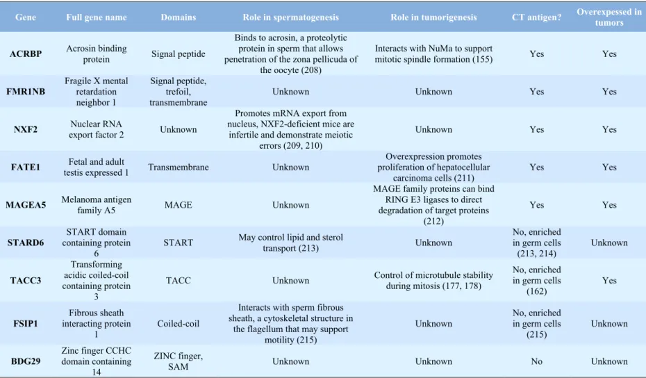
CT-antigens have diverse cellular functions in everything from modulating protein-protein interactions to regulating transcription (93). However, a function for the majority of the CT-antigens has not been assigned (93). It is therefore unclear whether most of the CT-CT-antigens are simply byproducts of dysregulated gene expression in tumor cells or whether they have a functional role in supporting tumorigenic phenotypes. This is an important distinction therapeutically because CT-antigens with no functional role are likely mainly useful as vaccine targets while CT-antigens with a functional role in supporting tumor phenotypes could be targeted with small molecules. Recent work demonstrated that the CT-antigen ACRBP functionally supports ovarian tumorigenesis by modulating expression of the microtubule-associated protein NuMa (155). Additionally, other CT-antigens have demonstrated functional relevance to tumor cell phenotypes (156, 157). This data suggests a functional role of at least some CT-antigens in tumorigenesis but the role of the vast majority of CT-antigens remains a black box. The known functions of the CT-antigens and gametogenic genes discussed in Chapter 3 of this dissertation are detailed in Table 3.1.
is strongly upregulated during gametogenesis and in tumor tissue but is also present in normal adult tissues, albeit at a lower level. This expression pattern and the rich functional knowledge about TACC3 compelled us to utilize it as a model for studying the gametogenic genes in cancer.
Transforming acidic coiled-coil containing protein 3
The TACC family - Transforming acidic coiled-coil containing protein 3 (TACC3) is a
member of the TACC protein family. This family shares a common TACC domain at the C-terminus that associates members of the family with microtubules and allows them to modulate microtubule stability (158, 159). There are three TACC proteins in humans (TACC1-3) but only one TACC protein in mouse (AINT), Drosophila (D-TACC), and Xenopus (Maskin) (160). All the human TACC genes are located in chromosomal regions that are commonly rearranged in cancer (55, 160, 161) but only TACC1 is directly transforming (161). Outside of the TACC domain, members of this family have distinct N-terminal signaling domains that allow TACC proteins to impact a range of processes including transcription and polyadenylation (160).
TACC3 expression pattern - TACC3 demonstrates a pattern of expression that suggests
controlled in a cell-cycle specific manner.
Role of TACC3 in mitosis - During mitosis TACC3, which is expressed in the nucleus
of interphase cells, is phosphorylated by aurora kinase A (AURKA) at three consensus AURKA phosphorylation sites (160, 172, 173). Phosphorylation at these sites directs TACC3 to a diffuse region around the centrosome in mitotic cells (158, 174). Mutation of the AURKA phosphorylation sites or treatment with the AURKA inhibitor VX-680 impairs TACC3 phosphorylation and inhibits its localization to the centrosome (172, 175). Once at the centrosome, TACC3 recruits and binds to the microtubule stabilizing protein CKAP5 (TOGp) (176). CKAP5 functions by opposing the depolymerizing activity of the protein kinesin family member 2C (MCAK) to allow for increased microtubule growth and stability (177, 178). This important role in stabilizing microtubules probably accounts for the synergy previously observed by other groups between TACC3 knockdown and paclitaxel treatment (179, 180). Therefore, TACC3 functions as a microtubule stabilizing protein and loss of TACC3 leads to decreased microtubule stability in vitro (176).
Role of TACC3 outside of mitosis - Aside from its most well studied role in mitosis,
polyadenylation, similarly to that observed in Xenopus. Second, TACC3 has been shown to have a role in driving cellular differentiation, potentially by controlling activity of transcription factors (163, 165, 181). This has relevance to this dissertation since a screen to identify small molecules that promote neuronal differentiation uncovered a TACC3 inhibitor that is used in Chapter 3 (182). Finally, the mouse TACC3 protein, AINT, may regulate the cellular response to hypoxia by binding to and altering the localization of the Ah receptor nuclear translocator protein (ARNT) (183). ARNT heterodimerizes with HIF-1alpha in the presence of low oxygen tension to induce transcription from hypoxia response element containing genes (183). This is relevant to this dissertation because the TACC3 inhibitor used in Chapter 3 likewise appears to influence response to hypoxia. Therefore, TACC3 can influence several important processes outside of its role in mitosis.
Thesis Summary
This dissertation will cover two processes that may impact responsiveness of tumor cells to mitotic stress: polyadenylation and gametogenesis. Both these projects arose from findings in genome-wide paclitaxel chemosensitizer screen (4). In particular, Chapter 2 will focus on the potent screen hit SYMPK while Chapter 3 will focus on the screen hit TACC3.
affect tumor, as opposed to normal cell lines, suggesting a unique dependence of tumor lines on polyadenylation for mitotic progression.
In Chapter 3 “TACC3 and multiple gametogenic genes support the cancer cell mitotic spindle” we study the links between several gametogenic genes and mitosis. We begin this work by depleting a panel of cancer-testis antigens and gametogenic genes from the NSCLC line H1155 in the presence of paclitaxel and showing how this impacts mitotic progression. Next, we focus on the gametogenic gene TACC3 and demonstrate TACC3 is uniquely required for mitosis in tumor cell lines. In particular, we perform immunofluorescence and live-cell imaging studies in a NSCLC progression model to determine the point during tumor evolution when TACC3 becomes required for mitosis. Finally, we use a TACC3 inhibitor to demonstrate that gametogenic genes can be targeted with small molecules and that doing so can increase the efficacy of current anti-mitotic drugs.
CHAPTER II
SYMPLEKIN IS REQUIRED FOR APPROPRIATE MICROTUBULE
FUNCTION AND MITOSIS
Elements of the work referenced in this chapter have been published in:
Summary
Introduction
Pangenomic loss of function screening is emerging as an effective tool for revealing the
components that support core biological processes including viral infection, DNA repair,
chemotherapeutic responsiveness, melanogenesis and endocytosis (4, 184-188). A number of
screening efforts have focused on identifying those gene products that are required for mitotic
progression in both the normal and tumorigenic setting (38, 41, 189-191). These screens have
successfully returned validated mitotic participants but also have isolated a diverse set of
unanticipated genes whose encoded proteins have no previously described role in mitotic
progression but instead have well established roles in processes such as transcription (189-191),
RNA splicing and translation (190, 191) and vesicle transport (190); thereby revealing an
unexpected diversity in the compendium of gene products supporting mitosis.
We have recently applied a genome-wide loss of function paclitaxel synthetic lethal strategy to identify genes that modulate chemoresponsiveness in non-small cell lung cancer cells (NSCLC) (4). This strategy returned a diverse set of gene products, including symplekin, whose depletion was the most potent for sensitizing NSCLC to a dose of paclitaxel that has no detectable impact on cell viability. Symplekin is a scaffold protein that supports the assembly of polyadenylation machinery on pre-mRNA transcripts; however no role for symplekin in drug sensitivity or mitosis has been reported (124). Polyadenylation is essential for the maturation of most pre-mRNAs and regulates mRNA nuclear export, stability and translation (100). In Xenopus laevis oocytes, the polyadenylation of specific meiotic transcripts is regulated such that their activation only occurs following meiotic maturation signals (120, 192, 193). In mammalian cells, the poly(A) tail length of specific transcripts changes in a cell cycle dependent manner (194), suggesting that cytoplasmic polyadenylation is a conserved mechanism for exerting translational regulation of gene expression prior to and during cell division.
the tumor cells. We find that symplekin is required to support bipolar spindle formation in multiple NSCLC derived tumor cells and that symplekin depletion impairs proliferation of NSCLC cells in vivo. The basis of symplekin’s contribution to mitotic progression appears to be at the level of microtubule function and expression of a critical component of the microtubule polymerization machinery, CKAP5 (TOGp). Depletion of other polyadenylation components causes similar alterations in CKAP5 expression and mitotic progression. Therefore, our results demonstrate that mitosis is acutely sensitive to perturbations of the polyadenylation machinery and suggest inhibition of polyadenylation may synergize with current anti-mitotic agents.
Results
Symplekin is required for mitotic spindle integrity - Symplekin was originally
Figure 2.1
Figure 2.1 SYMPK is required for spindle integrity after exposure to paclitaxel
aberrant mitosis. Indeed, symplekin depleted H1155 cells exposed to 10 nM paclitaxel displayed a high frequency of multipolar spindles characterized by disorganized tubulin and multiple centrosomes as compared to control-transfected cells (Figure 2.1E). These data suggest that symplekin function is directly coupled to the ability of cells to form a normal bipolar spindle.
Symplekin is required for high fidelity mitosis - Given our observations that symplekin
contributes to bipolar spindle formation, we directly assessed the consequence of symplekin depletion on mitotic progression in real-time by live imaging of H1155 cells stably expressing the chromatin marker GFP-H2B. By performing single-cell lineage tracing, we measured both the length and outcome of mitosis in symplekin and control siRNA transfected cells (Figure 2.2A). As expected, control or symplekin depletion alone had little effect on either mitotic fate or mitotic timing (Figure 2.2A and 2.2B). However, symplekin depleted cells exposed to 10 nM paclitaxel exhibited a significantly prolonged mitosis as compared to control transfected and paclitaxel treated cells (Figure 2.2B). The outcome of this prolonged mitosis was aberrant in 75% of the individual cells studied. In particular, instead of the formation of 2 daughter cells, symplekin depleted samples underwent either apoptosis, micronucleation or a multipolar mitosis following mitotic arrest (Figure 2.2A and 2.2C). Taken together, these observations suggest that symplekin supports mitotic spindle formation and mitotic progression in NSCLC.
Symplekin is necessary for mitosis in diverse NSCLC genetic settings - To determine
Figure 2.2
Figure 2.2 SYMPK is required for normal mitotic progression in tumor cells
Figure 2.3 SYMPK is necessary for mitosis in multiple tumor cell lines
We next sought to determine the impacts of prolonged suppression of symplekin expression in NSCLC cells. To this end, we stably repressed symplekin expression in H1299 cells using an shRNA-mediated system where we pooled two effective shRNAs targeting symplekin (Figure 2.3C). Stably repressing symplekin expression in the H1299 NSCLC line led to an increase in micronucleation in the absence of paclitaxel (Figure 2.3C). In our original screening cell line, H1155, stable repression of symplekin led to an increase in mitotic figures in the absence of paclitaxel (Figure 2.3D). Extending this analysis to two additional NSCLC lines and immortalized BJ fibroblasts revealed that the generation of micronucleated cells following prolonged symplekin depletion is a common phenomenon in NSCLC but not normal diploid fibroblasts immortalized with hTERT (Figure 2.3E). Thus, while the transient impacts of symplekin depletion are observable only in the presence of a microtubule disrupting agent, prolonged symplekin depletion alone increases the frequency of aberrant mitosis and mitotic arrest in tumor, but not normal, cells.
Loss of symplekin impairs tumor formation in a mouse xenograft model - To directly
Figure 2.4
Figure 2.4 Depletion of SYMPK impairs tumor growth in vivo
Symplekin modulates microtubule polymerization - The formation of a normal,
bipolar spindle apparatus is exquisitely dependent on proper microtubule function, which is significantly altered in the presence of chemotherapeutic drugs such as paclitaxel (43). Given the symplekin-paclitaxel synthetic lethal phenotype we observe in NSCLC, we probed microtubule polymerization efficiency in H1299 cells following transient depletion of symplekin using a microtubule regrowth assay. Here, H1299 cells transfected with control or symplekin siRNAs were exposed to a high dose of nocodazole to induce microtubule depolymerization. Microtubules were depolymerized to a similar degree in both control and symplekin transfected samples (Figure 2.5A and 2.5B). However, after 10 minutes of recovery, symplekin depleted cells displayed little microtubule regrowth from their centrosomes. A similar trend was observed in mitotic cells, where growth of microtubules from both the spindle poles and the kinetochores was significantly attenuated in symplekin depleted samples (Figure 2.5A and 2.5B). Similar phenotypes were observed in both H1299 and H1155 cells when we performed the experiment by an in vitro microtubule stability assay where polymerized tubulin is sedimented after the depolymerization and recovery steps and analyzed by immunoblotting (Figure 2.5C and 2.5D). Thus, depletion of symplekin significantly alters microtubule polymerization, a process that is essential for normal spindle formation.
SYMPK depletion leads to loss of CKAP5 - Symplekin is a multifunctional protein
Figure 2.5
Figure 2.5 Depletion of SYMPK reduces microtubule stability
Figure 2.6 SYMPK depletion leads to loss of CKAP5
we hypothesized that symplekin could be impacting expression of proteins required for mitotic spindle formation. To assess this possibility, we analyzed a panel of proteins required for proper centrosomal maturation and microtubule nucleation (176, 196-201). Symplekin depletion alone had no detectable effect on the localization and expression of almost all proteins studied (Figure 2.6B). However, symplekin depletion had a profound effect on the expression of CKAP5 as detected by both immunofluorescence (Figure 2.6C) and immunoblot analysis (Figure 2.6D and 2.6E). CKAP5 displays an elevated expression pattern in tumor cells (56) and localizes to the centrosome where it enhances microtubule polymerization and nucleation (57, 178). CKAP5 depletion significantly decreases viability of H1155 and H1299 cells (Figure 2.6F). This effect is likely due to the potent impact of CKAP5 on microtubule polymerization and stability in H1299 and other cell types (Figure 2.6G) (176, 202, 203).
SYMPK alters CKAP5 levels post-transcriptionally and independently of protein
degradation - Given symplekin’s role in transcription and translation, we evaluated CKAP5
transcript levels in symplekin-depleted cells. Symplekin depletion did not affect the level of CKAP5 mRNA, suggesting a post-transcriptional mode of regulation (Figure 2.7A). To determine whether the effects on CKAP5 were mediated by increased degradation, we evaluated CKAP5 protein levels in symplekin depleted cells exposed to the proteosome-inhibitor, MG-132. CKAP5 levels were globally increased in MG-132 treated cells, however, proteosome inhibition was not sufficient to rescue the reduced CKAP5 levels observed in symplekin depleted cells (Figure 2.7B). Since CKAP5 stabilizes microtubules primarily by opposing the depolymerizing activity of MCAK, we evaluated the impact of codepletion of SYMPK and MCAK on the microtubule network. In H1299 cells, MCAK depletion impairs microtubule depolymerization by nocodazole as has previously been reported (44). Importantly, co-depletion of MCAK and SYMPK results in the depolymerization of microtubules in the presence of nocodazole. (Figure 2.7C).
Multiple polyadenylation components collaborate with paclitaxel - Symplekin is a
Figure 2.7
Figure 2.7 SYMPK alters CKAP5 levels post-transcriptionally
regulating mitotic progression through phase-specific changes in poly(A) tail length (194). To determine if attenuation of the polyadenylation complex in general can collaborate with paclitaxel, we retrospectively examined the impact of the 14 core polyadenylation proteins in our original genome-wide paclitaxel sensitivity screen (4). In addition to symplekin, depletion of the polyadenylation proteins CPSF1, CSTF2 and CPSF3 all enhanced paclitaxel sensitivity to some degree in our primary screen. In H1299 cells, depletion of both CPSF1 and CPSF3 reduced CKAP5 protein levels as detected by immunoblot analysis (Figure 2.8A), suggesting that CKAP5 expression is exquisitely sensitive to perturbations of the polyadenylation complex. Additionally, H1299 cells depleted of CSTF2, CPSF1 or CPSF3 and exposed to paclitaxel demonstrated a significant increase in the occurrence of multi and micronucleated cells (Figure 2.8B). To determine if these subunits impacted mitotic progression in a similar manner to symplekin, we employed our time-lapse imaging system in H1155 GFP-H2B cells to evaluate mitotic outcomes. As with symplekin, CPSF1, CPSF3 and CSTF2 depletion increased the frequency of abnormal mitotic exits (Figure 2.8C). Thus, altered expression of multiple polyadenylation components has acute effects on mitotic fidelity.
Discussion
Figure 2.8
Figure 2.8 Polyadenylation is required for CKAP5 expression and mitosis
In particular, we find that the protein expression level of a critical mitotic component, CKAP5, is sensitive to depletion of polyadenylation machinery. While we have not yet determined if CKAP5 protein expression can be directly regulated by polyadenylation, we findthat symplekin depletion does not appear to affect CKAP5transcript abundance or protein turnover. Thus, the changes we observe in CKAP5 protein expression could be due to an alteration in translation initiation or mRNA stability, which could be a direct result of the depletion of key polyadenylation components. Alternatively, perturbations in polyadenylation machinery, which may be impacting a large set of set of transcripts (194), could alter endogenous mechanisms that regulate CKAP5 protein levels. In either case, we have revealed that mitosis, microtubule dynamics and CKAP5 levels are sensitive to alterations in the polyadenylation machinery.
while decreasing adverse events in normal tissues.
Experimental Procedures
Cell culture - H1155, H1299, HCC366 and HCC515 cells were a gift from John Minna.
All cells lines had recently been genotyped using SPR analysis. Cells were maintained in RPMI (Gibco) with 5% FBS as described (4). BJ fibroblasts immortalized with hTERT were a gift from Fred Grinnell (UT-Southwestern). BJs were maintained in DMEM + 10 % FBS.
Cell Titer Glo Assays - Cell Titer Glo assays were performed using independent siRNAs
from the siGENOME SMART pool targeting symplekin as previously described (4).
siRNA Transfections - Transfections were performed as described with siGENOME
SMART pools (ThermoFisher). Cells were transfected for either 72 or 96 hours as indicated in the figure legends. As a control, either a mismatch siRNA or an siRNA targeting DLNB14, which has no detectable impact in our assay system, was used.
High-Content Imaging - H1155 GFP-histone 2B-expressing cells were obtained by
retroviral transduction. Retrovirus was produced by Fugene (Roche) transfection of 293gp cells with pCLNCX-GFP-H2B (a gift from Gray Pearson, UT-Southwestern) and VSV-G and virus was harvested at 48 hours post-transfection. H1155 cells at 50% confluency were transduced with virus in 4 ug/mL Polybrene and stably expressing cells were selected using 600 ug/mL Geneticin (Gibco). For imaging, cells were reverse transfected with the indicated siRNAs, plated in a 96 well format and exposed to paclitaxel at 48 hours post-transfection. 24 hours post-paclitaxel treatment, the cells were imaged on a BD Pathway 855 bio-imager using a 40X or 20X high-NA objective. Images were taken every 15 minutes for the next 48 hours and an image sequence was generated using Image J. Manual quantification was used for the indicated parameters.
Flow cytometry - H1155 cells were fixed in 50% ethanol/PBS, washed and resuspended
software package (Verity Software House).
Lentivirus production - shRNA clones in the PLKO1 vector were obtained from The
RNAi Consortium (Open Biosystems). Lentivirus targeting SYMPK was produced by Fugene-mediated transfection of 293T cells with plasmids for VSV-G, ∆8.9 and shRNA’s targeting SYMPK or GFP (SYMPK clones TRCN0000141511 and TRCN0000144902 were effective). Virus was harvested at 48 hours post-transfection and used to infect cells at 50% confluency in conjunction with 5 ug/uL Polybrene. Infection rates based on GFP performed in parallel were over 90 %.
Quantitative real-time RT-PCR - Total RNA was collected from H1299 cells using the
GenElute Mammalian Total RNA Miniprep Kit (Sigma). cDNA was synthesized from 2 ug total RNA using the High-Capacity cDNA reverse transcription kit (Applied Biosystems). Real-time RT-PCR used inventoried TaqMan gene expression assays designed to detect mRNA exclusively and the 7500 Fast real-time PCR system (Applied Biosystems). Actin or GAPDH was used as the endogenous control and cells transfected with control siRNA were used for calculating differences in expression by the 2-∆∆CT method. For CKAP5 levels following SYMPK reduction, results are from pooling of 3 individual experiments performed in duplicate in which the average endogenous control Ct values between conditions never varied more than 0.3. For measurement of transcript knockdown, experiments were performed in triplicate with Ct values between conditions never varying more then 0.6.
Immunoblotting - Cells were lysed directly in boiling sample buffer (100 mM Tris-Cl,