Address for correspondence Mr. Atta Ur Rehman, Department of Zoology Hazara University, Mansehra Post Code: 21300, Pakistan
Email: [email protected]
Original Article
Cutaneous leishmaniasis in Dir Lower District,
North-West Pakistan: epidemiology and treatment response
Introduction
Leishmaniasis is not a single entity rather it refers to a group of diseases caused by a variety of protozoan parasites of the genus Leishmania. The clinical features of the disease vary from a localized cutaneous affection to a more generalized and life-threatening visceral disease. Clinically, leishmaniasis can be categorized as cutaneous, mucocutaneous and visceral
leishmaniasis, each caused by a distinct parasite. Cutaneous leishmaniasis (CL), caused by L.
tropica is usually a self-limiting disease,
however, it may take several months before complete healing occurs, with residual disfiguring scars. CL is a vector-borne disease and can be transmitted either from animals to human (zoonotic) or from humans to humans (anthroponotic) through the bite of sandflies from the subfamily Phlebotimidae. Moreover, the disease can also be transmitted directly from person to person through the sharing of needles, as is often the case among the intravenous drug users.1,2
Hidayat Ur Rahman, Atta Ur Rehman
Department of Zoology, Hazara University, Mansehra
Abstract Objective To assess the epidemiological and chemotherapeutic aspects of cutaneous leishmaniasis.
Methods This study was carried out during September, 2011 to December, 2011 and encompassed
a total of 1000 patients of both gender. All patients enrolled in this study were selected at random from four sub-divisions of Dir Lower district. Data regarding different variables were obtained on a designed questionnaire through face-to-face interviews with the patients after approval of their informed consent. Standard protocols were used for confirmation of Leishman-Donovan bodies using slit-skin smears.
Results The overall incidence of CL was found to be 21.1%. The prevalence was observed to be significantly higher among Afghans than the local population (32.5% vs. 20.3%, OR: 1.6, p<0.05). The distribution of CL showed significant difference with respect to all studied demographic parameters, except subject’s age and tehsil. More than half of the infected patients (56.8%) had wet lesions and majority of the infected patients (42.3%) had only one lesion. Moreover, face and arms were the most frequently inflicted body parts. It was further observed that among various antimonials used by the patients, sodium stibogluconate demonstrated the highest efficacy followed by glucantime® and stibotim® whereas thermotherapy showed no efficacy. Collectively, the efficacy significantly varied across different antimonials used in this study (p<0.05).
Conclusion This study provides detailed information regarding descriptive epidemiological aspects of cutaneous leishmaniasis in Dir Lower District.
Key words
Leishmaniasis is endemic in many tropical and subtropical regions affecting more than 12 million people in at least 88 countries of the world.3 According to the World Health Organization (WHO), the global annual incidence is estimated to be 1-1.5 million cases of CL and 0.5 million cases of the visceral form. About 90% cases of both visceral and cutaneous leishmaniasis have been reported alone from countries like Bangladesh Afghanistan, Saudi Arabia, Syria, Brazil, and Peru.4 Records are not available as to when and by whom the disease was first reported in Pakistan, however, CL has been found as one of the emerging diseases in parts of north-western Pakistan and the neighboring Afghanistan. The remarkable increase in the incidence of CL has been witnessed in areas of Afghanistan where civil unrest resulted in deterioration of the infrastructure and huge population displacements.5 In Pakistan, the first major outbreak of leishmaniasis was reported from Quetta, Baluchistan in 1935 following a severe earthquake. Afterwards another epidemic of the disease was witnessed in 1997 in an Afghan refugee camp in Dir district in north-west Pakistan along the Pak-Afghan border. Soon after, the biggest outbreak of leishmaniasis occurred in districts Dadu and Larkana of Sindh province in 2001. Since then, the status of leishmaniasis in Pakistan has been changing and both forms of the disease are being reported from various parts of the country.6,7
Although widespread but uneven in distribution, CL has always been considered as non-endemic in Pakistan8 except Baluchistan province where CL is considered endemic.9 In Pakistan, majority of the previous studies have focused on prevalence of either cutaneous or visceral leishmaniasis in the north-west and south-west populations of Khyber Pakhtunkhwa and Baluchistan provinces. These studies mainly focused on the suspected patients of
leishmaniasis and were restricted to the Afghan refugees rather than the local Pakistani populations.7,10-12 Nevertheless, the anticipated risk of potential spread of the disease to the local communities was always considered a big health concern. Dir Lower District has a long history of hosting the Afghan refugees during periods of civil unrest in Afghanistan. Even today, frequent migrations of Afghan refugees and Pakistani locals across the Durand line have been witnessed in this part of the country. Therefore, this study was carried out in Dir Lower district with the aim to investigate epidemiological and chemotherapeutic aspects of cutaneous leishmaniasis in local, as well as, Afghan refugees living in the region.
Methods
Study Area Dir Lower district is located
between coordinates 34°-38° North latitude and 71°-72° East longitudes in the temperate zone
about 1248 feet above sea level.
Administratively, the district is divided into four subdivisions, namely Timergara, Samar Bagh, Adenzai, and Maidan. Geographically, Dir Lower is bordered by district Chitral in the north, district Swat in the east, Bajaur Agency in the south and Afghanistan in the west (Figure 1). Dir Lower District has an area of 5282 km2 and home to a population of 1.4 million individuals.13
provided with a Basic Health Unit (BHU) which runs under the auspices of World Health
Organization, HealthNet International and
Pakistan Government. The Afghan refugees, as well as, the local Pakistani population living in the region, frequently visit this BHU for their treatment.
Sample collection A total of 1000 subjects who
visited the BHU during September-December, 2011 for their treatment were enrolled in this study. These subjects originated from different geographic locations, age groups, ethnicities, sexes, and occupations. Data regarding different variables were obtained on a designed questionnaire through face-to-face interview with the patient. Informed consent of each patient was taken into account prior to sample collection. Photographs of the lesion were taken using a digital camera (Sony, WX80®). Each subject was examined thoroughly for number of lesions, body parts involved, type of lesion and duration of the lesion. The lesions were classified as wet or dry on the basis of their clinical presentation. Duration of lesions was also confirmed by the patient itself and recorded in months. Participants in this study were divided into two groups, i.e.well-aware and less-aware, based upon their level of awareness about leishmaniasis. People who had prior information about leishmaniasis and its vector (sandfly) were classified as well-aware while those lacking such information were categorized as less-aware.
Slide preparation and microscopic examination
Slit-skin smears were prepared from the margins of the lesions using sterilized blood lancet. The smears were fixed with absolute ethanol followed by staining with 5% Giemsa stain and examined under a microscope. Detection of Leishman-Donovan (LD) bodies in the slit-skin smear was considered as a positive case. The nutrient broth (NB) medium was used for
culture. The nutrient broth was autoclaved at 121ºC for 15 minutes and by sterile wire loop slanted in tubes. Tubes were closed properly to avoid contamination. After 24 hours, the culture was studied to observe the growth of promastigote stage. Pictures of the promastigote stage were taken by using stereomicroscope.
Therapy A total of 93 patients were enrolled for
two types of treatment purposes i.e.
chemotherapy and thermotherapy. Of the total 93 patients enrolled for treatment purposes, 74
had received chemotherapy whereas the
remaining 19 patients had received
thermotherapy. The various antimonial drugs used in chemotherapy treatment included sodium stibogluconate (Albert David Limited,
Kolkata, India®), glucantime (Aventis,
France®), and stibotim (Star Laboratories, Pakistan®). In the case of chemotherapy (n=74), sodium stibogluconate (n=33) was injected intralesionally20 @ 50 mg per lesion whereas glucantime (n=24) and stibotim (n=17) were both injected intramuscularly @ 15mg/kg each as per body weight of patient. The patients treated with chemotherapy were followed for 21 days each. In the case of thermotherapy (n=19), the patients received the treatment on the surface of their lesions using Hand-Held Exothermic Crystallization Therapy for CL (HECT-CL) and the patients were followed for 30 days
Data analysis All the data were entered into
Microsoft Excel (Ver. 2007) and analyzed using Graphpad Prism software (Garstman, 2008). Chi-square (χ2) and Fisher’s exact test were used for checking statistical significance. P value <0.05 was considered significant. In order to
determine the correlation of different
Results
In the present study, a total of 1000 individuals comprising both sexes were randomly selected from the study area and analyzed for the presence of LD antibodies. Overall, 213 (21.3%) individuals were found positive for LD bodies. Gender-wise analysis of the data revealed significantly higher prevalence of cutaneous leishmaniasis among male subjects compared to female counterparts (24.5% vs. 16.1%; OR 1.5;
p=0.0017), (Table 1). Age-wise analysis of the data revealed the highest prevalence of CL in the age group 1-10 years whereas least prevalence was witnessed in the age group 31-40 years (25.1% vs. 14.3%; OR 1.7:1). Although, the collective prevalence rate was relatively higher in the lower age groups (>30 years) compared to higher age groups (>30 years), the difference was statistically not significant (p>0.05).
Furthermore, the prevalence of CL was checked across different subdivisions of Dir Lower District. In this regard, highest prevalence of CL was reported in Samar Bagh subdivision (24%) which was followed by subdivisions Adenzai and Timergara (21% each) and Maidan (18%)
(Table 1, Figure 1). Analyzing the data statistically; however, showed no significant difference across four subdivisions of Dir Lower district (p>0.05). When the data were analyzed with respect to the ethnic origin of the subject, significantly higher prevalence of CL was noted among the Afghans ethnicity compared to the local Pakistani population (32.5% vs. 20.3%; OR 1.6:1, p=0.0091), (Table 1). Additionally, when the data were analyzed with respect to subject’s occupational groups, the highest prevalence of CL was noticed among shepherds (51.9%; OR 5.6), followed by laborers (13.1%; OR 1.4), and merchants (11.1%; OR 1.2)
whereas least prevalence of CL was observed among teachers (9.2%). Analyzing the data
statistically showed significantly higher
difference across all occupational groups of the subjects (p=0.0001), (Table 1).
The data were further classified into two categories i.e. well-aware and less-aware, based upon level of awareness of each individual about CL and its transmission. When the data were analyzed in this respect, the prevalence of CL was found to be significantly higher (p=0.0001) among individuals with less awareness about the disease and its vector compared to individuals who were well aware of the disease and its vector (25.0% vs. 11.5%; OR 2.1:1). Similarly, the month-wise analysis of the data showed the highest prevalence in October (26.2%; OR 2.0) followed by September (23.4%; OR 1.7), November (17.6%; OR 1.3) and December (13.0%). Thus, the prevalence of CL showed statistically significant difference (p<0.05) across four months of the study period (Table 1).
The data were further analyzed with respect to types of lesion involved, the number of lesions per person and the body parts affected by the parasite (Figure 2). Of the 213 positive samples, 121 (57%) patients had developed wet type of lesions whereas the remaining 92 (43%) patients had only dry lesions. Although a great majority of the inflicted individuals had either one or two lesions (42% and 29%, respectively), a very small number of them had either three or more than three lesions on their body (11% and 18%, respectively). In the same way, majority of the patients had lesions on face (25.4%) which was followed by arms (21.6%), legs (15.5%), mixed (13.6%), nose (12.7%), and feet (11.3%),
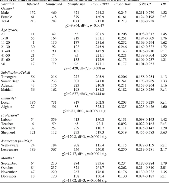
Table 1Prevalence of cutaneous leishmaniasis across different variables in Dir Lower District
Variable Infected Uninfected Sample size Prev. /1000 Proportion 95% CI OR Gender*
Male 152 469 621 244.8 0.245 0.211-0.279 1.52
Female 61 318 379 160.9 0.161 0.124-0.198 Ref.
Total 213 787 1000 213.0 0.213 0.188-0.238
χ2=9.864, df=1, p=0.0017 Age (years)
<1 11 42 53 207.5 0.208 0.098-0.317 1.45
1-10 55 164 219 251.1 0.251 0.194-0.309 1.76
11-20 41 136 177 231.6 0.232 0.169-0.294 1.62
21-30 30 92 122 245.9 0.246 0.169-0.322 1.72
31-40 15 90 105 142.9 0.143 0.076-0.210 Ref.
41-50 21 74 95 221.1 0.221 0.138-0.304 1.55
51-60 23 110 133 172.9 0.173 0.109-0.237 1.21
>61 17 79 96 177.1 0.177 0.101-0.253
χ2=5.428, df=7, p=0.608 ns Subdivisions/Tehsil
Timergara 56 216 272 205.9 0.206 0.158-0.254 1.13
Samar Bagh 74 233 307 241.0 0.241 0.193-0.289 1.33
Adenzai 47 176 223 210.8 0.211 0.157-0.264 1.16
Maidan 36 162 198 181.8 0.182 0.128-0.236 Ref.
χ2=2.677, df=3, p=0.444 ns Ethnicity*
Local 186 731 917 202.8 0.203 0.177-0.229 Ref.
Afghan 27 56 83 325.3 0.325 0.225-0.426 1.60
χ2=6.81, df=1, p=0.0091 sig. Profession*
Labour 54 359 413 130.8 0.131 0.098-0.163 1.42
Teacher 6 59 65 92.3 0.092 0.022-0.163 Ref.
Merchant 32 257 289 110.7 0.111 0.075-0.147 1.20
Shepherd 121 112 233 519.3 0.519 0.455-0.583 5.63
χ2=170.8, df=3, p<0.0001 sig. Awareness (n=964)*
Well-aware 24 184 208 115.4 0.115 0.072-0.159 Ref.
Less-aware 189 567 756 250.0 0.250 0.219-0.281 2.17
χ2=17.17, df=1, p<0.0001 sig. Months*
September 64 210 274 233.6 0.234 0.183-0.284 1.79
October 84 237 321 261.7 0.262 0.214-0.310 2.01
November 47 220 267 176.0 0.176 0.130-0.222 1.35
December 18 120 138 130.4 0.130 0.074-0.187 Ref.
χ2=13.02, df=3, p=0.0046 sig.
*:Statistically significant variable, sig:significant, ns:not significant, CI:Confidence Interval, OR=Odds Ratios Table 2Efficacy of different antimonials used against cutaneous leishmaniasis
Type of antimonial/therapy used
Drug route*
Drug dosage
Duration (weeks)
Total treated
Total recovered
Efficacy %
Sodium Stibogluconate IL 50 mg/day 3 33 27 81.8
Glucantime IM 15 mg/Kg/day 3 24 10 41.7
Stibotim IM 15 mg/Kg/day 3 17 6 35.3
Figure 1 Map of study area (A= Map of Pakistan with the study area shown in star, B=Map of Dir Lower district showing four subdivisions with their respective prevalence of cutaneous leishmaniasis).
The efficacy of different antimonials and types of therapy used in this study is shown in Table 2. Among the various antimonial drugs used as chemotherapy against cutaneous leishmaniasis, Sodium Stibogluconate resulted in the highest efficacy rate (81.8%) followed by Glucantime (41.7%) and Stibotim (35.3%). Thermotherapy, on the other hand, showed no efficacy. Overall, the efficacy rates of different antimonials and/or therapy used in this study were observed to show significant variation (p<0.0001).
Discussion
Our results show 21% incidence of CL in the human population of Dir Lower district which is significantly lower than a previous estimate (51%) by Ullah et al. (2009).7 A number of factors could possibly explain this observed disagreement between these two studies. Firstly, the difference may be a temporal variation in the incidence of CL as the population of Dir Lower district has undergone significant socio-demographic, geo-political and agro-climatic shifts over the years. For example, there is rapidly growing urbanization and industrialization in the area which is coupled with an increase in literacy level and public awareness about health. This may have collectively impacted the life standard of the people which could indirectly explain the reduced incidence of CL in the region. Moreover, the unprecedented incidence of CL as previously reported by Ullah et al.7 may be due to the fact that only clinically suspected patients with visible lesions/scars on their body were enrolled in their study. Understandably, enrollment of only clinically diagnosed patients could be the reason for such a high incidence of CL in their study. Conversely, our study encompassed patients who presented with lesions on their body clinically suggestive of leishmaniasis, confirmed by detection of LD bodies in their slit-skin smears. Thus, the
observed disparity between the two reports could reasonably be attributed in part to the different methodologies adopted during the two studies.
Before 1979, only sporadic cases of leishmaniasis were reported in the western territories of Pakistan. However, soon after the invasion of Soviet Union in Afghanistan 1979, marked increase in the prevalence of leishmaniasis was noticed in north-western and south-western territories of Pakistan along the Durand Line. It is, therefore, strongly believed that leishmaniasis was probably transmitted by the huge influx of Afghan refugees from Afghanistan, an endemic country, to Pakistan where the disease was non-endemic.14 Our study strongly supports this idea as we have found the significantly higher incidence of CL in Afghan refugees than the local Pakistani population (32.5% vs. 20.3%; OR 1.6:1.0; p=0.0091). This is strike contrast with a previous understanding that leishmaniasis has been equally affecting both Afghans and local Pakistani population without any significant difference.5,15 Our findings are also in close lines with a previous study by Nisar (2002) who reported almost similar results. Contrastingly; however, our results do not agree with several previous reports which suggest the higher incidence of CL in local population than the Afghan refugees.7,12,16
leishmaniasis was not significantly different (p>0.05) across different geographies (i.e. subdivisions) of district Dir Lower. The apparently higher incidence rate of leishmaniasis in Samar Bagh subdivision (24.1%) compared to other localities could possibly be attributed to the frequent human migrations between Dir lower district and the neighboring Afghanistan on one hand and Bajaur Agency on the other hand. Thus, Samar Bagh could be used as a corridor for human migrations not only between two districts but between two countries as well. Moreover, the incidence of leishmaniasis showed a declining trend from west to east within the district which corroborates the transmission of the parasite from western Afghanistan towards parts of north-eastern Pakistan as a result of frequent human migration across the border.
Like several previous studies,7,12,18 we have found the significantly higher incidence of leishmaniasis in male compared to female individuals (male: female OR: 1.52:1,
p=0.0017). Highest incidence in male than female subjects could probably be due to the fact that males remain outdoor for most of their time for making their livelihood as they are the only breadwinners, whereas female are mostly restricted to the household activity. Additionally, it is obligatory for female to be at home before the dusk and to use fully-covered dresses as part of their cultural habits. Moreover, males have been seen while sleeping in the open fields outside their homes especially during summer when the temperature is high and there is the limited power supply in the region. In a nutshell, males are more exposed to sandfly bites compared to their female counterparts thus increasing the risk of leishmaniasis. Nevertheless, our results do not agree with several other studies which suggest that there is no correlation between rates of leishmaniasis and gender of the patient.5,17
In addition to several other factors, agro-climatic conditions like temperature, rainfall and humidity play an important role in the transmission of this vector-borne disease. We have observed that incidence of CL has been significantly varying across different moths of the year (p<0.05), October being the peak month. This is in agreement with a previous study by Durrani et al.6 (2011) who reported October and November as the peak months for the incidence of CL. With respect to awareness, leishmaniasis was significantly higher in subjects with poor knowledge about leishmaniasis and its vector than subjects who had good knowledge of the disease and its vector (p<0.0001). Thus, it would be reasonable to argue that public awareness about this disease and its vector could be an effective tool to minimize the future prospects of leishmaniasis.
half of the patients had wet lesions and a great majority of them (42.3%) had only one lesion throughout their body surface. Similarly, face and arms were the most common body parts inflicted by this disease which is in close lines with a previous report by.12
Among the various antimonial drugs used as chemotherapy for leishmaniasis, sodium stibogluconate showed highest efficacy (81.8%) followed by glucantime (41.7%) and stibotim (35.3%). Thus, our findings are in close conformity with a similar previous study in which sodium stibogluconate when used against visceral leishmaniasis resulted in 100% efficacy rate.19 Use of thermotherapy, on the other hand, resulted in no efficacy. One reason for higher efficacy of sodium stibogluconate compared to other two antimonials could be the fact that sodium stibogluconate was administered intralesionally whereas glucantime and stibotim were both administered intra-muscularly. It has been previously known that administration of antimonials intralesionally greatly reduces the duration of treatment and amount of dose needed for full recovery.20 In our study, it is difficult to define whether the difference observed in the efficacy of the antimonials is due to the different types of antimonials used or due to the different routes of drug administration.
Acknowledgements
The volunteer participation of all the patients in this study is highly acknowledged. We are greatly indebted to Dr. Fazal Rahim and Muhammad Jamel (Technician) for their cooperation during the whole study period. Finally, World Health Organization and HealthNet International is gratefully acknowledged for their financial assistance, logistic support and giving permission to undertake this study.
References
1. Rahim F, Jamal S, Raziq F, Uzair M, Sarwar B, Ali Het al. An outbreak of cutaneous leishmaniasis in a village of district Dir lower. J Pak Med Assoc. 2002;17:343-5. 2. Svobodova M, Votypka J, Nicolas L, Volf
P. Leishmania tropica in the black rat (Rattusrattus): persistence and transmission from asymptomatic host to sand fly vector Phlebotomus sergenti. Microbes Infect. 2003;5:361-4.
3. Pourmohammadi B, Motazedian MH, Hatam GR, Kalantari M, Habibi P, Sarkari B. Comparison of three methods for diagnosis of cutaneous leishmaniasis. Iran J Parasitol. 2010;5(4):1-8.
4. Kumar R, Bumb R, Ansari NA, Mehta RD, Salotra P. Cutaneous leishmaniasis caused by Leishmania tropica in Bikaner, India, parasite identification and characterization using molecular and immunologic tools. Am J Trop Med Hyg. 2007;76:896-901.
5. Brooker S, Mohammed N, Adil K, Agha S, Reithinger R, Rowland Met al. Leishmaniasis in refugee and local Pakistani populations. Emerg Infect Dis. 2004;10:1681-4.
6. Durrani N, Rowland M, Muniti A, Noyes H, Reyburn H. An outbreak of cutaneous leishmaniasis in an Afghan refugee settlement in north-west Pakistan. Trans R Soc Trop Med Hyg. 1999;93(9):133-6. 7. Ullah S, Jan AH, Wazir SM, Ali N.
Prevalence of cutaneous leishmaniasis in Lower Dir District (N.W.F.P), Pakistan. J Pak Assoc Dermatol. 2009;19:212-5. 8. Massoom M, Marri S. Current status of
leishmaniasis in Pakistan. Bhaduri A, Basu M, Sen A, Kumar S, editors. Current Trends in Leishmaniasis Research. Calcutta, India: Council of Scientific and Industrial Research; 1993. P. 231-6.
9. Talat H, Attarwala S, Saleem M. Cutaneous leishmaniasis with HIV - Case report. J Coll Physicians Surg Pak. 2014;24(Special Suppl 2):S93-S5.
10. Kolaczinski J, Brooker S, Reyburn H, Rowland M. Epidemiology of anthroponotic cutaneous leishmaniasis in Afghan refugee camps in northwest Pakistan. Trans R Soc Trop Med Hyg. 2004;98:373-8.
Dargai Region in Pakistan. Pak J Zool. 2013;45:537-41.
12. Pakistan Bureau of Statistics. Pakistan Census Report, 1998. In: Pakistan Bureau of Statistics GoP. Islamabad; 1998.
13. Mujtaba G, Khalid M. Cutaneous leishmaniasis in Multan, Pakistan. Int J Dermatol. 1998;37:843-5.
14. Nawaz R, Khan AM, Khan SU, Rauf A. Frequency of cutaneous leishmaniasis in an Afghan refugee camp at Peshawar. Gomal J Med Sci. 2010;8(1):16-9.
15. Gul B. Incidence of leishmaniasis in District Loralai, Baluchistan. Peshawar: University of Peshawar; 2001.
16. Fazaeli A, Fouladi B, Sharifi I. Emergence of cutaneous leishmaniasis in a border area at south-east of Iran: an epidemiological survey. J Vector Borne Dis. 2009;46:36-42.
17. Shirzadi MR, Esfahania SB, Mohebalia M, Ershadia MRY, Gharachorlo F, Razavia MR et al. Epidemiological status of leishmaniasis in the Islamic Republic of Iran, 1983-2012. East Mediterr Health J. 2015;21:736-42.
18. Rahim F, Rehman F, Ahmad S, Zada B. Visceral leishmaniasis in District Dir, NWFP. J Pak Med Assoc. 1998;48:161-3. 19. Hanif MM, Akram K, Mustafa G.
Intralesional versus oral chloroquine in cutaneous leishmaniasis: comparison of outcome, duration of treatment and total dose of drug. J Coll Physicians Surg Pak. 2016;26:260-2.