0095-1137/10/$12.00
doi:10.1128/JCM.01796-09
Copyright © 2010, American Society for Microbiology. All Rights Reserved.
Multilocus Sequence Typing of
Clostridium difficile
䌤
David Griffiths,
1,5Warren Fawley,
3Melina Kachrimanidou,
1,5Rory Bowden,
8Derrick W. Crook,
1,5Rowena Fung,
1,5Tanya Golubchik,
8Rosalind M. Harding,
6Katie J. M. Jeffery,
7Keith A. Jolley,
6Richard Kirton,
7Tim E. Peto,
1,5Gareth Rees,
7Nicole Stoesser,
1,5Alison Vaughan,
1,5A. Sarah Walker,
1,5Bernadette C. Young,
7Mark Wilcox,
2,3and Kate E. Dingle
4,5*
Nuffield Department of Clinical Medicine, Oxford University, John Radcliffe Hospital, Oxford OX3 9DU, United Kindgom
1; Department of
Microbiology, Institute of Molecular and Cellular Biology, University of Leeds, Leeds LS2 9JT, United Kingdom
2; Department of
Microbiology, The General Infirmary, Old Medical School, Leeds LS1 3EX, United Kingdom
3; Nuffield Department of
Clinical Laboratory Sciences, Oxford University, John Radcliffe Hospital Oxford OX3 9DU, United Kingdom
4;
National Institute for Health Research, Oxford Biomedical Research Centre Programme, John Radcliffe Hospital,
Oxford OX3 9DU, United Kingdom
5; Department of Zoology, Oxford University, South Parks Road,
Oxford OX1 3PS, United Kingdom
6; Department of Microbiology, Oxford Radcliffe NHS Trust,
John Radcliffe Hospital, Oxford OX3 9DU, United Kingdom
7; and Department of Statistics,
University of Oxford, 1 South Parks Road, Oxford OX1 3TG, United Kingdom
8Received 11 September 2009/Returned for modification 24 October 2009/Accepted 19 December 2009
A robust high-throughput multilocus sequence typing (MLST) scheme for
Clostridium difficile
was developed
and validated using a diverse collection of 50 reference isolates representing 45 different PCR ribotypes and
102 isolates from recent clinical samples. A total of 49 PCR ribotypes were represented overall. All isolates were
typed by MLST and yielded 40 sequence types (STs). A web-accessible database was set up (http://pubmlst
.org/cdifficile/) to facilitate the dissemination and comparison of
C. difficile
MLST genotyping data among
laboratories. MLST and PCR ribotyping were similar in discriminatory abilities, having indices of
discrimi-nation of 0.90 and 0.92, respectively. Some STs corresponded to a single PCR ribotype (32/40), other STs
corresponded to multiple PCR ribotypes (8/40), and, conversely, the PCR ribotype was not always predictive
of the ST. The total number of variable nucleotide sites in the concatenated MLST sequences was 103/3,501
(2.9%). Concatenated MLST sequences were used to construct a neighbor-joining tree which identified four
phylogenetic groups of STs and one outlier (ST-11; PCR ribotype 078). These groups apparently correlate with
clades identified previously by comparative genomics. The MLST scheme was sufficiently robust to allow direct
genotyping of
C. difficile
in total stool DNA extracts without isolate culture. The direct (nonculture) MLST
approach may prove useful as a rapid genotyping method, potentially benefiting individual patients and
informing hospital infection control.
Clostridium difficile
is an obligate anaerobic Gram-positive
bacillus carried asymptomatically in the gut of approximately 2
to 7% of healthy human adults (28, 35). Nosocomial
acquisi-tion of
C. difficile
in humans is common, and symptoms ranging
from mild diarrhea to severe pseudomembranous colitis can
develop during antibiotic treatment or shortly afterwards (1,
28, 40). Symptoms are caused by toxins A and B encoded by
the
tcdA
and
tcdB
genes located within the pathogenicity locus
(PaLoc) and potentially an additional binary toxin (reviewed in
reference 7).
The rate and severity of nosocomial infections increased
between the years 2000 and 2008 (27, 46, 47), coincident with
the emergence of a hypervirulent fluoroquinolone-resistant
clone, designated PCR ribotype 027 (31, 34). Outbreaks of
C.
difficile
infection (CDI) caused by the 027 clone have been
reported in North America and throughout Europe (15, 18, 23,
31, 32). This strain is now endemic, causing 36% of cases in
England, United Kingdom, from April 2008 to March 2009
(10). Mortality can range from 6 to 15% (2), and the economic
burden of CDI is substantial, with an estimated direct cost of
over $6,000 per case in the United States (37).
Studies of global epidemiology require easily comparable
genotyping data for large numbers of bacterial isolates.
Geno-typing methods in common use for
C. difficile
include PCR
ribotyping, pulsed-field gel electrophoresis (PFGE), and
re-striction-endonuclease analysis (REA) (4, 10, 26, 41). These
techniques are generally labor- and resource-intensive, not
easily adapted to very high-throughput, and often restricted to
reference laboratories. Furthermore, interlaboratory
compar-ison of data can be difficult when they are based on gel banding
patterns. Multilocus sequence typing (MLST) is a microbial
genotyping method facilitating isolate discrimination using
nu-cleotide sequences of housekeeping gene fragments (24). Each
unique combination of alleles is assigned a sequence type (ST)
number. The MLST technique is scalable, according to the
question to be addressed or the resources available, and
ame-nable to automation using very-high-throughput robotic
sys-tems (12). Searchable Internet-accessible MLST databases (as
at http://pubmlst.org/) allow laboratories performing MLST to
maintain ownership of their data. However, having a single
laboratory as curator of the database avoids the potential
con-fusion that arises when new allele and ST numbers are
as-signed (14). MLST data are also a powerful tool for studying
* Corresponding author. Mailing address: Nuffield Department of
Clinical Laboratory Sciences, Oxford University, John Radcliffe
Hos-pital, Oxford OX3 9DU, United Kingdom. Phone: 44 1865 220870.
Fax: 44 1865 764192. E-mail: [email protected].
䌤
Published ahead of print on 30 December 2009.
770
on May 16, 2020 by guest
http://jcm.asm.org/
the population biology of bacterial species (25, 42). An MLST
scheme for
C. difficile
has been described (20) but has not been
widely adopted. This may be partly because the
ddl
locus was
a null allele and failed to amplify in certain strains (L. Lemee,
personal communication). Furthermore, a curated
Internet-accessible database was not available.
Our aim was to develop a robust MLST scheme for
C.
difficile
and to set up an Internet-accessible database to
allow the simple depositing, retrieval, and comparison of
data. The scheme was validated using two collections of
isolates, one representing a wide range of PCR ribotypes
and another consisting of isolates cultured from recent
clin-ical samples. The scheme was also sufficiently robust to
allow direct MLST typing of
C. difficile
in total DNA extracts
from stool samples, avoiding the need for isolate culture and
enabling rapid genotyping (3.5 days for 24 samples) to be
performed.
MATERIALS AND METHODS
C.difficilestools and culture.A total of 215 human stool specimens submit-ted to the Clinical Microbiology Laboratory, John Radcliffe Hospital, Oxford, United Kingdom, between 12 July and 17 October 2008 were included in this study. Stools were from both hospital and community patients. Stools were chosen so that half were sequential, enzyme-linked immunosorbent assay (ELISA) positive (n⫽107) with sufficient stool remaining, and half were ELISA negative (n⫽108) submitted during the same time period (Premier Toxins A&B Enzyme Immunoassay; Meridian Bioscience Europe, Villa Cortese, Italy). All stool samples underwent culture forC. difficile. Industrial methylated spirits ([IMS] 0.5 ml) was added to a 0.5-ml fecal sample (pea-sized portion if the stool was formed), and the sample was vortex mixed and incubated at room temperature for 1 h. A loopful was then cultured onto modified Brazier’s cycloserine-cefoxitin-egg yolk (CCEY) agar (CCEY agar base containing cycloserine-cefoxitin supplement and 5% defibrinated horse blood), and the plates were incubated anaerobically at 37°C for up to 7 days. A single colony was subcultured onto a Columbia blood agar (CBA) plate and incubated for 48 h, after which colonies giving the characteristic odor and fluorescence under UV illumination were obtained. For long-term storage, isolates were emulsified in nutrient broth containing 10% glycerol and stored at⫺80°C.
An additional 50 isolates were obtained from a collection held at Leeds General Infirmary (reference laboratory for theC. difficileRibotyping Net-work for England and Northern Ireland). They represented 45 different PCR ribotypes (plus five duplicates) chosen to represent the overall genetic diver-sity ofC. difficile(determined by PCR ribotyping) and were used to validate the MLST scheme.
Extraction of total stool DNA.Total DNA was extracted from stool samples using a FastPrep homogenizer (MP Biomedicals Europe, Illkirch, France) to lyse cells and spores, followed by DNA purification using a FastDNA Spin kit for soil (MP Biomedicals). The manufacturer’s protocol was followed with the following refinements. Stool samples (100l or equivalent volume if the stool was formed) were added to 978l of sodium phosphate buffer in impact-resistant 2.0-ml tubes containing matrix E, which comprises 1.4-mm ceramic spheres, 0.1-mm silica spheres, and one 4-mm glass bead in buffer. MT buffer (MP Biomedicals) (122
l) was added, and stools were homogenized for 40 s at a speed of 6.0 m per s. The lysate was clarified by centrifugation at 13,000 rpm for 15 min. Proteins were precipitated using 250l of protein precipitation solution (PPS) and removed by centrifugation for 10 min. The supernatant was mixed with 1 ml of silica binding matrix for 2 min to take up DNA, and then the matrix was allowed to settle for 5 min. The binding matrix was transferred to a SPIN filter (MP Biomedicals) and washed using 500l of SEWS-M (salt-ethanol wash solution). After the sample was air dried at room temperature, DNA was eluted from the matrix in 100l of DNA elution solution ([DES] DNase and pyrogen-free water).
Preparation of chromosomal DNA from culturedC. difficileisolates.Isolates were cultured onto CBA and incubated anaerobically for 48 h. A few colonies were emulsified in TE (Tris-EDTA) buffer (Sigma-Aldrich Co., Ltd., Gillingham, United Kingdom) and heated at 100°C for 10 min. Debris was removed by centrifugation at 13,500 rpm for 2 min, and the supernatant was removed for use in MLST. DNA was stored at⫺20°C.
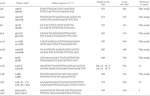
C. difficilenucleotide sequence alignment and choice of candidate loci for MLST. Ten publicly availableC. difficilegenome sequences, including six of PCR ri-botype 027, were aligned using Mauve alignment software (5). The annotatedC. difficile630 genome (36) was included as a reference. This alignment contained several large gaps and was refined using BLAST to position fragments of each genome that were left unaligned by Mauve. Candidate regions for MLST were determined from the refined alignment as follows. Using windows of 500 bp (10 to 60 variable sites), the numbers of variable sites and the numbers of gaps were calculated. MLST loci were chosen such that there was a significant degree of divergence across the 500 bp, and no gaps were present. Fragments were anno-tated according to their orthologues inC. difficile630 to ensure that they spanned housekeeping genes. Candidate fragments were testedin silicofor suitability for primer design. The standard BLASTn search for “short nearly exact matches” was used, and this search is equivalent to BLASTn with the following parame-ters: word size, 7; low-complexity filter (DUST) off; expect value, 1,000. The database was the entire GenBank nonredundant database (nr). Details are found at: http://www.ncbi.nlm.nih.gov/blast/producttable.shtml#shortn. PCR was car-ried out on a subset ofC. difficileisolates to verify amplification efficiency of the new MLST fragment. The seven loci and primers chosen for MLST are shown in Table 1.
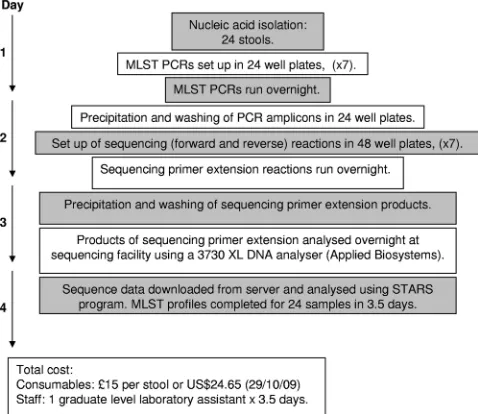
C. difficilehigh-throughput multilocus sequence typing.MLST was performed as described below by setting up PCR and sequencing reactions in 24-, 48-, or 96-well plates (Fig. 3 shows the procedure for 24 samples, but it can easily be scaled up to 48 samples). Seven PCR amplicons were obtained for each isolate using the primers shown in Table 1. Each 50-l PCR mixture contained 39.75l of molecular biology-grade water (Sigma-Aldrich Co., Ltd.), 5l of 10⫻PCR buffer (Qiagen Ltd., Crawley, United Kingdom), 1l of a 10M concentration of each forward and reverse primer, 1l of 10 mM deoxynucleoside triphosphate (dNTP) mix (Invitrogen Corp., Paisley, United Kingdom), 0.25l of HotStart
TaqDNA polymerase (Qiagen Ltd.), and 2l ofC. difficilechromosomal DNA (approximately 10 ng) or extracted total stool DNA. The amplification conditions were 95°C for 15 min, followed by 35 cycles of 94°C for 30 s, 50°C for 40 s, and 72°C for 70 s, with a final extension at 72°C for 5 min and storage at 15°C. The amplification products were purified by precipitation with 20% polyethylene glycol (molecular weight, 8,000) and 2.5 M NaCl, and their nucleotide sequences were determined on each DNA strand using the amplification primers and BigDye Ready Reaction Mix (Applied Biosystems, Warrington, United King-dom) as follows. Each 10-l sequencing reaction mixture comprised 2l of PCR amplicon, 4l of a 1:15 dilution of either forward or reverse PCR primer (0.66
M), 0.25l of BigDye Ready Reaction Mix, 1.875l of 5⫻sequencing buffer (20 ml of stock solution comprised 200l of 1 M MgCl2, 8 ml of 1 M Tris-HCl, pH 9, and 11.8 ml of molecular biology-grade water [all from Sigma-Aldrich Co., Ltd.]), and 1.875l of molecular biology-grade water. Dilution of the BigDye Ready Reaction Mix using 5⫻sequencing buffer reduces the cost of high-throughput sequencing without any compromise in sequence quality. The reac-tion condireac-tions were 30 cycles of denaturareac-tion at 96°C for 10 s, annealing at 50°C for 5 s, and extension at 60°C for 2 min. Unincorporated dye terminators were removed by precipitation of the termination products with 2 volumes of ethanol and 0.1 volume of 3 M sodium acetate (pH 5.2), followed by centrifugation, and the resulting pellet was then washed with 70% ethanol. The reaction products were separated and detected using a 3730 XL DNA analyzer (Applied Biosys-tems). For each sample, the program STARS (Sequence Typing Analysis Re-trieval System [http://pubmlst.org/software/assembly/]) was used to rapidly col-late paired reads, determine sequences, and identify alleles. The data forC. difficilealleles and STs were deposited in a newly developedC. difficileMLST database, which is accessible at http://pubmlst.org/cdifficile. Phylogenetic analysis was performed using the program MEGA, version 4 (Molecular Evolutionary Genetics Analysis [http://www.megasoftware.net/]).
Detection of PaLoc genes by PCR.The oligonucleotide primers used to detect thetcdA(encoding toxin A),tcdB(encoding toxin B), andtcdC(encoding a negative regulator of toxins A and B) sequences found within the pathogenicity locus operon (PaLoc) are summarized in Table 1. ThetcdAassay was published by Lemee et al. (21) and amplifies a 369-bp amplicon for toxin A-positive B-positive (A⫹B⫹) strains and a 110-bp amplicon for A-negative (A⫺) B⫹ strains, which contain a deletion in thetcdAgene. The reaction conditions were 95°C for 15 min, followed by 35 cycles of 94°C for 30 s, 52°C for 30 s, and 72°C for 40 s, with a final extension at 72°C for 5 min and storage at 15°C. ThetcdB
primers amplify a 688-bp amplicon under the same reaction conditions used for
tcdA, except an annealing temperature of 50°C for 40 s and extension of 72°C for 70 s were used. ThetcdCprimers (33) amplify the 5⬘region of thetcdCgene, giving a 475-bp amplicon under the same conditions used fortcdA. The absence of the PaLoc was demonstrated using primers lok1 and lok3 (3), which amplify
on May 16, 2020 by guest
http://jcm.asm.org/
a 769-bp amplicon in strains without the PaLoc. The reaction conditions were the same as those fortcdBabove.
PCR ribotyping. All PCR ribotyping of reference isolates and cultured isolates described in the present study was performed at the reference labo-ratory for theC. difficile Ribotyping Network for England and Northern Ireland, Leeds General Infirmary. PCR ribotyping was performed as de-scribed previously, with modifications (30). Briefly, bacterial growth was harvested from cultures raised on modified Brazier’s CCEY agar with the omission of egg yolk and addition of 5 mg/liter lysozyme (CCEYL) (Bio-Connections, Wetherby, United Kingdom) for 48 h at 37°C (44). Template DNA was prepared using a QIAxtractor automated nucleic acid extraction system (Qiagen Ltd). Amplification reactions were performed in 50-l vol-umes containing 50 pmol of both forward and reverse primers, 25l of HotStartTaqPlus PCR Master Mix (Qiagen Ltd.), 19l of water, and 5l of DNA template. The reaction mixtures were activated by heating to 95°C for 5 min and then subjected to 30 cycles of 92°C for 1 min, 55°C for 1 min, and 72°C for 1.5 min. A final cycle of 95°C for 1 min, 55°C for 45 s, and 72°C for 5 min was added. The resultant amplimer was concentrated to a final volume of approximately 20l by heating the opened reaction tubes at 75°C for 30 min. Amplification products were subjected to electrophoresis using 3% Metasieve agarose (Flowgen Bioscience, Nottingham, United Kingdom) at a field strength of 7.5 V/cm for approximately 2.5 h. Agarose gels were imaged using a GeneGenius camera system (Syngene, Cambridge, United Kingdom) after ethidium bromide staining. DNA profiles were analyzed and identified against a library of known PCR ribotypes using BioNumerics, version 4.6, software (Applied Maths, Belgium).
ID. The index of discrimination (ID) for MLST and PCR ribotyping was calculated according to Hunter and Gaston (11). The ID expresses the average probability that two individuals in the collection will have the same MLST type.
RESULTS
C
.
difficile
MLST.
The MLST scheme was designed to be
technically robust, generating high amplicon yields for all
C.
difficile
genotypes, under the same PCR conditions for all
seven loci. Loci were chosen that were widely distributed
around the chromosome (Fig. 1), had demonstrable
discrimi-natory ability, and lacked insertions or deletions that caused
changes in length. The longest possible sequence was used at
each locus to maximize resolution, trim sites were located so
that primer sequences were removed, and null alleles were
avoided (as far as possible, given current information on the
species) to ensure 100% typeability.
Validation of the
C. difficile
MLST scheme. (i) Typeability of
all genotypes.
The ability of the MLST primers to amplify and
sequence all
C. difficile
genotypes was assessed using DNA
from 152 cultured isolates. These included 50 isolates of 45
different PCR ribotypes (from a collection held at Leeds
Gen-eral Infirmary, the reference laboratory for the
C. difficile
Ri-TABLE 1. Nucleotide sequences of the oligonucleotide primers used in the study
aLocus Primer nameb Primer sequence (5⬘–3⬘) Amplicon size
(bp)
Locus size (bp)c
Reference or source
adk
adk1F
TTACTTGGACCTCCAGGTGC
635
501
This study
adk1R
TTTCCACTTCCTAAGGCTGC
atp
A
atpA1F
TGATGATTTAAGTAAACAAGCTG
674
555
This study
atpA1R
AATCATGAGTGAAGTCTTCTCC
dxr
dxr3F
GCTACTTTCCATTCTATCTG
525
411
This study
dxr4R
CCAACTCTTTGTGCTATAAA
gly
A
glyA1F
ATAGCTGATGAGGTTGGAGC
625
516
This study
glyA1R
TTCTAGCCTTAGATTCTTCATC
rec
A
recA2F
CAGTAATGAAATTGGGAGAAGC
705
564
This study
recA2R
ATTCAGCTTGCTTAAATGGTG
sod
A
sodA5F
CCAGTTGTCAATGTATTCATTTC
585
450
This study
sodA6R
ATAACTTCATTTGCTTTTACACC
tpi
tpi2F
ATGAGAAAACCTATAATTGCAG
640
504
This study
tpi2R
TTGAAGGTTTAACACTTCCACC
tcd
A
tcdA-F
AGATTCCTATATTTACATGACAATAT
369 (A
⫹B
⫹)
dNA
21
tcdA-R
GTATCAGGCATAAAGTAATATACTTT
110 (A
⫺B
⫹)
dtcdB
tcdB1
TGATGAAGATACAGCAGAAGC
688
NA
This study
tcdB2
TGATTCTCCCTCAAAATTCTC
tcd
C
tcdC-F(
⫺
17)
AAAAGGGAGATTGTATTATGTTTTC
479
NA
33
tcdC-R(
⫹
462)
CAATAACTTGAATAACCTTACCTTCA
cdd1
lok1(
cdd1
)
AAAATATACTGCACATCTGTATAC
769
NA
3
cdu1
lok3 (
cdu1
)
TTTACCAGAAAAAGTAGCTTTAA
aPrimers were used (i) to performC. difficileMLST, (ii) to detect the presence of three loci within the pathogenicity locus (tcdA,tcdB, andtcdC), and (iii) to confirm the absence of the pathogenicity locus in nontoxigenic strains (lok1/lok3).
bIn the primer names F indicates forward, and R indicates reverse. cNA, not applicable.
dThe size of the amplicon varies with the strain genotype.
on May 16, 2020 by guest
http://jcm.asm.org/
[image:3.585.44.542.82.400.2]botyping Network for England and Northern Ireland)
repre-senting the known diversity of the species (Fig. 2) and an
additional 102 isolates from recent clinical samples (91 from
107 ELISA-positive stools and 11 from 108 ELISA-negative
stools) (Tables 2 and 3). The clinical isolates were also PCR
ribotyped, and consequently four additional PCR ribotypes
were included, giving a total of 49 in the study as a whole (Fig.
2 and Tables 2 and 3). All isolates were successfully typed by
MLST, and a total of 40 STs were identified (Fig. 2). These
data have been made available at http://pubmlst.org/cdifficile/.
The MLST primers amplified DNA from both toxigenic and
nontoxigenic strains. The 91 isolates from ELISA-positive
stools were presumed to be toxigenic, and all were typed by
MLST. The 11 isolates from ELISA-negative stools were
as-sessed by PCRs designed to amplify fragments of three genes
located within the PaLoc (Table 3). Nine of 11 isolates were
toxigenic. The other two were nontoxigenic and were assigned
a unique genotype, ST-15. They both yielded a 769-bp
ampli-con with the lok1/lok3 primer pair (Tables 1 and 3), ampli-confirming
the absence of the PaLoc. The MLST primers have been
fur-ther validated using a collection of 37 isolates (including 26
nontoxigenic strains) from healthy infants aged under 2 years
and a large collection of
⬃
1,600 clinical isolates collected at
the John Radcliffe Hospital, Oxford, United Kingdom,
be-tween September 2006 and April 2009 (molecular
epidemiol-ogy data are beyond the scope of this paper).
(ii) Discrimination among isolates.
The level of
discrimina-tion achieved with MLST was compared to PCR ribotyping.
The total number of isolates with both ST and PCR ribotype
data available was 152, comprising 50 in the Leeds PCR
ri-botype collection, 91 from ELISA-positive stools (Table 2),
and 11 from ELISA-negative stools (Table 3). Among the 152
isolates, 32/40 STs occurred with a single PCR ribotype, but 8
STs were associated with two or more PCR ribotypes (Fig. 2).
Conversely, the PCR ribotype was not always predictive of the
ST, with three PCR ribotypes being associated with more than
one ST (Fig. 2). The multiple PCR ribotypes associated with
single STs had a high degree of similarity in DNA profile. For
example, when PCR ribotypes share an ST (n
⫽
8), 6/8 STs
were PCR ribotypes that differed by a single band. The other
two were PCR ribotypes 023 and 063 (both ST-5) (Fig. 2),
which differ by four bands, and PCR ribotype 009 (ST-3),
which differs from the other PCR ribotypes associated with
ST-3 by four bands.
The set of 102 clinical isolates was used to compare the
discriminatory ability of the two methods. The IDs (11) for
MLST and PCR ribotyping were 0.90 and 0.92, respectively, a
difference that is unlikely to be statistically significant.
(iii) MLST performed directly on total stool DNA.
The
ro-bustness of the MLST scheme was validated by typing strains
contained in total DNA extracted from stool samples.
C.
dif-ficile
DNA isolated from cultured cells is a straightforward
template for PCR amplification. However, when the same
tar-get is mixed in human feces, successful amplification requires
efficient lysis of
C. difficile
cells and spores, PCR inhibitor
removal (19, 29), and specific amplification from a complex
mixture of DNAs (8, 22). To test the approach, MLST data
were generated from both cultured isolates and total stool
DNA extracts for 95 clinical samples (89/107 ELISA-positive
and 6/108 ELISA-negative samples) (Tables 2 and 3). The
direct and culture MLST data were generated by two different
people, each without prior knowledge of the other’s results.
Direct MLST data were examined carefully for evidence of
primer cross-reaction with other clostridial species in stools
and coinfections with multiple
C. difficile
genotypes. Among 93
of the 95 stools, there was 100% agreement of the direct and
culture MLST data. The two exceptions yielded occasional
mixed peaks in the sequence chromatograms (2.1%),
indicat-ing a coinfection of more than one
C. difficile
genotype. This
compares to 3/23 (13%) stools reported by Wroblewski et al.
(45) using PCR detection of binary toxin gene(s) to detect
heterogeneous isolate populations, and 2/23 (9%) patients with
a first episode of CDI detected by van den Berg et al. (43) using
PCR ribotyping. One additional stool DNA extract (Table 2,
sample 104
⫹
) yielded very low levels of PCR amplicons,
in-sufficient for sequencing at all seven loci.
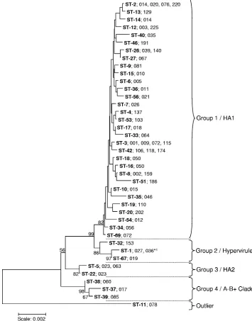
Genetic variation and phylogenetic relationships among
iso-lates.
The relationships among the 40 STs were examined using
the concatenated sequences of the seven MLST loci to
con-struct a neighbor-joining tree (Fig. 2). The total number of
variable nucleotide sites was 103/3,501 (2.9%), falling to 59/
3,501 (1.7%) if the outlier ST-11 (078) was excluded. The
comparable data for amino acid sites were 22/1,167 (1.9%) and
17/1,167 (1.5%). Despite the relatively low level of genetic
diversity, the STs clustered into four groups with one outlier
(ST-11) (Fig. 2). The majority of sequence types clustered in
group 1 with very low internal bootstrap values. Group 2
con-tained ST-1 (027), the hypervirulent clone; group 3 concon-tained
two STs, both of which were associated with PCR ribotype 023;
and group 4 contained the toxin A
⫺B
⫹ST-37 (PCR ribotype
017). The outlier ST-11 is associated with PCR ribotype 078,
which has been reported as an emergent hypervirulent clone.
DISCUSSION
MLST is a proven technology for understanding the
molec-ular epidemiology and population biology of bacterial species
FIG. 1. The relative positions (to scale) of the seven housekeeping
loci chosen for MLST on the
C. difficile
strain 630 chromosome,
to-gether with three genes of the pathogenicity locus (PaLoc) detected by
PCRs referred to in this study and the genes encoding the binary toxin
cdtA
and
cdtB
.
on May 16, 2020 by guest
http://jcm.asm.org/
(25). Although it has been applied to a diverse collection of
C.
difficile
isolates (20), MLST has not been widely adopted for
this organism, in contrast to the majority of clinically important
bacterial species (25). Our aim was to further develop MLST
for
C. difficile, setting up a more robust method by the
follow-ing steps: (i) replacfollow-ing the null allele employed at one of the
loci included in the previously published scheme (20) with an
allele present in all strains, (ii) improving discrimination by
using longer sequences for MLST, and (iii) establishing an
Internet-accessible MLST database to allow straightforward
accumulation of data over time and to simplify the comparison
of data among laboratories. This MLST scheme for
C. difficile
was also sufficiently robust to allow typing to be performed
directly on DNA extracted from stool, without culture. This
could potentially be used to generate actionable genotyping
data close to real time since the entire process can be
com-pleted for a batch of 24 isolates in 3.5 days (Fig. 3), at a
consumables cost of £15 per stool (or $24.65 as of 29 October
2009) and the cost of one graduate-level member of staff.
[image:5.585.109.464.73.526.2]The MLST scheme was sufficiently discriminatory to give
FIG. 2. Neighbor-joining tree constructed using the concatenated sequences (3,501 nucleotides) of the seven loci used in MLST. Bootstraps were
generated using 1,000 replicates, and low values were removed for clarity. STs are shown in bold. The PCR ribotype(s) found in association with each
ST are indicted. The STs cluster into four groups, designated 1 to 4, with bootstraps greater than 80 and one outlier (ST-11). These correspond to groups
defined previously by microarray analysis of whole genomes (39) which were designated HA1 (human and animal 1), hypervirulent (containing the 027
strain), HA2, and the A
⫺B
⫹clade (also containing other toxigenic types). The total number of variable sites was 103/3,501 (2.9%), and if the outlier ST-11
was excluded, the number was 59/3,501 (1.7%). ST-1 (indicated by *
1) was associated with PCR ribotype 027 and also with a single PCR ribotype 036
isolate. DNA profiles for PCR ribotypes 036 and 027 are very similar and differ by only a single band.
on May 16, 2020 by guest
http://jcm.asm.org/
TABLE 2. MLST of
C. difficile
from ELISA-positive stool samples determined both directly using total stool DNA and
using isolates cultured from the same stools
Sample
no.a ELISA
ODb
MLST PCR result (direct)
Direct STc Culture
resulti Culture
STd PCR
ribotype
Sample
no.a ELISA
ODb
MLST PCR result (direct)
Direct STc Culture
resulti Culture
STd PCR
ribotype
23
⫹
⫹⫹⫹⫹⫹
⫹
1
⫹⫹⫹
1
027
77
⫹
2.427
⫹
8
⫹⫹⫹
8
002
17
⫹
0.353
⫹
1
⫹⫹
1
027
96
⫹
2.282
⫹
8
⫹⫹⫹
8
002
2
⫹
2.251
⫹
1
⫹
1
027
97
⫹
2.379
⫹
8
⫹⫹⫹
8
002
8
⫹
⫹⫹⫹⫹⫹
⫹
1
⫹⫹⫹
1
027
105
⫹
2.136
⫹
8
⫹⫹⫹
8
002
37
⫹
1.216
⫹
1
⫹
1
027
73
⫹
0.499
⫹
8
⫹⫹⫹
8
002
43
⫹
0.663
⫹
1
⫹
1
027
79
⫹
1.163
⫹
9
⫹
9
081
72
⫹
2.5
⫹
1
⫹⫹⫹
1
027
7
⫹
0.35
⫹
9
⫹
9
081
75
⫹
2.5
⫹
1
⫹⫹
1
027
80
⫹
2.5
⫹
10
⫹⫹⫹
10
015
85
⫹
0.388
⫹
1
⫹
1
027
99
⫹
0.796
⫹
10
⫹⫹⫹
10
015
88
⫹
5.584
⫹
1
⫹⫹⫹
1
027
15
⫹
0.202
⫹
10
⫹
10
015
89
⫹
5.392
⫹
1
⫹⫹
1
027
82
⫹
2.5
⫹
11
⫹⫹
11
078
94
⫹
7.098
⫹
1
⫹⫹⫹
1
027
4
⫹
2.13
⫹
11
⫹⫹⫹
11
078
98
⫹
2.5
⫹
1
⫹⫹⫹
1
027
61
⫹
0.567
⫹
11
⫹
11
078
101
⫹
1.208
⫹
1
⫹⫹⫹
1
027
20
⫹
⫹⫹⫹⫹⫹
⫹
12
⫹
12
003
107
⫹
2.5
⫹
1
⫹⫹⫹
1
027
103
⫹
0.657
⫹
12
⫹⫹⫹
12
225
60
⫹
⫹⫹⫹⫹⫹
⫹
1
⫹⫹
1
027
108
⫹
2.5
⫹
13
⫹⫹⫹
13
129
53
⫹
1.8
⫹
2
⫹
2
014
22
⫹
⫹⫹⫹⫹⫹
⫹
14
⫹⫹⫹
14
014
58
⫹
0.261
⫹
2
⫹⫹
2
020
5
⫹
1.658
⫹
14
⫹
14
014
27
⫹
⫹⫹⫹⫹⫹
⫹
2
⫹⫹⫹
2
020
33
⫹
1.658
⫹
14
⫹⫹
14
014
11
⫹
1.614
⫹
2
⫹
2
020
42
⫹
0.317
⫹
14
⫹⫹
14
014
12
⫹
0.968
⫹
2
⫹⫹⫹
2
020
46
⫹
0.385
⫹
14
⫹
14
014
13
⫹
7.718
⫹
2
⫹⫹⫹
2
020
62
⫹
2.5
⫹
14
⫹⫹⫹
14
014
14
⫹
3.648
⫹
2
⫹⫹
2
020
19
⫹
2.257
⫹
14
⫹
14
014
31
⫹
5.648
⫹
2
⫹
2
014
36
⫹
1.09
⫹
14
⫹⫹
14
014
41
⫹
0.843
⫹
2
⫹
2
020
28
⫹
0.447
⫹
19
⫹⫹
19
110
44
⫹
0.29
⫹
2
⫹⫹⫹
2
020
6
⫹
⫹⫹⫹⫹⫹
⫹
22
⫹
22
023
48
⫹
1.003
⫹
2
⫹⫹
2
076
49
⫹
0.41
⫹
37
⫹⫹
37
017
63
⫹
2.5
⫹
2
⫹⫹⫹
2
014
9
⫹
⫹⫹⫹⫹⫹
⫹
42
⫹⫹⫹
42
106
66
⫹
2.5
⫹
2
⫹⫹⫹
2
020
24
⫹
0.182
⫹
42
⫹
42
174
67
⫹
2.5
⫹
2
⫹⫹
2
014
40
⫹
⫹⫹⫹⫹⫹
⫹
42
⫹⫹
42
106
84
⫹
0.563
⫹
2
⫹⫹⫹
2
020
56
⫹
3.296
⫹
54
⫹⫹
54
012
1
⫹
⫹⫹⫹⫹⫹
⫹
2
⫹⫹
2
076
104
⫹
0.387
(
⫹
)
hND
e⫹⫹⫹
1
027
106
⫹
1.049
⫹
2
⫹⫹⫹
2
020
100
⫹
0.67
⫹
Mixed
g⫹
5
023
52
⫹
⫹⫹⫹⫹⫹
⫹
3
⫹⫹
3
072
71
⫹
2.322
⫹
Mixed
g⫹⫹⫹
11
078
55
⫹
⫹⫹⫹⫹⫹
⫹
3
⫹⫹
3
001
45
⫹
2.127
⫹
1
⫺
57
⫹
0.285
⫹
3
⫹
3
072
47
⫹
6.186
⫹
1
⫺
16
⫹
⫹⫹⫹⫹⫹
⫹
3
⫹⫹
3
001
50
⫹
0.278
⫺
⫹
2
220
64
⫹
0.434
⫹
3
⫹⫹⫹
3
072
86
⫹
0.369
⫺
⫹
7
026
65
⫹
2.5
⫹
4
⫹⫹
4
137
91
⫹
3.578
⫺
⫹⫹⫹
8
002
68
⫹
1.322
⫹
4
⫹⫹
4
137
90
⫹
1.155
⫺
⫹
ND
fND
f69
⫹
2.294
⫹
5
⫹⫹⫹
5
023
102
⫹
0.885
⫺
⫹
ND
fND
f76
⫹
2.5
⫹
5
⫹⫹
5
023
54
⫹
0.26
⫺
⫺
78
⫹
2.5
⫹
5
⫹⫹
5
023
21
⫹
7.792
⫺
⫺
95
⫹
2.5
⫹
5
⫹⫹⫹
5
023
25
⫹
0.67
⫺
⫺
59
⫹
4.083
⫹
6
⫹
6
005
29
⫹
3.475
⫺
⫺
51
⫹
⫹⫹⫹⫹⫹
⫹
6
⫹⫹⫹
6
005
30
⫹
0.875
⫺
⫺
26
⫹
1.155
⫹
6
⫹
6
005
18
⫹
0.158
⫺
⫺
70
⫹
2.5
⫹
6
⫹⫹
6
005
34
⫹
1.487
⫺
⫺
92
⫹
1.896
⫹
6
⫹⫹⫹
6
005
35
⫹
0.435
⫺
⫺
93
⫹
0.7
⫹
6
⫹⫹⫹
6
005
38
⫹
0.270
⫺
⫺
3
⫹
0.679
⫹
7
⫹
7
026
81
⫹
0.537
⫺
⫺
32
⫹
0.499
⫹
7
⫹
7
026
83
⫹
0.475
⫺
⫺
74
⫹
2.5
⫹
7
⫹⫹⫹
7
026
87
⫹
0.391
⫺
⫺
39
⫹
0.27
⫹
8
⫹⫹⫹
8
002
aThe plus sign indicates an ELISA-positive result. bOD, optical density.⫹⫹⫹⫹⫹, value above the scale. cMLST was performed directly from stool samples. dMLST was performed from cultured isolates.
eND, not done due to very low PCR amplicon yields or no amplicon.
fND, not done because sample was negative on reculturing from frozen isolate stock.
gEvidence of more than one genotype present in stool due to mixed peaks in the nucleotide sequence chromatogram. hPoor amplicon yield.
iCrude estimate of viableC. difficileload.⫹,⬍100 colonies;⫹⫹, 100 to 1,000 colonies;⫹⫹⫹,⬎1,000 colonies.
on May 16, 2020 by guest
http://jcm.asm.org/
typing data which can be interpreted with confidence;
accord-ing to Hunter and Gaston (11) an ID greater than 0.90 is
desirable to meet this requirement. For our 102 clinical
iso-lates, MLST and PCR ribotyping had comparable
discrimina-tory abilities (ID of 0.90 for MLST and of 0.92 for PCR
ri-botyping). The differences between the methods were
generally consistent with a simple genetic explanation; multiple
ribotypes for the same ST usually had very similar profiles, and
multiple STs for the same ribotype generally had very closely
related STs. Capillary gel electrophoresis-based PCR
ribotyp-ing is a promisribotyp-ing tool to study subtypes within ribotypes, and
it may assist the explanation of such observations (13). They
may also be consistent with limited recombination that may be
characteristic of
C. difficile.
We calculated the ID as 0.958 for the previously published
MLST scheme (34 STs, 62 PCR ribotypes, and 72 isolates) (20)
and as 0.983 for PCR ribotyping for the same collection.
How-ever, this is not an entirely robust comparison since all the
isolates were specifically chosen for their genetic diversity, and
a true ID should reflect the capacity of a typing method to
discriminate epidemiologically unrelated isolates within a
pop-ulation.
Pulsed-field gel electrophoresis is another genotyping
tech-nique widely used to characterize
C. difficile. The IDs for
PFGE, the previously published MLST scheme of Lemee et al.
(20), and PCR ribotyping were found by Killgore et al. (17) to
be 0.843 (PFGE), 0.699 (MLST), and 0.700 (PCR ribotyping)
for a collection of 42 isolates from four countries representing
epidemic strains and the next most commonly isolated strain
types.
[image:7.585.42.542.90.227.2]Despite the relatively low overall genetic diversity detected
within these housekeeping loci, it was still possible to identify
four different phylogenetic groups of
C. difficile
STs (Fig. 2).
The majority of STs clustered in group 1, group 2 contained
ST-1 (PCR ribotype 027), group 3 contained two STs
associ-ated with PCR ribotype 023, and group 4 contained toxin A
⫺B
⫹ST-37 (PCR ribotype 017). A single outlier, ST-11, was
associated with PCR ribotype 078. The previously described
MLST scheme for
C. difficile
identified three divergent
lin-eages, one containing the A
⫺B
⫹isolates, which corresponds
to our group 4 (20). Stabler et al. (39) used comparative
genomics to identify four clades, and these appear to correlate
with the four groups we have identified by MLST in the present
study (Fig. 2). In that previous study HA1 (human and animal
1) (39) contained mainly human isolates with just a few
ani-mals, and this clade probably corresponds to our group 1,
which contained the majority of our human isolate STs. HA2
probably corresponds to our group 3 as this contained mainly
animal isolates (pig and bovine), with few isolates from
hu-mans. These data suggest that genotypes clustered by MLST
may correlate with groups derived from whole-genome
com-parisons using DNA microarrays (39), implying that MLST
TABLE 3. MLST of
C. difficile
from ELISA-negative stool samples determined both directly using total stool DNA and
using isolates cultured from the same stools
Sample no.a
MLST PCR result (direct)
Direct
STd Culture
resultg Culture
STe PCR
ribotype
PaLoc PCR result
lok1/lok3 PCR
22
⫺
⫹
6
⫹
6
005
⫹
⫺
23
⫺
⫹
15
⫹
15
010
⫺
⫹
f36
⫺
⫹
6
⫹
6
005
⫹
⫺
43
⫺
⫹
8
⫹
8
002
⫹
⫺
69
⫺
⫹
2
⫹
2
020
⫹
⫺
79
⫺
⫹
2
⫹
2
020
⫹
⫺
15
⫺
⫺
⫹
69
072
⫹
⫺
55
⫺
b⫺
⫹
1
027
⫹
⫺
85
⫺
c⫺
⫹
1
027
⫹
⫺
99
⫺
⫺
⫹⫹
15
010
⫺
⫹
f108
⫺
⫺
⫹
8
002
⫹
⫺
aThe minus sign indicates an ELISA-negative result.
bPatient provided a subsequent sample not included in the study (⫹7 days) that was ELISA positive and ST-1 by culture (sample not typed by direct MLST). cPatient provided a subsequent sample not included in the study (⫹39 days) that was ELISA positive and ST-1 by culture (sample not typed by direct MLST). dMLST was performed directly from stool samples.
eMLST was performed from cultured isolates.
fPCR yielded a769-bp amplicon, confirming the absence of the PaLoc.
gCrude estimate of viableC. difficileload.⫹,⬍100 colonies;⫹⫹, 100 to 1,000 colonies;⫹⫹⫹,⬎1,000 colonies.
FIG. 3. Flow diagram summarizing the time required to perform
the laboratory work and sequence data analysis in high-throughput
MLST.
on May 16, 2020 by guest
http://jcm.asm.org/
[image:7.585.43.282.487.694.2]may be an accurate proxy for whole-genome analysis. The
newly emergent ST-11 (PCR ribotype 078) hypervirulent clone
was a genetically distinct outlier. This genotype causes
infec-tion in humans, pigs, and calves (6, 16) and has been found in
cooked and raw meat products (38). Multilocus
variable-num-ber tandem-repeat analysis (MLVA) data confirmed a strong
degree of genetic relatedness between human and animal
iso-lates belonging to this genotype in The Netherlands (9). ST
data presented here suggest that ST-11 (078) has emerged
from a single, genetically distinct clade. The other four ST
groups may represent different
C. difficile
clonal complexes,
with the level of nucleotide sequence divergence between STs
representing each group ranging from 11/3,501 (0.3%) to 60/
3,501 (1.7%).
A robust MLST scheme can now be applied to studies of
C.
difficile
epidemiology and population structure. Direct MLST
of
C. difficile
in stool provides a rapid genotyping method
which generates data that are easily compared among
labora-tories using an Internet-accessible database. It will now be
possible to test in a clinical setting the utility of MLST for
outbreak identification, detection of transmission events
among patients, and the identification of emergent
hyperviru-lent clones, thereby assessing the potential benefits of MLST to
individual patients and hospital infection control.
ACKNOWLEDGMENTS
This study was supported by the NIHR Biomedical Research
Cen-tre, Oxford, United Kingdom. Melina Kachrimanidou was supported
by the Society of Biopathology of Northern Greece.
We thank the staff of the Clinical Microbiology Laboratory and
Infection Control, John Radcliffe Hospital, Oxford, and Infection
Con-trol Laboratory staff, Leeds General Infirmary, for their assistance
throughout this work.
REFERENCES
1.Bartlett, J. G. 2002. Clinical practice. Antibiotic-associated diarrhea. N. Engl. J. Med.31:334–339.
2.Bartlett, J. G., and D. N. Gerding.2008. Clinical recognition and diagnosis ofClostridium difficileinfection. Clin. Infect. Dis.46:S12–S18.
3.Braun, V., T. Hundsberger, P. Leukel, M. Sauerborn, and C. von Eichel-Streiber.1996. Definition of the single integration site of the pathogenicity locus inClostridium difficile. Gene181:29–38.
4.Clabots, C. R., S. Johnson, K. M. Bettin, P. A. Mathie, M. E. Mulligan, D. R. Schaberg, L. R. Peterson, and D. N. Gerding.1993. Development of a rapid and efficient restriction endonuclease analysis typing system forClostridium difficileand correlation with other typing systems. J. Clin. Microbiol.31: 1870–1875.
5.Darling, A. C., B. Mau, F. R. Blattner, and N. T. Perna.2004. Mauve: multiple alignment of conserved genomic sequence with rearrangements. Genome Res.14:1394–1403.
6.Debast, S. B., L. A. van Leengoed, A. Goorhuis, C. Harmanus, E. J. Kuijper, and A. A. Bergwerff.2009.Clostridium difficilePCR ribotype 078 toxinotype V found in diarrhoeal pigs identical to isolates from affected humans. En-viron. Microbiol.11:505–511.
7.Dene`ve, C., C. Janoir, I. Poilane, C. Fantinato, and A. Collignon.2009. New trends inClostridium difficilevirulence and pathogenesis. Int. J. Antimicrob. Agents33(Suppl. 1):S24–S28.
8.Fujisawa, T., K. Namba, K. Hirayama, W. K. Lee, and T. Mitsuoka.1995. New selective media for isolation of clostridia from faecal specimens. J. Appl. Bacteriol.78:481–486.
9.Goorhuis, A., D. Bakker, J. Corver, S. B. Debast, C. Harmanus, D. W. Notermans, A. A. Bergwerff, F. W. Dekker, and E. J. Kuijper.2008. Emer-gence ofClostridium difficileinfection due to a new hypervirulent strain, polymerase chain reaction ribotype 078. Clin. Infect. Dis.47:1162–1170. 10.Health Protection Agency.2009.Clostridium difficileRibotyping Network for
England and Northern Ireland report 2008/09. Health Protection Agency, Lon-don, United Kingdom. http://www.hpa.org.uk/web/HPAwebFile/HPAweb_C /1258560554236.
11.Hunter, P. R., and M. A. Gaston.1988. Numerical index of discriminatory ability of typing systems: an application of Simpson’s index of diversity. J. Clin. Microbiol.26:2465–2466.
12.Ibarz Pavo´n, A. B., and M. C. Maiden.2009. Multilocus sequence typing. Methods Mol. Biol.551:129–140.
13.Indra, A., S. Huhulescu, M. Schneeweis, P. Hasenberger, S. Kernbichler, A. Fiedler, F. Wewalka, G. Allerberger, and E. J. Kuijper.2008. Characteriza-tion ofClostridium difficileisolates using capillary gel electrophoresis-based PCR ribotyping. J. Med. Microbiol.57:1377–1382.
14.Jolley, K. A., M. S. Chan, and M. C. Maiden.2004. MlstdbNet—distributed multi-locus sequence typing (MLST) databases. BMC Bioinformatics5:86. 15.Kazakova, S. V., K. Ware, B. Baughman, O. Bilukha, A. Paradis, S. Sears, A.
Thompson, B. Jensen, L. Wiggs, J. Bessette, J. Martin, J. Clukey, K. Gen-sheimer, G. Killgore, and L. C. McDonald.2006. A hospital outbreak of diarrhea due to an emerging epidemic strain ofClostridium difficile. Arch. Intern. Med.166:2518–2524.
16.Keel, K., J. S. Brazier, K. W. Post, S. Weese, and J. G. Songer.2007. Prevalence of PCR ribotypes amongClostridium difficileisolates from pigs, calves, and other species. J. Clin. Microbiol.45:1963–1964.
17.Killgore, G., A. Thompson, S. Johnson, J. Brazier, E. Kuijper, J. Pepin, E. H. Frost, P. Savelkoul, B. Nicholson, R. J. van den Berg, H. Kato, S. P. Sambol, W. Zukowski, C. Woods, B. Limbago, D. N. Gerding, and L. C. McDonald. 2008. Comparison of seven techniques for typing international epidemic strains ofClostridium difficile: restriction endonuclease analysis, pulsed-field gel electrophoresis, PCR-ribotyping, multilocus sequence typing, multilocus variable-number tandem-repeat analysis, amplified fragment length poly-morphism, and surface layer protein A gene sequence typing. J. Clin. Mi-crobiol.46:431–437.
18.Kuijper, E. J., F. Barbut, J. S. Brazier, N. Kleinkauf, T. Eckmanns, M. L. Lambert, D. Drudy, F. Fitzpatrick, C. Wiuff, D. J. Brown, J. E. Coia, H. Pituch, P. Reichert, J. Even, J. Mossong, A. F. Widmer, K. E. Olsen, F. Allerberger, D. W. Notermans, M. Delme´e, B. Coignard, M. Wilcox, B. Patel, R. Frei, E. Nagy, E. Bouza, M. Marin, T. Akerlund, A. Virolainen-Julkunen, O. Lyytika¨inen, S. Kotila, A. Ingebretsen, B. Smyth, P. Rooney, I. R. Poxton, and D. L. Monnet.2008. Update ofClostridium difficileinfection due to PCR ribotype 027 in Europe, 2008. Euro Surveill.31:18942.
19.Lantz, P. G., M. Matsson, T. Wadstro¨m, and P. Rådstro¨m.1997. Removal of PCR inhibitors from human faecal samples through the use of an aqueous two-phase system for sample preparation prior to PCR. J. Microbiol. Meth-ods28:159–167.
20.Lemee, L., A. Dhalluin, M. Pestel-Caron, J. F. Lemeland, and J. L. Pons. 2004. Multilocus sequence typing analysis of human and animalClostridium difficileisolates of various toxigenic types. J. Clin. Microbiol.42:2609–2617. 21.Lemee, L., A. Dhalluin, S. Testelin, M. A. Mattrat, K. Maillard, J. F. Leme-land, and J. L. Pons.2004. Multiplex PCR targetingtpi(triose phosphate isomerase),tcdA(toxin A), andtcdB(toxin B) genes for toxigenic culture of
Clostridium difficile. J. Clin. Microbiol.42:5710–5714.
22.Ley, R. E., M. Hamady, C. Lozupone, P. J. Turnbaugh, R. R. Ramey, J. S. Bircher, M. L. Schlegel, T. A. Tucker, M. D. Schrenzel, R. Knight, and J. I. Gordon.2008. Evolution of mammals and their gut microbes. Science320: 1647–1651.
23.Loo, V. G., L. Poirier, M. A. Miller, M. Oughton, M. D. Libman, S. Michaud, A. M. Bourgault, T. Nguyen, C. Frenette, M. Kelly, A. Vibien, P. Brassard, S. Fenn, K. Dewar, T. J. Hudson, R. Horn, P. Rene´, Y. Monczak, and A. Dascal.2005. A predominantly clonal multi-institutional outbreak of Clos-tridium difficile-associated diarrhea with high morbidity and mortality. N. Engl. J. Med.353:2442–2449.
24.Maiden, M. C., J. A. Bygraves, E. Feil, G. Morelli, J. E. Russell, R. Urwin, Q. Zhang, J. Zhou, K. Zurth, D. A. Caugant, I. M. Feavers, M. Achtman, and B. G. Spratt.1998. Multilocus sequence typing: a portable approach to the identification of clones within populations of pathogenic microorganisms. Proc. Natl. Acad. Sci. U. S. A.95:3140–3145.
25.Maiden, M. C.2006. Multilocus sequence typing of bacteria. Annu. Rev. Microbiol.60:561–588.
26.McDonald, L. C., G. E. Killgore, A. Thompson, R. C. Owens, Jr., S. V. Kazakova, S. P. Sambol, S. Johnson, and D. N. Gerding.2005. An epidemic, toxin gene-variant strain ofClostridium difficile. N. Engl. J. Med.353:2433– 2441.
27.McFarland, L. V.2009. Renewed interest in a difficult disease:Clostridium difficile infections—epidemiology and current treatment strategies. Curr. Opin. Gastroenterol.25:24–35.
28.McFarland, L. V., M. E. Mulligan, R. Y. Kwok, and W. E. Stamm.1989. Nosocomial acquisition ofClostridium difficileinfection. N. Engl. J. Med. 320:204–210.
29.Monteiro, L., D. Bonnemaison, A. Vekris, K. G. Petry, J. Bonnet, R. Vidal, J. Cabrita, and F. Me´graud.1997. Complex polysaccharides as PCR inhib-itors in feces:Helicobacter pylorimodel. J. Clin. Microbiol.35:995–998. 30.O’Neill, G. L., F. T. Ogunsola, J. S. Brazier, and B. I. Duerden.1996.
Modification of a PCR ribotyping method for application as a routine typing scheme forC. difficile. Anaerobe2:205–209.
31.Pe´pin, J., L. Valiquette, M. E. Alary, P. Villemure, A. Pelletier, K. Forget, K. Pe´pin, and D. Chouinard.2004.Clostridium difficile-associated diarrhea in a region of Quebec from 1991 to 2003: a changing pattern of disease severity. CMAJ171:466–472.
32.Pe´pin, J., L. Valiquette, and B. Cossette. 2005. Mortality attributable to
on May 16, 2020 by guest
http://jcm.asm.org/
nosocomial Clostridium difficile-associated disease during an epidemic caused by a hypervirulent strain in Quebec. CMAJ173:1037–1042. 33.Persson, S., M. Torpdahl, and K. E. Olsen. 2008. New multiplex PCR
method for the detection ofClostridium difficiletoxin A (tcdA) and toxin B (tcdB) and the binary toxin (cdtA/cdtB) genes applied to a Danish strain collection. Clin. Microbiol. Infect.14:1057–1064.
34.Razavi, B., A. Apisarnthanarak, and L. M. Mundy.2007.Clostridium difficile: emergence of hypervirulence and fluoroquinolone resistance. Infection35: 300–307.
35.Rolfe, R. D.1988. Asymptomatic intestinal colonization byClostridium dif-ficile, p. 201–225.InR. D. Rolfe and S. M. Finegold (ed.),Clostridium difficile: its role in intestinal disease. Academic Press, Inc., New York, NY. 36.Sebaihia, M., B. W. Wren, P. Mullany, N. F. Fairweather, N. Minton, R. Stabler, N. R. Thomson, A. P. Roberts, A. M. Cerden˜o-Ta´rraga, H. Wang, M. T. Holden, A. Wright, C. Churcher, M. A. Quail, S. Baker, N. Bason, K. Brooks, T. Chillingworth, A. Cronin, P. Davis, L. Dowd, A. Fraser, T. Felt-well, Z. Hance, S. Holroyd, K. Jagels, S. Moule, K. Mungall, C. Price, E. Rabbinowitsch, S. Sharp, M. Simmonds, K. Stevens, L. Unwin, S. Whithead, B. Dupuy, G. Dougan, B. Barrell, and J. Parkhill.2006. The multidrug-resistant human pathogenClostridium difficilehas a highly mobile, mosaic genome. Nat. Genet.38:779–786.
37.Song, X., J. G. Bartlett, K. Speck, A. Naegeli, K. Carroll, and T. M. Perl. 2008. Rising economic impact ofClostridium difficile-associated disease in adult hospitalized patient population. Infect. Control Hosp. Epidemiol.29: 823–828.
38.Songer, J. G., H. T. Trinh, G. E. Killgore, A. D. Thompson, L. C. McDonald, and B. M. Limbago.2009.Clostridium difficilein retail meat products, USA, 2007. Emerg. Infect. Dis.15:819–821.
39.Stabler, R. A., D. N. Gerding, J. G. Songer, D. Drudy, J. S. Brazier, H. T. Trinh, A. A. Witney, J. Hinds, and B. W. Wren.2006. Comparative phylog-enomics ofClostridium difficilereveals clade specificity and microevolution of hypervirulent strains. J. Bacteriol.188:7297–7305.
40.Starr, J.2005.Clostridium difficileassociated diarrhoea: diagnosis and treat-ment. BMJ331:498–501.
41.Stubbs, S. L., J. S. Brazier, G. L. O’Neill, and B. I. Duerden.1999. PCR targeted to the 16S-23S rRNA gene intergenic spacer region ofClostridium difficileand construction of a library consisting of 116 different PCR ri-botypes. J. Clin. Microbiol.37:461–463.
42.Urwin, R., and M. C. Maiden.2003. Multi-locus sequence typing: a tool for global epidemiology. Trends Microbiol.11:479–487.
43.van den Berg, R. J., H. A. Ameen, T. Furusawa, E. C. Claas, E. R. van der Vorm, and E. J. Kuijper.2005. Coexistence of multiple PCR-ribotype strains of Clostridium difficile in faecal samples limits epidemiological studies. J. Med. Microbiol.54:173–179.
44.Wilcox, M. H., W. N. Fawley, and P. Parnell.2000. Value of lysozyme agar incorporation and alkaline thioglycollate exposure for the environmental recovery ofClostridium difficile. J. Hosp. Infect.44:65–69.
45.Wroblewski, D., G. E. Hannett, D. J. Bopp, G. K. Dumyati, T. A. Halse, N. B. Dumas, and K. A. Musser.2009. Rapid molecular characterization of Clos-tridium difficile, and assessment of the population ofC. difficilein stool specimens. J. Clin. Microbiol.47:2142–2148.
46.Zilberberg, M. D.2009.Clostridium difficile-related hospitalizations among US adults, 2006. Emerg. Infect. Dis.15:122–124.
47.Zilberberg, M. D., A. F. Shorr, and M. H. Kollef.2008. Increase in adult
Clostridium difficile-related hospitalizations and case-fatality rate, United States, 2000–2005. Emerg. Infect. Dis.14:929–931.