Published online November 20, 2013 (http://www.sciencepublishinggroup.com/j/sjcm) doi: 10.11648/j.sjcm.20130206.15
The efficacy of diffusion weighted imaging and apparent
diffusion coefficient mapping for meniscal tears in the
knee
Volkan Kızılgöz, Hasan Aydın, Baki Hekimoğlu
Radiologist-Radiology Department
,
Dışkapı Y.B. Eğitim ve Araştırma Hastanesi İrfan Baştuğ Caddesi Altındağ, Ankara-TurkeyEmail address:
[email protected](V. Kızılgöz), [email protected](H. Aydın), [email protected](B. Hekimoğlu)
To cite this article:
Volkan Kızılgöz, Hasan Aydın, Baki Hekimoğlu. The Efficacy of Diffusion Weighted Imaging and Apparent Diffusion Coefficient Mapping for Meniscal Tears in the Knee. Science Journal of Clinical Medicine. Vol. 2, No. 6, 2013, pp. 171-175.
doi: 10.11648/j.sjcm.20130206.15
Abstract:
Objective : The aim of this study was to analyse the efficacy of Diffusion Weighted MR Imaging (DWI) and Apparent Diffusion Coefficient (ADC) mapping in the diagnosis of meniscal tears in the knee. Conclusion : DWI and ADC mapping technique are fast and easily applicable with routine MRI sequences and these new MRI techniques (for the knee) depicts very important information for menisci and cartilages of the knee.Key words:
Diffusion, Knee, Imaging, MRI, Weighted1. Introduction
MR imaging (MRI) is a reliable method of evaluating the meniscal tears and injuries. MRI provides important datas about bone and soft tissue disorders in and around the knee joint. This imaging method is also useful to avoid unnecessary diagnostic arthroscopy procedures and guides for artroscopy before the surgery. T1 and PD-T2W SE (proton density – T2 weighted spin echo), FSE (fast spin echo), Gradient Echo (GE) sequences (with transverse, sagittal and coronal images) with or without Fat-Saturation (FS) are still used as a routine knee MR imaging protocol in radiology departments (1,2). The accuracy of 3D-MR imaging for meniscal tears and other knee pathologies was shown to be more than 90% in several papers (1-4). New MRI sequences, using different physical principles of MR imaging, have been introduced lately to be an alternative of routine MRI sequences for different purposes. Herein the efficacy of 3D-DWI and ADC mapping with different b-values is going to be analysed and the results will be compared with routine MRI sequences for meniscal tears.
2. Basic Physical Principles of DWI and
ADC Mapping
Diffusion-weighted magnetic resonance imaging measures differences in water mobility within different
tissue microstructures and characterizes tissues based on the random Brownian displacement of water molecules. In biological tissues, water molecules are restricted within tissue spesific barriers. Blood vessels, macromolecules and cell membranes provides restricted areas for water molecules in the tissues. There are three major compartments which water molecules displace from one compartment to another. These compartments are intracellular space, extracellular extravascular space and extracellular intravascular space. Extracellular extravascular space is more important than the mobility in intracellular space and displacement between the compartments in currently most used DWI sequence applications.
Diffusion studies are performed with field gradients in addition to the radiofrequency and gradient pulses used for conventional MRI. During the time of echo (TE), a pair of field gradients is used to perform “diffusion-encoding.” Each gradient in this gradient pair will last a time δ, with strength G (usually in units of mT/m), and the pair is separated by a time (5). And the intensity of the signal will depend these parameters, by the formula;
S=S0exp(-b ADC), (6)
expressed in mm²/s), b is the gradient factor (simply called the b-factor) and S0 is the signal intensity acquired when no diffusion gradients are used. The lasting time of gradient pair gradient strength (G) and the time needed for gradient pair to seperate are the parameters that affect on the b-factor (5). The b-b-factor can be set by the applicator. The equation above suggests that signal intensity is reducted when diffusion weighted sequence is applied. The diffusing spins move inside the field gradient and each spin is affected differently by the field. This difference destroys the alignment of spins with each other. This misalignment or “dephasing” results as signal dropping (5). The motion-probing gradients provides sensitivity to water proton mobility when used with T2 weighted sequence. As the amount or speed of the proton movement increases the more signal loss is induced by these gradients. Besides, the signal loss will be higher with increasing b-values. The average mobility can be quantified as apparent diffusion coefficient (ADC) indicates the amount of signal loss with different b-values.
3. General Information about the
Menisci
The menisci handles very important biomechenical functions in the knee since they can absorb shock and distribute load during dynamic loading. Also they assist in joint lubrication (4,7,8). The menisci provide stability to the knee. In weight-bearing or non-weight-bearing body positions, (in extension or flexion) 50–90% of body weight is transmitted through the menisci (4,7). They provide a nearly frictionless articulation that can perform extensive biomechanical maneuvers which minimize the stress effect on the joint by distributing the stress forces over a large area of articular cartilage and over the underlying bone, enhance the ability of articular cartilage (7,8).
Meniscal tears increase stress on tibial and femoral articular cartilages, disable the load-bearing function of meniscus, ultimately leading to degeneration. Exaggerated meniscal tears may even cause spontaneous osteonecrosis (7-9).
MRI provides information that can be used to grade pathology, guide therapy, prognosticate conditions for a wide spectrum of orthopedic conditions in the knee. MR imaging has emerged as the primary imaging method for the knee since it is the most sensitive and also a non-invasive technique for the diagnosis of meniscal tears. MRI can be performed on high field or low field systems with a knee coil Use of a local coil is mandatory to maximize signal-to-noise ratio (1,2,4,7,10). MR images are acquired in sagittal, coronal and transverse planes, with a field of view (FOV) of about 16 cm, imaging matrix of about 192*256, 3–4 mm slice thickness, an adequate signal-to-noise ratio and an appropriate receiver bandwidth (1,2,7). Some authors reported that sensitivity of 3D-MR imaging with a standard knee protocol was 86–96% and specificity
was 84–94% for diagnosing the medial meniscal tears and they reported 68–86% sensitivity and 92–98% specificity for the lateral meniscal tears (1,2,11,12). With developing advances in MRI systems, technology and RF coils, new sequences are applicable both in the studies and in a daily practice nowadays.
4. Using DWI and ADC Mapping in
General Clinical Pratice
The acquisition of diffusion weighted images and evaluation of the images with this sequence still lacks standardization, but this technique is rapidly developing and continuing to progress into our clinical practice. Measuring the diffusion properties of water within tissues, either unidirectional (isotropic) or multidirectional (anisotropic) is the basic principle to provide image contrast by DW imaging (12,13). The majority of DW imaging studies has focused on the measurement of extra-cellular water diffusion (both the magnitude and direction of diffusion) in order to indicate free diffusion of water protons (13).
DWI and ADC mapping can be adversely affected by artifacts from motion (vascular pulsations, voluntary movements and respiration etc.) rather than from the diffusion of molecules. Motion artefacts can easily interfere with the quality of images (12,13). There are some MR sequences as spin echo (SE), gradient echo (GE) and single-shot EPI (echoplanar imaging) which are used as a standard technique that can provide complete images fast (within a second) and which are robust to
motion and resistance to magnetic field inhomogeneities. DWI sensitivity to diffusion (as mentioned above, characterised by its b-value) can be adjusted by altering the combination of gradient pulse amplitude. The higher the b-factor, the more sensitive is the image to the effects of diffusion (12). ADC map is used to remove the effects different than diffusion (as T2 weighting). ADC mapping is created by combining two images (with and without DWI). ADC mapping can also be created by using two b-values (in order to create an ADC map free of all contrast influences other than the displacement of water during the application of the diffusion gradients) (12,13). ADC mapping with range of b-values from 0 to 1000 s/mm2 can be applied. Besides ADC mapping has been shown to be more sensitive than DWI, as it is only influenced by the magnitude of the diffused water molecules (12).
5. Evaluation of Meniscal Tears
An increased meniscal signal was regarded as a meniscal tear when it could communicate with the inferior, superior or free edge of meniscal surface (or more than one of those) on two consecutive images and planes (sagittal and coronal) in MR imaging (2,23,24). Tears were categorised as horizontal, longitudunal, oblique, radial, and root tears. Horizontal tear is regarded as a tear parallel to the tibial plateau separating the meniscus into the upper and the lower parts; longitudinal tear is a vertical tear perpendicular to the tibial plateau extending parallelly to the main axis of the meniscus; oblique tear is a tear extending obliquely to
the main axis of the meniscus; radial, a vertical tear that begins in the central free margin perpendicular both to the tibial plateau and the free edge of the meniscus; complex, multiple tears of more than one configuration and one cleavage orientation lacking continuity; root, defined as a tear in the posterior or anterior meniscal attachments (4,23,25). The criterias for meniscal tears in DWI and ADC mapping were the same criterias as in routine MR imaging. All types of tears also classified as: medial meniscal tears and lateral meniscal tears in the study. The absence of meniscus or exaggerated tears causing great damage to meniscus tissue due to the tear was recorded as maceration. One of our cases had maceration in this study.
Figure 1. Medial meniscal tear presented with Proton Density Weighted (PDW) coronal fat saturated images (A) and DW image with b-value 50 mm2/sec
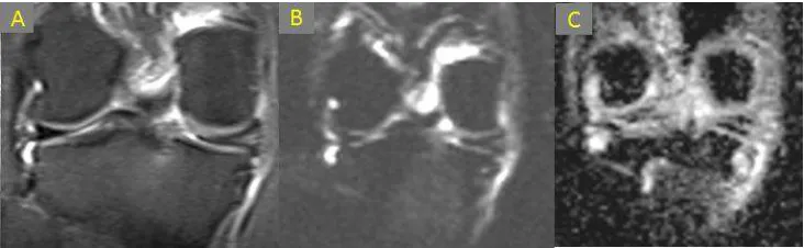
Figure 2. Medial meniscal tear shown by PDW (A), DW imaging with b-value 400 mm2/sec (B) and ADC mapping with b-value 400 sec/mm2 in coronal plane images.
Residual meniscal fragments after surgery and tears remaining after arthroscopy and were excluded from this study. Increased meniscal signal, located in the meniscal bodies, without any relationship to the inferior, superior or free edges of meniscal surfaces, was regarded as degeneration. Horizontal or triangular hyperintensity limited to meniscal bodies, was regarded as degenerated meniscal fragments. Oblique or vertical signals in articular surfaces and increased meniscal signals in meniscal attachment sites were never classified as degeneration.These criterias which indicated degeneration, were used for both routine and DWI-ADC mapping images.
6. DWI-ADC Mapping in the
Evaluation of Knee Joint, Including
Menisci and Cartilage Injuries
Aydin et al. reported a research about DWI and ADC mapping in the diagnosis of meniscal tears (26). He studied 74 consecutive(30 men and 44 women) patients with
meniscal tear under 1.5 T magnet, 11 of the patients had lateral meniscal tears, and 63 of them had medial meniscal tears which were consisted of 38 horizontal tears, 18 longitudinal tears, 2 radial tears, 2 root tears, and 7 complex and oblique tears. Lateral meniscal tears were of root, oblique and horizontal type, whereas medial meniscal tears included all types of tears. He had performed DWI by 3D-SE echo-planar imaging (EPI) in coronal and sagittal planes and ADC mapping in coronal planes with b-factors of 50, 400, and 800 sec/mm2.
He reported 100% specificity and 86% sensitivity with NPV of 0.09 and PPV of 1 for all meniscal tears according to the DWI results, regarded 100% specificity and 38% sensitivity by ADC mapping with b-value of 50 sec/mm2 with NPV of 0.02 and PPV of 1, 100% specificity and 78% sensitivity with NPV of 0.06 and PPV of 1 by ADC mapping with b-value of 400 sec/mm2 , and 100% specificity and 68% sensitivity with NPV of 0.04 and PPV of 1 for ADC mapping with b-value of 800 sec/mm2 with regard to routine MRI sequences.
revealed no significant statistical differences when compared to routine MRI sequences, DWI and ADC mapping with b-value of 400 sec/mm2 revealed higher sensitivity and specificity than other imaging modalities(26).
As a result, he claimed that “DWI and ADC mapping, especially with b-factor of 400 sec/mm2, might be an alternative imaging modality to the routine MR imaging sequences for visualisation of meniscal tears especially for the medial meniscus. Despite their low spatial resolution, these scans were relatively fast and they should be easily used by an experienced musculoskeletal radiologist for detailed knee MR imaging”.
Xu et al. (27) studied T2 mapping and diffusion weighted imaging in the diagnosis of early injury of knee cartilage under 3.0 T scanner. In this study, 72 subjects, including healthy group (n=30) and early cartilage injury group (n=42), were tested on MR scans with T2-mapping and DWI.
The normal group (n=30) showed no abnormality in rutine MR imaging. 10 cases of 42 patients underwent conventional MR examination, in which the routine MR images depicted significant abnormal cartilages excluded from this study. 32 patients (the remaining patient group) with no cartilage abnormalities but different amounts of joint effusion were included in the study of early injury group. The result indicated that the average T2 values and ADC values of cartilage in early cartilage injury group were significantly higher than the normal group. The remarkable differences showed statistical significance.
7. Conclusion
There are not much studies mentioned about DWI and ADC mapping of knee in the literature so far. However, the studies in the literature about this subject indicates that these sequences are fast and easily applicable with routine MRI sequences and these new MRI techniques (for the knee) depicts very important information for menisci and cartilages of the knee, especially ADC mapping via b-value of 400 sec/mm2 for menisci of the knee moreover T2-mapping and DW imaging are valuable imaging sequences in the diagnosis of early articular cartilage injury (26,27).
Author Contributions
Guarantor of integrity of entire study : H.A,V.K. Study design : V.K,H.A
Study concepts : V.K,H.A,B.H. Data analysis : H.A.
Manuscript drafting : V.K
Literature research : H.A,V.K, B.H. Clinical studies : H.A.
Statistical analysis : H.A. Manuscript editing : V.K İnterpretation : H.A,V.K
Financial Disclosure
All authors state no financial relationship to disclose.
References
[1] Wolff AB, Pesce LL, Wu SJ et al: Comparison of spin echo T1W sequences versus fast spin-echo proton density-weighted sequences for evaluation of meniscal tears at 1.5 T. Skeletal Radiol, 2009; 38(1): 21–29
[2] Ohishi T, Takahashi M, Abe M et al: The use of axial reconstructed images from three-dimensional MRI datasets for morphological diagnosis of meniscal tears of the knee. Arch Orthop Trauma Surg, 2005; 125(2): 622–27
[3] Kocabey Y, Tetik O, Isbell WM et al: The value of clinical examination versus Magnetic resonance imaging in the diagnosis of meniscal tears and anterior cruciate ligament rupture. Arthroscopy, 2004; 20(7): 696–700
[4] Boxheimer L,Lutz AM, Zanetti M et al: Characteristics of displaceable and non-displaceable meniscal tears at kinematic MR imaging of the knee. Radiology, 2006; 238(1): 221–31
[5] Moritani T, Ekholm S, Westesson PL. Diffusion-Weighted MR Imagingof theBrain. Berlin: Springer-Verlag; 2004; 1-5.
[6] Brown R (1928) A brief account of microscopical observations made in the months of June, July, and August, 1827, on the particles contained in the pollen of plants; and on the general existence of active molecules in organic and inorganic bodies. Phil Mag 4:161–173
[7] Rubin DA, Palmer WE: Imaging of the knee. IDKD, 2005: 26–38
[8] Harper KW, Helms CA, Lambert HS et al: Radial meniscal tears: Significance, incidence and MR appearance. AJR, 2005; 185(4): 1429–34
[9] MR Quinn SF, Brown TR, Szumowski J: Menisci of the knee: Radial imaging correlated with arthroscopy in 259 patients. Radiology, 1992; 185(11): 577–80
[10] Rubin DA, Kneeland JB. MR imaging of the musculoskeletal system: technical considerations for enhancing image quality and diagnostic yield. AJR Am J Roentgenol 1994; 163:1155-1163
[11] Rubin DA, Paletta GA: Current concepts and controversies in meniscal imaging. Magn Reson Imaging Clin N Am, 2000; 8(3): 243–70
[12] Quinn SF, Brown TF: Meniscal tears diagnosed with MR imaging versus Arthroscopy: How reliable a standard is arthroscopy? Radiology, 1991; 181(12): 843–47
[13] Mori S, Barker BP: Diffusion magnetic resonance imaging: Its principle and applications. The anatomical record (New Anat), 1999; 257: 102–43
[15] Nakajo M, Nakajo M, Kajiya Y et al. FDG PET/CT and diffusion-weighted imaging of head and neck squamous cell carcinoma: comparison of prognostic significance between primary tumor standardized uptake value and apparent diffusion coefficient. Clin Nucl Med. 2012 May;37(5):475-80.
[16] Verhappen MH, Pouwels PJ, Ljumanovic R. et al. Diffusion-weighted MR imaging in head and neck cancer: comparison between half-fourier acquired single-shot turbo spin-echo and EPI techniques. AJNR Am J Neuroradiol. 2012 Aug;33(7):1239-46.
[17] Choi BB, Kim SH, Kang BJ. et al. Diffusion-weighted imaging and FDG PET/CT: predicting the prognoses with apparent diffusion coefficient values and maximum standardized uptake values in patients with invasive ductal carcinoma. World J Surg Oncol. 2012 Jun 28;10:126.
[18] Thomassin-Naggara I, De Bazelaire C, Chopier J. et al. Diffusion-weighted MR imaging of the breast: advantages and pitfalls. Eur J Radiol. 2013 Mar;82(3):435-43.
[19] Curvo-Semedo L, Lambregts DM, Maas M. et al. Diffusion-weighted MRI in rectal cancer: apparent diffusion coefficient as a potential noninvasive marker of tumor aggressiveness. J Magn Reson Imaging. 2012 Jun;35(6):1365-71.
[20] Jang KM, Kim SH, Choi D et al. Pathological correlation with diffusion restriction on diffusion-weighted imaging in patients with pathological complete response after neoadjuvant chemoradiation therapy for locally advanced rectal cancer: preliminary results. Br J Radiol. 2012 Sep;85(1017):e566-72.
[21] Aydin H, Hekimoglu B, Kızılgöz V. A brief review of the combined use of T2-weighted MRI and diffusion-weighted imaging for prostate cancer diagnosis. AJR Am J Roentgenol. 2013 Feb;200(2):W219.
[22] Aydin H, Kizilgöz V, Tatar IG. et al. Detection of prostate cancer with magnetic resonance imaging: optimization of T1-weighted, T2-weighted, dynamic-enhanced T1-weighted, diffusion-weighted imaging apparent diffusion coefficient mapping sequences and MR spectroscopy, correlated with biopsy and histopathological findings. J Comput Assist Tomogr. 2012 Jan-Feb;36(1):30-45.
[23] Englund M, Guermazi A, Gale D et al: Incidental meniscal findings on knee MRI I middle-aged and elderly persons. N Engl J Med, 2008; 359(11): 1108–15
[24] Quinn SF, Brown TF: Meniscal tears diagnosed with MR imaging versus Arthroscopy: How reliable a standard is arthroscopy? Radiology, 1991; 181(12): 843–47
[25] Koenig JH, Ranawat AS, Umans HR et al: Meniscal root tears: Diagnosis and treatment. Arthroscopy, 2009; 25(9): 1025–32
[26] Aydin H, Kızılgöz V, Hekimoğlu B. Is the quantitative Diffusion-Weighted MR Imaging and ADC mapping with b-values of 50, 400, and 800 sec/mm2 a reliable method for evaluation of meniscal tears in the knee? Pol J Radiol, 2011; 76(1): 30-40