Retinal Disease Detection Using LBP Technique
Manojkumar S B, Shama Firdose U, Dr. M.B Anandaraju, Dr. H S Sheshadri
Asst. Professor, PG Student, Professor & HOD, Professor , Dept. of ECE, BGSIT, BG Nagar
[email protected], [email protected], [email protected], [email protected]
Abstract-This work explores discrimination capabilities in the texture of fundus images to differentiate between pathological and healthy images. For this reason, the execution of Local Binary Patterns (LBP) as a surface descriptor for retinal pictures has been investigated and contrasted and different descriptors.The objective is to separate between diabetic retinopathy (DR) and typical fundus pictures investigating the surface of the retina foundation and keeping away from a past sore division organize. Experiments were designed and validated with the proposed procedure obtaining promising results. For each experiment, several classifiers were tested. An average sensitivity and specificity higher than 0.86 in all the cases and almost of 1 and 0.99 respectively, for DR detection were achieved. These outcomes recommend that the strategy exhibited in this paper is a powerful calculation for portraying retina surface and can be valuable in an analysis help framework for retinal sickness screening.
Keywords: Local Binary Patterns (LBP), diabetic retinopathy (DR) Non-Proleferative Diabetic Retinopathy(NPDR), Proliferative Diabetic Retinopathy(PDP).
1. INTRODUCTION
Diabetic Retinopathy is the term that is used to describe the retinal damage causing the vision loss. Some people are born with it, whilst others acquire the disease in later life. The body can’t store the sugar from food, and it courses through the bloodstream. This sugar reacts with the walls of the blood vessels as it does so, causing them to break down over time.
Diabetic Retinopathy will be harmful to the retina. As light beams come into the eye, through the viewpoint, it arrives on the retina to be transform into electrical signs to be sent to the mind. Courses and veins take oxygen and supplements to your retina and diabetes harms and devastates these veins
The WHO assesses that in 2010 there were 285 million individuals outwardly hindered nearby the globe. Despite the way that the quantity of visual deficiency cases has been essentially decreased as of late, the situation is assessed that 80% of the instances of optical disability are avoidable or curable. DR and age related mascular degeneration is these days two of the greatest continuous reasons of visual deficiency and apparition misfortune. Furthermore, these maladies will encounter a high development later on because of diabetes occurrence increment and maturing populace in the present society.
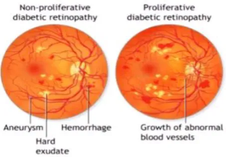
Figure 1: Non-proliferative and Proliferative Diabetic Retinopathy
Their initial determination permits, through fitting action, to lessen costs produced the minute they are in cutting edge situations and may end up unending. This reality legitimizes screening efforts.DR and AMD can be described by the contiguity of particular kinds of retinal injuries, for example, microaneurysms, exudates or druses, among others. Figure 1 delineates a few cases of these maladies in examination with the fundus picture from a solid patient.
1. Non Proliferative diabetic retinopathy (NPDR) spillage may incite swelling to the macula.
2. Proliferative Diabetic Retinopathy (PDR) is the primary condition based on the disease. The present stage stream issues preclude the retina from securing the oxygen. In this manner new fragile vein can start to develop in the retina is as shown in the figure 1.2. 2. LITERATURE SURVEY
Sandra Morales et al. [1], Retinal Disease Screening through Local Binary Patterns. This paper researches segregation capacity in the surface of fundus to separate amongst neurotic and solid pictures. Specifically, the fundamental concentration lies in investigating the execution of Local Binary Patterns (LBP) as a surface descriptor for retinal pictures.
T. Ojala et al. [2], A comprehensive local binary pattern operator for multiresolution gray scale and rotation invariant texture classification. Presents a hypothetically exceptionally straightforward, yet effective, multi determination way to deal with dark scale and revolution invariant surface grouping in light of neighborhood parallel examples and nonparametric separation of test and model dispersions.
T. Ahonen et al. [3] ,Face description with local binary patterns: Application to face recognition . This paper exhibits a novel and proficient facial picture portrayal in view of neighborhood paired example (LBP) surface highlights. The face picture is separated into a few locales from which the LBP include appropriations are removed and connected into an upgraded highlight vector to be utilized as a face descriptor.
Kjersti Engan et al. [4], Detection of diabetic retinopathy and age-related macular degeneration from fundus images through local binary patterns and random forests, This paper centers around investigating the execution of Local Paired Pattern (LBP) as a surface descriptor for retinal pictures to separate amongst neurotic and sound pictures. In spite of the fact that LBP has been generally used to order surfaces, not much is accounted for on the investigation of the retina surface.
L. Nanni et al. [5], Face description with local binary patterns: Application to face recognition. This work centers around the utilization of picture based
machine learning methods in therapeutic picture investigation.
Mahendran Gandhi et al. [6], Diagnosis of Diabetic Retinopathy Using Morphological Process and SVM Classifier. This paper additionally centers around exudates for the reason that it gives data about prior diabetic retinopathy. The proteins and lipids getting spilled from the circulatory system into the retina through harmed veins is the main source of exudates.
3. MATERIALS AND METHODS
A. Materials
The material of this work are pictures beforehand analyzed as should be expected , DR or AMD.The related macular degeneration (AMD) subjects (n=23), sound control-bunch subjects (n=61), and diabetic subjects (n=59). Prepared picture examination specialists have followed out the veins, and furthermore the optic circle and fovea where pertinent. Gaze database is a full arrangement of 402 pictures (700 605 pixels) where thirteen distinct findings were considered. From this dataset, three subsets were created: age-related macular degeneration (n=47), typical (n=37), and diabetic retinopathy (n=89). E-OPHTHA is a database of fundus pictures particularly intended for diabetic retinopathy screening. It contains 257 pictures with no injury, 47 pictures with exudates and 148 with microaneurysms or little hemorrhages making a sum of 174 pictures with diabetic retinopathy. At last, DIAGNOS is a private database, property, made out of 45 fundus pictures, 22 tormented with AMD and 23 solid. Both E-OPHTHA and DIAGNOS have a range of distinctive picture resolutions. The four databases encounter a huge fluctuation in shading, brightening, determination, quality, and so forth both inside and, considerably more, among the databases.
B. Local Binary Patterns
estimation of the focal pixel of the area producing a twofold string or, at the end of the day, a double example. The LBP is given as follow:
Where gp and gc are the dark estimations of the area and focal pixel, separately. P speaks to the quantity of tests going on the symmetric round neighborhood of sweep R. 2P diverse double examples can be created in every area.
Figure 2: LBP computation perform on input image Grey values of a circular neighbourhood of radius 1 and 8 samples. Thresholding between the grey value of the region and the central pixel. The rotation invariant local binary pattern generated is 00101101 (the arrows indicate the order in which the string is formed).
When LBP are used for texture description, it is common to include a contrast measure by defining the rotational invariant local variance as follows:
The LBP and VAR measures are complementary and are combined to enhance the performance of the LBP operator.
4. SYSTEM METHADOLOGY
This paper researches separation capacities in the surface of fundus to separate amongst neurotic and sound pictures. Specifically, the primary concentration lies in investigating the execution of Local Binary Patterns (LBP) as a surface descriptor for retinal pictures.
The objective of this paper is to recognize DR, and typical fundus pictures in the meantime and maintaining a strategic distance from any past division phase of retinal sores. The surface of the retina foundation is specifically broke down by methods for LBP, and just this data is utilized to separate solid patients and these two pathologies. An exhaustive report about what sort of classifier gets the best outcomes is likewise attempted.
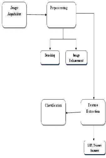
Figure 3: Block Diagram of Detection of Retinal Disease using local binary patterns
The block diagram of Detection of Retinal Disease using local binary patterns is as shown in the figure 3.having the following functional units:
1. Image Acquisition: Images are acquired from Gallery.
2. Preprocessing: The objective of the preprocessing phase is to apply possible image enhancement techniques to obtain the required visual quality of the images. Image enhancement techniques,
RGB Channel Extraction Filtered Image
Histogram Equalization Image
RGB Channel Extraction: In that R, G and B channels are extracted from color image. Filtered Image: A median filter does a very
good job at reducing the noise in image. This filter is applied in each channel of image for reduce the noise.
Histogram Equalization Image: The contrast enhancement of the image can be observed by applying histeq (enhance contrast using histogram equalization).
3. Feature Extraction: In feature extraction stage, local binary pattern is used for extraction of features in each channel separately. Then extract the texture features for each channel of LBP image. After that, take average of all channel features for construct the final features. The final features such as mean, standard deviation, Entropy, Kurtosis and Skewness.
4. Classification: The classification process is done from extraction of final features. The main novelty here is the adoption of Random Forest. RF classifier is applied over the features and the classification is done.
Figure 4: Activity Diagram
The Activity Diagram of its proposed system having the function as follows:
Smoothed rectangles symbolize events. Diamonds symbolize decisions. Gash or stop of simultaneous behavior. A blank loop represents the beginning of the
work flow.
A bordered blank sphere represents the closing stages.
A Preprocessing
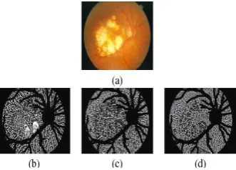
The red, green and blue components of each image are independently analysed. The aspect of the LBP and VAR images obtained from an AMD fundus is represented in Figure 5. Only the pixels of the retina background are considered significant for the texture analysis. The optic disc and blood vessels are detected by our own methods and subsequently, the LBP and VAR labels from these areas are removed by masking. The same mask for optic disc and blood vessels are used for three RGB components.
B Feature Extraction
final features such as mean, standard deviation, Entropy, Kurtosis and Skewness.
Figure 5: Feature extraction using P = 8 and R = 5. (a) AMD fundus image,(b-d) LBP images calculated on R , G and B components, respectively. Optic disc and vessel segments are removed (black).
C Classification:
The classification procedure is done from extraction of conclusive highlights. The primary oddity now the appropriation of Random forest (RF). RF classifier is connected over the highlights also the arrangement is finished.
5. RESULTS AND ANALYSIS
Figure 6: Process perform for Detection of Retinal Disease Using LPB Technique
Figure 6 shows the Process steps perform for the Detection of Retinal Disease using LBP. The first step is to Test an input Image which is Acquire from gallery. The second step is to preprocessing an image by using R G B channel extraction in which image has
to be Denoised and enhancementing an image by using Histogram Equation. The third step is Feature Extraction by using R G B component in which Mean, Standard Deviation,
Entropy, Kurtosis and Skewness values can be obtained for each channel.
Figure 7: Final Features and Classification of Test image
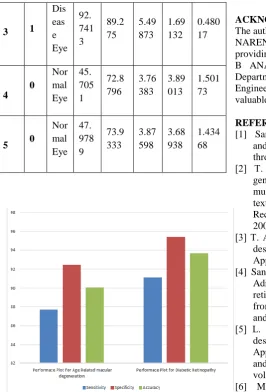
Figure 7 shows the Final Features and Classification of Test Image having an Average of R G B Features and classification result if the obtained Class is 1 the output is Disease Eye, if it is 0 the output is Normal Eye.
Table 1: Final Average Features of RGB Channel
3 1 degeneration and diabetic retinopathy
6. CONCLUION
In this paper, a new approach for DR diagnosis was presented. It is based on analyzing texture discrimination capabilities in fundus images to differentiate healthy patients from DR images. The performance of LBP along with different classifiers was tested and compared with other texture descriptors. The most important finding is that the proposed method is capable of discriminating the classes based on analysing the texture of the retina background, avoiding previous segmentation of retinal lesions. Such lesion segmentation algorithms might be time consuming, thus avoiding the segmentation is beneficial. The obtained results demonstrate that using LBP as texture descriptor for fundus images provides useful features for retinal disease screening.
ACKNOWLEDGEMENT
The authors would like to sincerely thanks to DR. B K NARENDRA, Principal, BGSIT, BG Nagar for providing the necessary facilities and support, DR. M B ANANDARAJU, Professor and Head of the Department, Electronics and Communication Engineering BGSIT, BG Nagar, for their support and valuable suggestion to complete this paper.
REFERENCES
[1] Sandra Morales, Kjersti Engan, Valery Naranjo and Adri´an Colomer, “Retinal Disease Screening through Local Binary Patterns”.
[2] T. Ojala, M. Pietikinen, and T. Menp “A generalized local binary pattern operator for multiresolution gray scale and rotation invariant texture classification”. in Advances in Pattern Recognition, 2nd International Conference on, 2001, pp. 397–406.
[3] T. Ahonen, A. Hadid, and M. Pietikainen “Face description with local binary patterns: Application to face recognition”.
[4] Sandra Morales, Kjersti Engan, Valery Naranjo, Adri´an Colomer “Detection of diabetic retinopathy and age-related macular degeneration from fundus images through local binary patterns and random forests”.
[5] L. Nanni, A. Lumini, and S. Brahnam, “Face description with local binary patterns: Application to face recognition,” Pattern Analysis and Machine Intelligence, IEEE Transactions on, vol. 28, no. 12, pp. 2037– 2041, 2006.
BIOGRAPHIES
Shama Firdose U obtained her BE degree in Electronics and communication Engineering from BGSIT, BG Nagar in 2016. Currently she is pursuing M.Tech in VLSI Design and Embedded System in BGS Institution of Technology, BG Nagar, Mandya, and Karnataka.
Manoj Kumar S.B obtained his BE degree in Electronics and communication Engineering from Shridevi Institute of Engineering and Technology, Tumkur in 2007, M.Tech in VLSI Design and Embedded System from PESCE, Mandya and pursuing his PhD on medical image processing in PET Research Center, Mandya at University of Mysore. He is currently working as Assistant Professor in Department of Electronics and communication Engineering BGSIT, BG Nagar, and Mandya from past 9 years, Karnataka.