Heather A Minges Wols,
Barat College of DePaul University, Lake Forest, Illinois, USA Plasma cells are terminally differentiated B lymphocytes that provide protective immunity through the continuous secretion of antibodies. Antibody-secreting cells develop in secondary lymphoid tissue following antigen stimulation and may enter a short-lived plasma cell population that reside primarily in the nonlymphoid area of the spleen or lymph nodes, or instead may migrate to the bone marrow where the majority enter a long-lived population of plasma cells.Introduction
The humoral branch of the immune system is critical for providing protective, circulating antibodies. In humans, antigen-specific antibody has been detected in the serum for decades following antigen encounter. The antibody pool is maintained by plasma cells, which secrete antibod-ies following antigen exposure.
Since soluble antibody is necessary for providing pro-tective immunity to an individual or animal, it must be able to circulate readily throughout the body as a means of surveillance. Because plasma cells themselves are not known to migrate from tissue to tissue, it is important that plasma cells are situated in tissues such that antibody can easily enter the circulation. In the spleen and lymph nodes, plasma cells are detected among the reticular sinus-oidal cells in the red pulp and medullary cords, respec-tively, which are rich in vasculature facilitating antibody circulation. The exact location of plasma cells in the bone marrow is not known, however, it is hypothesized that plasma cells interact with the reticular stromal cells sur-rounding the sinusoidal endothelial cells, again facilitating antibody secretion directly into the bloodstream.See also: Lymph nodes; Spleen
The plasma cell literature is often confusing as plasma cells are repeatedly misclassified. It is important when interpreting data that one is clear as to precisely what population of cells is being examined. Plasmablasts are precursor cells of short- and long-lived plasma cells and are generally described as a proliferating fraction of antibody-secreting cells, often found in the bloodstream emigrating to organs such as the bone marrow. Plasma cells are simply terminally differentiated noncycling antibody-secreting cells. Plasma cells are not normally found in the circulation, but rather remain residents in their organ of choice for life; any antibody-secreting cells in the blood en route to, for example the bone marrow, are plasmablasts. With this often ambiguous terminology clear, readers can more effectively evaluate scientific literature regarding plasma cells and their precursors.
Plasma cell morphology
Plasma cells are easily distinguished from mature B cells by their morphological appearance. Mature B cells exhibit a high nucleus to cytoplasm ratio, little rough endoplasmic reticulum (RER), and an uncondensed nucleus. In con-trast, plasma cells exhibit a small, dense, eccentric nucleus, voluminous cytoplasm containing prominent amounts of RER and enlarged Golgi (Figure 1).
Development, Differentiation and
Migration
Parallels between B-cell development and
plasma cell support in the bone marrow
The majority of long-lived plasma cells are detected in the bone marrow. Notably, B cells, the precursors of plasma cells, also undergo much of their early development in the bone marrow. A critical component of B-cell development in the bone marrow is the reticular stromal cell. It provides
Article Contents
. Introduction
. Development, Differentiation and Migration
. Short- and Long-lived Populations of Plasma Cells
. Antibody Secretion by Plasma Cells
. Cytokine Production by Plasma Cells
doi: 10.1038/npg.els.0004030
Figure 1 Characteristic plasma cell morphology shown of bone marrow plasma cells stained with haematoxylin and eosin.
both the contact and growth factors B cells need to progress through stages of maturation. Both plasma cells and developing B cells are likely to interact with stromal cells in the bone marrow. Therefore, an important under-standing of the components required for B-cell develop-ment may elucidate similar requisites for plasma cell survival. Studies have begun to identify such parallels.
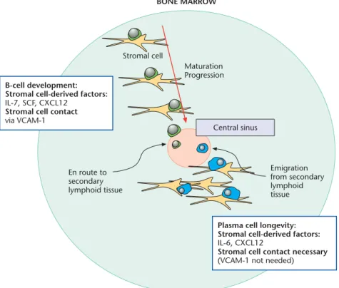
As B cells develop within the marrow, the location of the cells change such that maturation proceeds from the outer region of the bone marrow to the inner region where the central sinus is located (Osmond and Park, 1987). The early stages of B-cell development are dependent on interaction with stromal cells via vascular cell adhesion molecule-1 (VCAM-1), as well as the growth factors stromal cells pro-duce, such as interleukin-7 (IL-7), stem cell factor (SCF) and C-X-C chemokine ligand-12,(CXCL12) among others (Dorshkind, 1990). Without these developmental cues, B cells cease maturation. Concurrent with maturation and migration within the marrow, the B-cell precursors lose dependency for stromal cell-derived cytokines as well as the need for stromal cell contact. To ensure emigration from the bone marrow does not occur prematurely, B-cell precursors are retained in the marrow by chemokines, spe-cifically CXCL12 (Maet al., 1999). Immature B cells, the final stage of development in the marrow and located clos-est to the central sinus, lose expression of CXCR4 (C-X-C chemokine receptor-4), the receptor for CXCL12, and are no longer retained in the marrow. They enter the central sinus and migrate to the spleen (Figure 2).See also: Bone marrow; Chemokines; Cytokines; Interleukins; Lympho-cyte development
Many of the requirements needed for plasma cell sur-vival in the bone marrow are parallel to those needed for B-cell development. First, plasmablasts and plasma B-cells ap-pear to regain their dependency for stromal cell interaction and stromal cell-derived factors in the bone marrow. Sim-ilar to that observed of developing B cells, contact is nec-essary for plasma cell survival: plasma cell numbers wane quicklyin vitroif stromal cells are absent (Minges Wols et al., 2002). Although the growth factors indispensable for B-cell development are dispensable for plasma cell sur-vival, stromal cell-derived factors are still a necessity. Stromal cells produce IL-6, which is critical for plasma cell survival. Thus, while the players are different, the interac-tive dependencies of plasma cells and B-cell precursors in terms of stromal cells parallel each other in several aspects (Figure 2).
Just as chemokines are responsible for retaining B-cell precursors in the marrow, chemokines appear responsible for plasmablast entry into and plasma cell retention within the bone marrow. CXCL12 is not only produced by stromal cells in the marrow, but also by the sinusoidal endothelial cells in the marrow – the cells plasmablasts are likely to encounter upon exit from the peripheral blood (Cyster, 2003). Plasma cells express CXCR4 on their sur-face and exhibit migration towards CXCL12, suggesting
plasma cells possess factors necessary to localize within the bone marrow (Cyster, 2003). Thus, the same chemokine that retains B-cell precursors in the marrow before they are developmentally ready to enter the periphery also plays a role in retaining terminally differentiated B cells (plasma cells) in the marrow later in the B-cell lifespan (Figure 2).
Plasma cell differentiation
Immature B cells leave the circulation and enter the spleen to complete development into a naive mature B cell. De-pending on the events that follow, naive B cells may leave the tissue and reenter the bloodstream to continue scav-enging for antigen. Alternatively, B cells may encounter antigen within the tissue during which time the progeny of activated B cells will differentiate into memory B cells, plasma cells or plasmablasts that will emigrate to the bone marrow.See also: B lymphocytes
Severalin vitrostudies have attempted to identify the key components of plasma cell generation. The addition of IL-3 and IL-10 to B-cell blasts co-cultured with bone marrow stromal cells stimulates immunoglobulin G (IgG) secretion and differentiation into nonproliferating plasma cells (Me-rvilleet al., 1995). An essential role for tumour necrosis factor-a(TNF-a) in the differentiation of antibody-secret-ing cells has also been demonstrated (Rodriguez et al., 1993). It appears that the cytokine is critical early in differ-entiation as the presence of TNF-ais required within the first 24 h of culture for subsequent Ig secretion. The neces-sity of IL-6 in generating antibody-secreting cells is enig-matic and appears to vary depending on the immunogen and the immunization protocol. Further complicating studies is the detection of plasma cells in IL-6 deficient mice suggesting that IL-6 is dispensable and/or that compensa-tion mechanisms exist (Kopfet al., 1994). Taken together, these studies only begin to elucidate the plethora of soluble factors that may be necessary for the commitment to an antibody-secreting cell.
Molecular and cellular events of plasma cell
differentiation
An intricate molecular programme governs plasma cell transition and commitment. A transcriptional repressor called B lymphocyte-induced maturation protein-1 (Blimp-1) is a master regulator of plasma cell differentia-tion (Calameet al., 2003). Cytokine activation of the B-cell lymphoma line (BCL-1) induces Blimp-1 expression, whereas removing cytokine help results in the halt of differentiation, decreasedBlimp-1message, and decreased IgM secretion. Blimp-1 is detected in antibody-secreting cells following both T-dependent and -independent anti-gen challenge, in plasma cells in the bone marrow, as well as in a subset of cells in the germinal centre displaying a
preplasma phenotype. However, Blimp-1 is not detected in memory B cells (Figure 3).
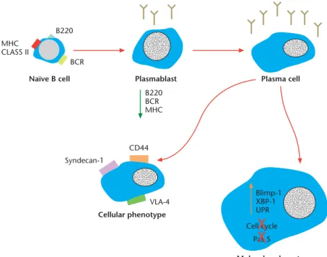
Terminally differentiated plasma cells are noncycling and thus phenotypically different from their predecessors. Expression of Blimp-1 protein results in concomitant re-pression of the B-cell-specific transcription and survival factorsBCL-6andPax5,the cell cycle proteinc-myc, the antigen presentation gene CIITA (necessary for MHC Class-II protein synthesis), and the B-cell receptor (BCR) signalling molecules Spi-BandId3,among many others (Calameet al., 2003). Conversely, Blimp-1 also indirectly upregulatesJ-chainmessenger ribonucleic acid (mRNA) expression and Syndecan-1 protein synthesis (Calame et al., 2003) (Figure 4).
X-box-binding protein-1 (XBP-1) is also a transcription factor essential for plasma cell differentiation (Calame et al., 2003). XBP-1 is sufficient to induce the generation of antibody-secreting cells in the BCL-1 cell line. Further-more, the absence of XBP-1 does not affect Blimp-1 ex-pression suggesting that XBP-1 may promote plasma cell differentiation through an unique pathway or downstream of Blimp-1.
Plasma cells are committed to the synthesis and secretion of antibody. Therefore, their cell surface phenotype differs greatly from memory B cells. The various stages in plasma cell development can also be distinguished based on the expression of cell surface molecules. Because plasmablasts no longer need to bind or present antigen, the expression of the BCR (surface Ig) and MHC Class II are present at decreased levels on the cell surface and are ultimately ab-sent on the surface of plasma cells (Calameet al., 2003). Another quintessential B-cell marker is surface expression of B220. The function of this protein is unknown, however its expression is intermediary on plasmablasts and is absent from the surface of plasma cells (Figure 3).
Plasma cells are best distinguished from other cell pop-ulations based on the membrane expression of Syndecan-1 (Cluster of differentiation, CD138), which is widely used as a means for isolation and detection (Minges Wolset al., 2002). Syndecan-1 has been shown to bind fibronectin, collagen and basic fibroblast growth factor; however, the consequence of Syndecan-1 ligation is unknown. In addi-tion to Syndecan-1, plasma cells exhibit surface expression of the adhesion molecules CD44 and very late antigen-4
Maturation Progression Stromal cell Emigration from secondary lymphoid tissue En route to secondary lymphoid tissue B-cell development:
Stromal cell-derived factors: IL-7, SCF, CXCL12
Stromal cell contact via VCAM-1
Plasma cell longevity: Stromal cell-derived factors: IL-6, CXCL12
Stromal cell contact necessary (VCAM-1 not needed) Central sinus
BONE MARROW
Figure 2 Maturation and migration of developing B cells. B cells develop from haematopoietic stem cells found in the periphery of the bone marrow. Differentiation progresses from the periphery of the marrow towards the central sinus. Development requires specific cytokines produced by and contact provided by stromal cells as indicated. Developing B cells are retained in the bone marrow (and restricted from early release) by chemokines produced primarily by stromal cells, namely CXCL12. Developing B cells express CXCR4, the receptor for CXCL12 on their surface. Once the immature B-cell stage is reached, the cells lose expression of CXCR4 and are released into the central sinus where they migrate to secondary lymphoid tissue. Plasmablasts entering the bone marrow from the secondary lymphoid tissue similarly find stromal cells to support their longevity. Again, stromal cells provide soluble factors necessary for retention and survival in the marrow, such as IL-6 and CXCL12. While contact is needed for plasma cell survival, the adhesion event is unclear; however, VCAM-1 is dispensable.
(VLA-4) (Figure 3) suggesting that there may be various means by which plasma cells can contact stromal elements in the marrow, keeping the antibody-secreting cells in close proximity to stromal cell-derived survival factors. Fur-thermore, an increase in overall size, as detected by forward light scatter by flow cytometry, is also observed.
The maturation progression of plasma cells
Human antibody-secreting cells isolated from various or-gans differ in cell surface phenotype. Cell surface molecule expression suggests a maturation progression from tonsil to blood to bone marrow (Medinaet al., 2002). For the most part, bone marrow plasma cells exhibit the following phenotypes (in comparison to tonsillar plasma cells): the gain of a Syndecan-1 plasma cell phenotype and the loss of a B-cell phenotype (CD19, CD20, CD22, human leukocyte antigen-DR (HLA-DR), Pax-5, among others), the gain of the survival factor Bcl-2 and the loss of the death receptor CD95, the gain of adhesion molecules such as VLA-4 and the gain of the chemokine receptor CXCR4 (Medinaet al., 2002). In addition, antibody-secreting cells from tonsil and blood secrete peak levels of antibody after 3 days ofin vitro culture whereas bone marrow plasma secrete antibody in a linear fashion for 3 weeks (Brieva et al., 1994). These MHC CLASS II B220 BCR Naïve B cell Pas 5 Blimp-1 XBP-1 UPR Cell cycle Molecular phenotype Cellular phenotype Syndecan-1 CD44 VLA-4 Plasma cell Plasmablast B220 BCR MHC
Figure 3 Schematic of plasma cell development and resultant phenotype. Naive B cells stimulated with antigen form antibody-secreting, proliferation-capable plasmablasts. Provided with the appropriate cues, plasmablasts develop into terminally differentiated plasma cells. Plasma cells express Syndecan-1 (CDSyndecan-138), CD44 and VLA-4 on their surface (among others); they downregulate expression of MHC Class II, B220 and the BCR complex. Further, plasma cells express Blimp-1 and XBP-1, which results in the inhibition of proliferation and ofPax-5expression, among others (seeFigure 4).
BCL-6 Blimp-1 Pax5 XBP-1 J-chain GERMINAL CENTRE BCL-6 Blimp-1 Pax5 XBP-1 J-chain PLASMA CELL c-myc Spi-B Id3 CIITA Ig synthesis B cell receptor signalling cell cycle MHC II
Figure 4 A simplified model of the regulatory cascades initiated during plasma cell differentiation. Targets activated by a particular factor are indicated by arrows; targets repressed are indicated by bars.
studies highlight the differences between tissue-tropic plasma cells and suggest that antibody-secreting cells pro-ceed in maturation from secondary lymphoid tissue to blood to their final destination in the marrow.
Studies performed in mice also support a population of precursor plasma cells found in the marrow. These cells continue to express many B-cell surface markers (such as BCR, MHC Class II and B220, among others) and retain their proliferative capacity (O’Connoret al., 2003). These cells do not appear to be memory B cells as they continue to secrete antibody. Further, these cells express VLA-4, CD44 and IL-6 receptor, which likely facilitate their development into terminally differentiated plasma cells when provided with the correct cues in the bone marrow.
Short- and Long-lived Populations of
Plasma Cells
Processed antigen is presented to T cells in the secondary lymphoid tissue. Once interactions between antigen-spe-cific B and T cells occur, a tight synapse between the two cells is formed resulting in subsequent B-cell activation and proliferation. Two fates arise for the activated B cells at this juncture: (1) differentiation into short-lived plasma cells or (2) the formation of a germinal centre and gener-ation of long-lived plasma cells. As a generalizgener-ation, the preponderance of short-lived plasma cells is found in the secondary lymphoid tissue, while the majority of long-lived plasma cells are detected in the bone marrow. However, some long-lived plasma cells remain in secondary lymph-oid tissue.
Generation of short-lived plasma cells
Short-lived plasma cells secrete nonmutated IgM or IgG, peak in numbers at days 8–10 postimmunization, and are found primarily at the B/T zone borders of the red pulp in the spleen or in the medullary cords of the lymph nodes. During a primary immune response, the low-affinity an-tibody secreted by these plasma cells is the early defence against the immunogen, while B cells possessing higher affinity antibodies are generated. Following secondary an-tigen challenge, the cellular response is several orders of magnitude greater than after a primary immunization. The short-lived plasma cell population contributes largely to the secondary antigen response. Studies suggest that the precursors of the short-lived plasma cells in a secondary response are the antigen-binding B cells located in the marginal zone of the spleen: B cells that have already un-dergone affinity maturation and selection in the germinal centre during the primary immune response (McHeyzer-Williams, 1997). Thus, the protective antibody from short-lived plasma cells has a greater affinity for the antigen after secondary immunization.
The germinal centre reaction and generation
of long-lived plasma cells
A possible fate of B cells following B–T cell interaction in secondary lymphoid tissue is the formation of germinal centres. The details will not be described here, however, as a result of germinal centre formation, B cells displaying high-affinity BCR on their surface are selected to survive. Selected clones may either reenter the germinal centre for further rounds of diversification or contribute to the mem-ory pool as either plasmablasts or memmem-ory B cells (McHeyzer-Williams, 1997).See also: Germinal centres
One cellular fate of a B cell that has completed the mul-tiple selection checkpoints in a germinal centre is to differ-entiate into an antibody-secreting plasmablast and ultimately a plasma cell. Currently, it is believed that the plasma cells that do not undergo affinity maturation (non-germinal centre) are short-lived, while the B cells that enter the germinal centre and undergo affinity maturation are long-lived plasma cells (McHeyzer-Williams, 1997). Long-lived plasma cells secrete high-affinity antibody. This is beneficial for the host, because the high-affinity antibody then remains for extended periods of time. See also: Antibodies; Antibody classes
Evidence to support the extended longevity of
bone marrow plasma cells
The bone marrow becomes a major site of antibody pro-duction. It has become apparent that the population of plasma cells that reside in the bone marrow is long-lived. This hypothesis is supported by a study in mice showing that antigen-specific bone marrow plasma cells survive >90 days postimmunization without cell division (Manzet al., 1997). This finding suggested that bone marrow plasma cells are not a dynamic, dividing population but instead are long-lived cells constitutively producing antibody. Antigen-spe-cific bone marrow plasma cells are also detected for >300 days after viral infection (Slifkaet al., 1998). In these same studies, plasma cells transferred to naive mice maintain se-rum antibody levels for >120 days posttransfer, supporting the idea that plasma cells elicited by a single antigen are long-lived and not a constantly replenishing population. Immunization of naive recipients with the original immuno-gen does not affect antibody secretion rates from the trans-ferred plasma cells; nor is necessary to maintain plasma cell longevity (Manzet al., 1998). However, this view of plasma cell longevity is not universally accepted. Data obtained by Ochsenbeinet al., (2000) suggest repeated antigen exposure is needed to maintain long-lasting antibody protection.
Effects of ageing
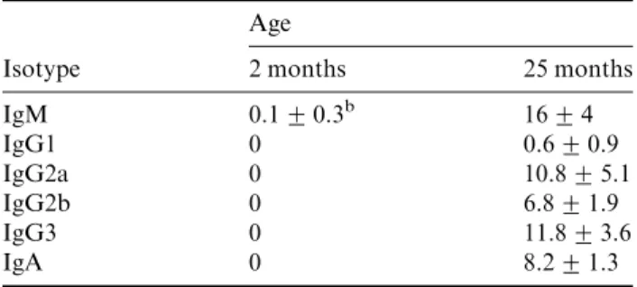
Data indicate that the frequency of plasma cells (regardless of isotype specificity) in the bone marrow is greater in older
animals (25 months) as compared to younger animals (59 months) (Table 1) suggesting that plasma cells accumulate in the marrow of an aged animal. Freshly isolated bone marrow cells from aged mice secrete Ig (without stimula-tion) but bone marrow cells from young mice do not. Fur-ther, long-term bone marrow cultures that support B-lymphopoiesis initiated from aged mice contain detectable amounts of antibody in their supernatants ‘six weeks’ after initiation, suggesting that plasma cells are present in the cultures at setup and survive for the duration of the culture period. In contrast, neither plasma cells nor detectable amounts of antibody are found in cultures initiated from young mice. These findings together suggest the plasma cells are long-lived and may be maintained by cells in the bone marrow culture microenvironment. Whether the in-creased numbers of plasma cells detected in the bone mar-row of aged mice is due to an accumulation of plasma cells over the lifetime of the animal or due to an aberrant mi-croenvironment in the aged animal is unclear.See also: Ageing and the immune system
While human bone marrow has been found to be a major source of plasma cells and antibody as well, studies that parallel those described above have not been performed, likely due in part to the heterogeneic composition of hu-mans. It is anticipated that similar increases in bone mar-row plasma cell numbers occur as humans’ age. Indeed multiple myeloma, a malignant plasma cell disorder, tends to afflict primarily older populations.
The role of the bone marrow
microenvironment
One critical element of the bone marrow microenviron-ment is the bone marrow reticular stromal cell. The role of stromal cells in maintaining plasma cell longevity remains largely unexplored. However, recent studies have begun to
unravel the interaction between plasma cells and bone marrow stromal cells. Data indicate that plasma cells cul-tured in the presence of stromal cells survived and contin-ued to secrete antibodyin vitrofor at least 3 weeks, whereas antibody could only be detected for 7 days in the absence of stromal cells, suggesting stromal cell contact and/or se-creted factors are necessary components for longevity (Minges Wolset al., 2002). Further, the addition of soluble IL-6 enhanced plasma cell survival in the absence of stromal cells, and plasma cells cultured with stromal cells lacking 6 resulted in a loss of antibody. The need for IL-6 by plasma cells to maintain longevity was further sup-ported by the observation that IL-6 mRNA message is upregulated in stromal cells following plasma cell co-cul-ture (Minges Wolset al., 2002). Further, stromal cells pro-duce CXCL12, which appears to have a role not only in attracting plasmablasts into the marrow, but also in the retention of antibody-secreting cells. CXCL12 may also be necessary for maintaining plasma cell survival within mi-croenvironmental niches (Cassese et al., 2003). Finally, VLA-4 must be engaged on plasma cells and may represent one of the mechanisms through which stromal cells and plasma cells interact. Although stromal cells express VCAM-1, one of the ligands for VLA-4, VCAM-1 is not necessary to maintain plasma cell survival. Taken together, the studies provide an initial glimpse of the basic elements necessary for maintaining an extended lifespan of plasma cells in the bone marrow (Figure 5). Exactly how IL-6, CXCL12 and VLA-4 interact, if at all, to support plasma cell longevity is presently unknown.See also: Immunolo-gical adhesion and homing molecules
Antibody Secretion by Plasma Cells
The function of a plasma cell is clear: secrete antibody. In fact, the purpose of a nonsecreting plasma cell is such an
IL-6 mRNA CXCL12
VLA-4 IL-6
Figure 5 A model of the elements necessary for maintaining plasma cell survival. Plasma cells and stromal cells interact in the bone marrow via VLA-4 on the plasma cell and an unknown ligand on the stromal cell. Contact induces the expression of IL-6 mRNA and consequently IL-6 secretion, which is critical to maintain plasma cell longevity. Further, stromal cells secrete CXCL12, which is important for attracting plasmablasts to the marrow, for retention of plasma cells in the marrow, and possibly for maintaining plasma cell survival.
Table 1 Changes in the numbers of bone marrow plasma cells with agea
Age
Isotype 2 months 25 months
IgM 0.1+0.3b 16+4 IgG1 0 0.6+0.9 IgG2a 0 10.8+5.1 IgG2b 0 6.8+1.9 IgG3 0 11.8+3.6 IgA 0 8.2+1.3 a
Bone marrow cells were isolated from mice at the indicated ages. Cells were cytocentrifuged on to slides, fixed, and stained with the indicated isotype-specific antibodies.
b
The value shown indicates the average number of isotype positive cells/1000 cells counted+s.d. The value ‘‘0’’ is indicative of detecting51 isotype positive cell/1000 total bone marrow cells.
enigma, its rarely considered. Supporting its sole respon-sibility to secrete antibody, the ratio of secretory Ig heavy chain mRNA to membrane Ig heavy chain mRNA in-creases greatly in a plasma cell. Further, plasma cells lose expression of membrane Ig and BCR, ablating activation potential. Since the primary role of plasma cells is to secrete antibody, many requirements for plasma cell survival and for antibody secretion appear to be synonymous. There-fore, the survival elements required by murine plasma cells also maintain antibody secretion. Additionally, several studies assessing antibody secretion from human mono-nuclear cells have found a role for IL-6, TNF-a, IL-1b, VLA-4, and fibronectin in enhancing or maintaining an-tibody titresin vitro(Brievaet al., 1994).
Further, several studies have begun to elucidate molec-ular mechanisms regulating antibody secretion. Transgenic mice possessing a truncated form of Blimp-1 (known to block Blimp-1 functions) exhibit an increased number of IgM-secreting cells and elevated and sustained IgM levels (Calameet al., 2003). The increased numbers of antibody-secreting cells appear due to increased proliferation and prolonged survival, suggesting that Blimp-1 has a role in cell cycle arrest and lifespan. Blimp-1 also indirectly regulates ‘J-chain’ mRNA expression and thus Ig synthesis. Further, XBP-1 expression appears to regulate antibody secretion; it was demonstrated that mice withXBP-12/2lymphocytes have extremely low levels of serum Ig, cannot control viral infection or respond properly to T-independent or QJ;T-dependent antigens, and show a general absence of Syndecan-1+ cells, despite having normal numbers of B cells and formation of germinal centres (Calameet al., 2003).
Cytokine Production by Plasma Cells
Most cytokine production by antibody-secreting cells has been studied in the malignant form of plasma cells: mul-tiple myeloma cells. The literature on mulmul-tiple myeloma cells is extensive and will not be reviewed here (although interested parties should review Jelinek, 1999 in the Fur-ther Reading). Unfortunately, little is known about cyto-kine production by plasma cells, likely because pure populations are difficult to obtain in adequate quantities to perform such experiments. Further, plasma cells are synthesizing large amounts of Ig and may not exert much energy in secreting cytokines readily available to them in the surrounding microenvironment. However, if plasma cells do secrete cytokines, a likely candidate would be IL-6, based on its necessity for survival and known autocrine production by myeloma cells. Studies found no IL-6 mRNA message in nonmalignant murine plasma cells, suggesting IL-6 is not produced by plasma cells themselves (Minges Wolset al., 2002). Thus, although IL-6 is a key element in maintaining plasma cell survival, nonmalignant plasma cells do not produce their own IL-6 but rather
in-duce its production in cells of the surrounding microenvi-ronment. Obviously, this is an area of plasma cell research still unexplored by researchers, and further knowledge is needed to fully appreciate the intricacies of a plasma cell. See also: Tumours of immune system
References
Brieva JA, Roldan E, Rodriguez C and Navas G (1994) Human tonsil, blood and bone marrowin vivo-induced B cells capable of spontaneous and high-rate immunoglobulin secretionin vitro: differences in the requirements for factors and for adherent and bone marrow stromal cells, as well as distinctive adhesion molecule expression.European Journal of Immunology24: 362–366.
Calame KL, Lin K-I and Tunyaplin C (2003) Regulatory mechanisms that determine the development and function of plasma cells.Annual Review of Immunology21: 205–230.
Cassese G, Arce S, Hauser AEet al.(2003) Plasma cell survival is me-diated by synergistic effects of cytokines and adhesion-dependent sig-nals.Journal of Immunology171: 1684–1690.
Cyster JG (2003) Homing of antibody secreting cells.Immunological Reviews194: 48–60.
Dorshkind K (1990) Regulation of hemopoiesis by bone marrow stromal cells and their products.Annual Review of Immunology8: 111–137. Kopf M, Baumann H, Freer Get al.(1994) Impaired immune and
acute-phase responses in interleukin-6-deficient mice.Nature368: 339–342. Ma Q, Jones D and Springer TA (1999) The chemokine receptor CXCR4 is required for the retention of B lineage and granulocytic precursors within the bone marrow microenvironment.Immunity10: 463–471. Manz RA, Lohning M, Cassese G, Thiel A and Radbruch A (1998)
Survival of long-lived plasma cells is independent of antigen. Inter-national Immunology10: 1703–1711.
Manz RA, Thiel A and Radbruch A (1997) Lifetime of plasma cells in the bone marrow [letter].Nature388: 133–134.
McHeyzer-Williams MG (1997) Immune response decisions at the single cell level.Seminars in Immunology9: 219–227.
Medina F, Segundo C, Campos-Caro A, Gonzalez-Garcia I and Brieva JA (2002) The heterogeneity shown by human plasma cells from tonsil, blood, and bone marrow reveals graded stages of increasing maturity, but local profiles of adhesion molecule expression.Blood99: 2154– 2161.
Merville P, Dechanet J, Grouard G, Durand I and Banchereau J (1995) T cell-induced B cell blasts differentiate into plasma cells when cultured on bone marrow stroma with IL-3 and IL-10.International Immunol-ogy7: 635–643.
Minges Wols HA, Underhill GH, Kansas GS and Witte PL (2002) The role of bone marrow-derived stromal cells in the maintenance of plasma cell longevity.Journal of Immunology169: 4213–4221. Ochsenbein AF, Pinschewer DD, Sierro Set al.(2000) Protective
long-term antibody memory by antigen-driven and T help-dependent differentiation of long-lived memory B cells to short-lived plasma cells independent of secondary lymphoid organs.Proceedings of the Na-tional Academy of Sciences of the USA97: 13263–13268.
O’Connor BP, Gleeson MW, Noelle RJ and Erickson LD (2003) The rise and fall of long-lived humoral immunity: terminal differentiation of plasma cells in health and disease.Immunological Reviews194: 61–76. Osmond DG and Park YH (1987) B lymphocyte progenitors in mouse
bone marrow.International Reviews in Immunology2: 241–261. Rodriguez C, Roldan E, Navas G and Brieva JA (1993) Essential role of
tumor necrosis factor-alpha in the differentiation of human tonsilin vivoinduced B cells capable of spontaneous and high-rate immuno-globulin secretion.European Journal of Immunology23: 1160–1164.
Slifka MK, Antia R, Whitmire JK and Ahmed R (1998) Humoral im-munity due to long-lived plasma cells.Immunity8: 363–372.
Further Reading
Gass JN, Gunn KE, Sriburi R and Brewer JW (2004) Stressed-out B cells? Plasma-cell differentiation and the unfolded protein response. Trends in Immunology25: 17–24.
Iwakoshi NN, Lee AH, Vallabhajosyula Pet al.(2003) Plasma cell differentiation and the unfolded protein response intersect at the tran-scription factor XBP-1.Nature Immunology4: 321–329.
Jelinek DF (1999) Mechanisms of myeloma cell growth control. He-matology/Oncology Clinics of North America13: 1145–1157. Reimold AM, Etkin A, Clauss Iet al.(2000) An essential role in liver
development for transcription factor XBP-1.Genes and Development 14: 152–157.
Tew JG, DiLosa RM, Burton GFet al.(1992) Germinal centers and antibody production in bone marrow.Immunological Reviews126: 99–112.
Turner CA Jr, Mack DH and Davis MM (1994) Blimp-1, a novel zinc finger-containing protein that can drive the maturation of B lymph-ocytes into immunoglobulin-secreting cells.Cell77: 297–306.