International Journal of Innovative Technology and Exploring Engineering (IJITEE) ISSN: 2278-3075, Volume-9 Issue-1, November 2019
Abstract: Medical images do contain important and unimportant spatial regions. Compression methods which are capable of reconstructing the image with high quality are required to compress the medical images. For these images, only a portion of it is useful for diagnosis hence a region based coding techniques are significant for compressing and transmission. Extracting a significant region is of great demand since a slighter mistake may leads to wrong diagnosis. This paper is focused on investigating multiple image processing algorithms for medical images. All the images may not contain the same region of interest, so different approaches are supposed to apply for different images. In this three types of medical images were considered like magnetic resonance (MR) brain images, computer tomography (CT) abdomen images and X-ray lung images. In this paper three automatic region of interest extraction algorithms were proposed for different types of images.
Keywords: Medical image compression, Region of interest, CT, MRI, abdominal image compression
I. INTRODUCTION
Medical images are acquired in multiple forms like computer tomography (CT), Ultra sound (US), Magnetic resonance imaging (MRI), positron emission tomography (PET), Single photon emission computed tomography (SPECT) and others that generate image data which require huge storage space. These images are stored in Hospital information system (HIS) or picture archiving and communication systems (PACS) and it was observed that a semi demanding hospital may produce a data around 10~15GB per on average per day which becomes critical demand for them store such a huge data [16] [5]. On other hand, to transmit this huge data for telemedicine applications require a dedicated high end network involving huge cost of maintenance [9].
There are various issues involved in transmission of the image data to rural areas, according to survey conducted by a French agency it was mentioned that only 25 % of the doctors‟ conduct practice in rural areas where the major portion of the people reside [25]. So exchange of information to these rural destinations requires dedicated networks which are practically cumbersome. One of the alternate is to suppress the data without losing the meaningful information in it and transmitting them though low speed networks.
Medical image compression is one of the solutions for reducing the storage space and handles the transmission
Revised Manuscript Received on November 05, 2019.
B. P. Santosh Kumar*, Department of Electronics and Communication Engineering, YSR Engineering College of Yogi Vemana University, Proddatur, India, Email: [email protected]
Dr. K. Venkata Ramanaiah, Department of Electronics and Communication Engineering, YSR Engineering College of Yogi Vemana University, Proddatur, India.
bandwidth requirements [20] Image compression methods are broadly categorized into lossy and lossless methods. For medical images, a lossy compression with 10 % compression is not acceptable as they distort the texture content in the images leading to the improper location and orientation of abnormality.
Storage of these medical images is too problematic for instance a 3D MRI contains a stack slices (images) that usually requires a huge space for storage. To overcome such limitations a region of interest (ROI) based compression is introduced that intends to preserve the quality of certain important regions while compression others. In image processing it can be termed as hybrid mechanism of integrating both lossy and lossless compression methods for single image
A. ROI image Compression
The concept of ROI based image compression is introduced to overcome the limitation of Lossy and Lossless compression modes. In Lossy mode, higher compression ratios can be achieved but it may lead to the loss of important clinical information on other hand in lossless mode the data is not compressed largely thereby occupies a large storage space and bandwidth. So there is a need for hybrid ROI image compression method that aims to compress for higher ratios and also preserve the important information [8].
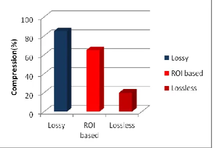
[image:1.595.327.542.569.717.2]The figure 1 depicts the compression spectrum from which it can be observed that in lossy mode the image can be compressed up to 85 % retaining 15-40% of the original file size. In loss less mode it can retain 80% of the original size, compressing only up to 20 %, the intermediate ROI based compression can compress up to 65 %, hence it can be concluded that ROI based image compression is more suitable for medical images.
Fig 1: Compression mode spectrum
Efficient Region of Interest Extraction Methods
for Multiple Medical Images
1555
Published By:
Blue Eyes Intelligence Engineering & Sciences Publication
Retrieval Number: A4526119119/2019©BEIESP DOI: 10.35940/ijitee.A4526.119119
In general, different medical images have different important regions and a single extraction approach is not suitable for all the images. So there is a need to develop multiple extraction algorithms which are suitable for particular images. Most of the researchers proposed ROI based compression for MR or CT brain images which is not sufficient and may not work with other parts of human body. Many researchers have focused on extracting the abnormality region and treating the region as ROI and the rest others as Non ROI but the method fails to apply compression when there is no presence of abnormality. This paper concentrates on proposing extraction approach for MR brain image, CT abdomen image and Lung images.
II. LITERATUREREVIEW
In order to extract a particular ROI from medical image four main methods are been proposed so far [1]. The first, is the segmentation that aims to partition into multiple segments for easier analysis of every region. But manual segmentation is time consuming and tedious process and hugely depends upon the user experience [2]. Second is the mathematical approach where three points from left, right and top are selected to form a sector of circle. These points are interpolated to form a contour for ROI. Third is interactive segmentation approach that requires annotations from the user to detect the ROI. Fourth is the mask based approach, in which a binary mask is generated using image processing methods that indicate the ROI.
Several methods were proposed so far which fall into one of the category, Canadian radiologists (CAR,2011) found that K-means clustering could able to produce a size reduction of 21%~22 % when compared with respect to original image. Another algorithm names as Chan vase method could achieve average reduction of 23%~24%. Vikas and Manpreet in [10] performed fractal lossy compression for non ROI and also introduced an approach for removing noise. Liu and others in [27], proposed a lossy to loss-less ROI compression scheme, this scheme relies on SPIHT and EBCOT coding where the images are partitioned into foreground and background. To code the ROI information a chain code is used. As it involves complex algorithms, this method consumes more time for processing. Ravi kiran et.al, in [18] [19], proposed a block based PCA algorithm for implementing lossy and lossless compression for non ROI and ROI. This method uses canny edge operator for extraction of mask region which produces segmentation blobs and fails to neglect the skull region specifically for MR brain images. Most of the methods concentrate on a single medical image and in particular MR brain, which brings the scope for new methods to be proposed for images projecting different parts of human body
III. PROPOSEDMETHODOLOGY
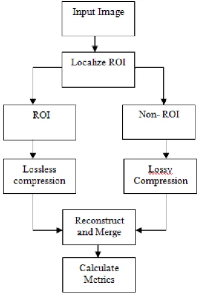
The block diagram of the ROI based image compression is depicted in figure 3, the input image is first processed for ROI localization which is a crucial step in ROI based image compression. In this work three types of medical images were considered as shown in figure 3.
For every type of medical image that represent a certain organ in human body has different region of interest and a single approach cannot be used to extract it.
[image:2.595.308.548.50.162.2](a)
(b)
(c)
Fig 2: Samples of different types of medical images that wereused in this work
A. ROI Extraction for MR brain Image
[image:2.595.313.507.290.577.2]In general, a brain image consists of white matter, Gray matter, skull and background regions [28]. The important clinical information required for diagnosis is usually not relied in skull and background region, so appropriate techniques may be incorporated to remove these regions. The ROI and NROI regions which are considered in this work are shown in figure 4. In this work a sequence of morphological operations was applied to remove the non ROI from MR brain image.
Fig 3: Block diagram of ROI based image compression
Fig 4: ROI and NROI for different medical images Algorithm for ROI extraction
Read the input image and do necessary pre processing Apply morphological erosion process with octagon
[image:2.595.311.543.614.675.2]International Journal of Innovative Technology and Exploring Engineering (IJITEE) ISSN: 2278-3075, Volume-9 Issue-1, November 2019
Convert the eroded image into binary with thresholding Apply dilation followed by filling process for binary
image
Retain the binary mask for ROI, and extract it respective elements from input image
[image:3.595.56.286.240.418.2]The present approach is compared against Kalaiselvi and somasundaram work mentioned in [31].This approach includes bi-level thresholding for generating the head mask and with the use of morphological operations at the post processing results an extracted brain image. The authors have attained an accuracy of 90%~92% when compared with manual segmented images. The visual and qualitative comparison between the method and our work is tabulated below.
Fig 5: Extraction of ROI from MR brain image (a1-a2)
Original Image (b1) Generated mask for ROI with our approach (b2) Generated mask with [31] approach (c1) Extracted ROI with our approach (c2) Extracted ROI with [31] approach (d1) Non ROI/ Difference Image obtained for our approach (d2) Difference obtained for [31] approach The segmentation efficiency of the works is compared in terms of accuracy and total segmentation errors mentioned in [31]. The numerical results attained for different images are tabulated below in table 1.
Table 1: Performance analysis of the proposed work in comparison with [31] for brain extraction process
S.NO Proposed Kalaiselvi [31]
ACC TSE ACC TSE
Patient 1 0.9164 0.182 0.88 0.218
Patient 2 0.9174 0.179 0.891 0.217
Patient 3 0.9108 0.1957 0.865 0.219
Patient 4 0.9092 0.1999 0.84 0.2199
Patient 5 0.9301 0.1230 0.91 0.2130 From the above analysis it can be observed that the present approach could able to attain an average accuracy of 3% more than [31] work, this is due to the inappropriate selection of thresholds and model parameters which was not in the case of present approach, hence the present work could able to extract more accurately than [31] in very short span of time.
B. ROI extraction from CT abdomen image There are several organs located in the abdominal CT image; it was observed that there are 12 major regions that are marked in the CT image. In [25] proposed a segmentation approach with prior information of the orientation and location of the organs and a prediction based assessment was employed to mark the region for latter images but this method proves to be mathematically complex and marking of prior require ample experience and more over the regions are overlapped with each other in the image so there is a requirement to extract all regions apart from skin and fat. In this work the ROI extraction from CT abdomen image can be extracted with the sequence morphological operations. The algorithm for the extraction process is presented below
Algorithm
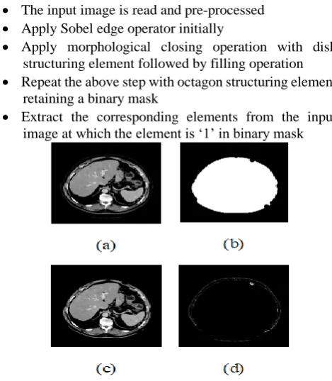
The input image is read and pre-processed Apply Sobel edge operator initially
Apply morphological closing operation with disk structuring element followed by filling operation Repeat the above step with octagon structuring element
retaining a binary mask
[image:3.595.310.545.255.524.2] Extract the corresponding elements from the input image at which the element is „1‟ in binary mask
Fig 6: Extraction of ROI from CT abdomen image (a) Original Image (b) Generated mask for ROI (c)
Extracted ROI (d) Non ROI C. ROI extraction from lung image
Morphological operations are not sufficient to extract the lung portion from chest x-ray /CT image since it contains the lung region, ribs, shoulders and background. Presence of the heterogeneous regions makes any method difficult in segmenting the lung image [21]. So appropriate edge based segmentation algorithms like active contour models are to be employed for segmenting and extracting the lung portion. Keita et.al in [11] proposed a graph cut method which consider multiple shape constraints and segment the lung portion.
In this work, structured edge detector (SED) in [26] is employed to segment the lung portion. A lung boundary map is detected initially using ultra metric contour map (UCM) [14] and then transformed with marker controlled watershed algorithm [17]. It was observed
[image:3.595.49.289.579.674.2]1557
Published By:
Blue Eyes Intelligence Engineering & Sciences Publication
Retrieval Number: A4526119119/2019©BEIESP DOI: 10.35940/ijitee.A4526.119119
could able to perform with efficiency greater than 90%.
Fig 7: Extraction of ROI from lung image (a) Original Image (b) Edge Contour mapping (c) Generated mask for
ROI (d) Extracted ROI
IV. EXPERIMENTALRESULTS
[image:4.595.309.546.44.842.2]The extracted ROI are compressed with lossless approaches like run length encoding and Non ROI are compressed with JPEG [15], EZW [7] and SPIHT [3] methods. Experiments are conducted on T1-weighted MR brain images collected from BRATS image database [29] and Lung images were collected from JSRT public database available at [30]. The compressed image is reconstructed by merging of the ROI and non ROI regions and evaluation metrics are calculated with respect to original Image. Multiple metrics like compression ratio (CR), peak signal to noise ratio (PSNR), mean square error (MSE), mean absolute error (MAE), mean structural similarity index (MSSIM), normalization cross correlation coefficient (NCC), structural content (SC) and image fidelity (IF) were calculated [22], [4], [12], [13].
Fig 8: Average PSNR comparison analysis for proposed work and [31] work.
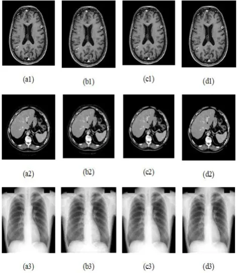
Reconstructed images with different methods are presented in figure 8 and the performance evaluation metrics are tabulated in tables 2, 3, and 4. From the tabulated data which are calculated on average for 50 images of each category and it can be observed that the SPIHT algorithm outperforms in terms of quality and compression
Table2: Average metrical values for MR brain image with current approach
Method RLE+
JPEG
RLE+
JPEG
RLE+
JPEG
CR 7.347 6.69 6.60
PSNR 38.16 49.99 39.23
MSSIM 0.995 0.999 0.996
MAE 1.41 0.285 1.23
NC 0.993 0.999 0.994
SC 1.002 1.002 1.007
[image:4.595.79.258.73.253.2]IF 0.92 0.99 0.94
Table 3: Average metrical values for [31] approach
Method RLE+
JPEG
RLE+
JPEG
RLE+
JPEG
CR 7.47 6.72 6.62
PSNR 37.36 44.50 38.36
MSSIM 0.994 0.999 0.995
MAE 1.71 0.55 1.44
NC 0.994 0.997 0.993
SC 0.999 1.004 1.007
IF 0.90 0.98 0.92
Table 4: Average metrical values for CT abdomen image
Method RLE+
JPEG
RLE+
JPEG
RLE+
JPEG
CR 7.64 8.07 8.05
PSNR 51.07 40.35 40.35
MSSIM 0.985 0.989 0.988
MAE 1.28 0.74 0.76
NC 1.005 0.996 0.995
SC 0.982 1.003 1.0032
IF 0.994 0.936 0.935
Table 5: Average metrical values for Lung images
Method RLE+ JPEG
RLE+ JPEG
RLE+ JPEG
CR 8.72 8.86 8.23
PSNR 36.74 48.54 38.57
MSSIM 0.963 0.998 0.984
MAE 4.02 0.54 2.87
NC 1.003 1.002 0.998
SC 0.9964 0.9994 0.9994
[image:4.595.71.264.509.651.2]International Journal of Innovative Technology and Exploring Engineering (IJITEE) ISSN: 2278-3075, Volume-9 Issue-1, November 2019
Fig 9: Reconstruction images with different methods (a1-a3) original images (b1-b3) compressed with RLE+JPEG (c1-c3) compressed with RLE+SPIHT
(d1-d3) compressed with RLE+EZW
Fig 10: PSNR performance analysis for brain images
Fig 11: PSNR performance analysis for abdomen images
Fig 12: PSNR performance analysis for Lung images It is observed from the experimental analysis that the method RLE+SPIHT approach retain higher PSNR values at different bits per pixels and from tables 1 to table 4 the method RLE+SPIHT has high MSSIM values, mean that the reconstructed image is of high quality which is extremely preferred for medical image compression.
V. CONCLUSION
This paper presents ROI extraction for different types of medical images projecting different parts of the human body. Since many algorithms concentrated only on single MR brain images this work provides a clear solution to compress images of different regions. On other hand, most of the techniques concentrated on extracting abnormal region treating it as ROI but fails for the images that doesn‟t possess abnormality here the main objective of this work is to provide a solution to store any medical image and from the experiments and evaluation this objective was achieved. It is observed that RLE with SPIHT gives more précised outcome compared with JPEG and EZW.
REFERENCES
1. Ansari M. A., Anand R. S., "Context based medical image compression for ultrasound images with contextual set partitioning in hierarchical trees algorithm", Advances in Engineering Software, vol. 40 no.7, pp.487-496, Jan. 2009.
2. AssafHoogi, Christopher F. Beaulieu, Guilherme M. Cunha, ElhamyHeba, Claude B. Sirlin, Sandy Napel, and Daniel L. Rubin , “Adaptive Local Window for Level Set Segmentation of CT and MRI Liver Lesions”, Medical Image Analysis ,2017, doi: 10.1016/j.media.2017.01.002
3. Said and W. A. Pearlman, "A new, fast, and efficient image codec based on set partitioning in hierarchical trees," in IEEE Transactions on Circuits and Systems for Video Technology, vol. 6, no. 3, pp. 243-250, June 1996.
4. Ahmet M Eskicioglundpaul S Fisher , “ Image quality measures and their performances “, IEEE transactions on communications Vol 43, No 12
5. Baeza. I, Verdoy. A, “ROI-based procedures for progressive transmission of digital images: a comparison” Journal of Math. Comput. Model, 2009, 50
6. Kumar, S. B. Kumar and C. Kumar, "Development of improved SSIM quality index for compressed medical images," 2013 IEEE Second International Conference on Image, Information Processing (ICIIP-2013), Shimla, 2013
7. D. Creusere, "Robust image coding using the embedded zerotree wavelet algorithm," Proceedings of Data Compression Conference - DCC '96, Snowbird, UT, USA, 1996
[image:5.595.314.540.51.215.2]1559
Published By:
Blue Eyes Intelligence Engineering & Sciences Publication
Retrieval Number: A4526119119/2019©BEIESP DOI: 10.35940/ijitee.A4526.119119
"Wavelet-Based Compression With ROI Coding Support for Mobile Access to DICOM Images Over Heterogeneous Radio Networks," in IEEE Transactions on Information Technology in Biomedicine, vol. 13, no. 4, pp. 458-466, July 2009.
9. JavidAli.T, Akhtar, P, “Significance of region of interest applied on MRI and CT images in tele-radiology-telemedicine”, Springer-Verlag, Berlin, Heidelberg, 2008
10. Kaur, Manpreet, and Vikas Wasson. ”ROI Based Medical Image Compression for Telemedicine Application.” Procedia Computer Science 70, 2015
11. Keita Nakagomi, Akinobu Shimizu, HidefumiKobatake, Masahiro Yakami, Koji Fujimoto, Kaori Togashi,” Multi-shape graph cuts with neighbor prior constraints and its application to lung segmentation from a chest CT volume”, Medical Image Analysis, Volume 17, Issue 1,2013,
12. Martin bernas ,” Image quality evaluation “, VIpromCom 2002, 4th Eurisp-IEEE region 8th international symposium on video image processing and multi- media communication 16-19 june 2002 13. M. Kass, A. Witkin, and D. Terzopoulos, “Snakes: Active contour
models,” International Journal of Computer Vision, vol. 1, no. 4, 1988.
14. P. Arbelaez, “Boundary Extraction in Natural Images Using Ultra metric Contour Maps,” in IEEE Conference on Computer Vision and Pattern Recognition Workshop (CVPRW'06) 2006
15. Pennebaker, William B., and Joan L. Mitchell, JPEG: Still Image Data Compression Standard, Van Nostrand Reinhold, 1993.
16. Placidi. G, “Adaptive compression algorithm from projections: application on medical grayscale images”, Journal of Comput. Biol. Med.2009, 39.
17. P. Soille, Morphological Image Analysis: Principles and Applications, 2nd ed.: Springer, Berlin Heidelberg, 2004
18. Ravi Kiran and Chandrasekhar kamargaonkar, "Region Separation Techniques for Medical Images", International Journal of Innovative Research in Science, Engineering and Technology, vol. 5, issue 2, February 2016.
19. Ravi Kiran and Chandrasekhar kamargaonkar, “Region Based Medical Image compression using Block-Based PCA”, nternational Conference on Computation of Power, Energy Information and Communication (ICCPEIC)-2016
20. S. Miaou, F. Ke and S. Chen, "A Lossless Compression Method for Medical Image Sequences Using JPEG-LS and Inter frame Coding," in IEEE Transactions on Information Technology in Biomedicine, vol. 13, no. 5, pp. 818-821, Sept. 2009.
21. Shuo Wang, Mu Zhou, Zaiyi Liu, Zhenyu Liu, DongshengGu, YaliZang, Di Dong, Olivier Gevaert, Jie Tian,” Central focused convolutional neural networks: Developing a data-driven model for lung nodule segmentation”, Medical Image Analysis, Volume 40, 2017,
22. Sonja Grgic, MislavGrgic , Marta mrak , “ reliability of objective picture quality measures”, journal of electrical engineering Vol 55 No 1-2, 2004
23. Standard Lossy Compression”, Canadian Association of Radiologists, Ottawa, ON, 2011
24. Toshiko Kaneda and Kristin Bietsch, "2016 World population data sheet with a special focus on human needs and sustainable resources," Population reference bureau, 1875- Connecticut Ave, NW, Suite 520, Washington DC 20009, ISSN 0085-8315, 2016.
25. Toshiyuki Okada, Marius George Linguraru, Masatoshi Hori, Ronald M. Summers, Noriyuki Tomiyama, Yoshinobu Sato,” Abdominal multi-organ segmentation from CT images using conditional shape–location and unsupervised intensity priors” ,Medical Image Analysis, Volume 26, Issue 1, 2015,
26. W. Yang et al., "Lung Field Segmentation in Chest Radiographs From Boundary Maps by a Structured Edge Detector," in IEEE Journal of Biomedical and Health Informatics, vol. 22, no. 3, pp. 842-851, May 2018.
27. Z. Liu, J. Ha, Z. Xiong, Q. Wu, and K. Castleman, “Lossy-to-lossless ROI coding of chromosome images using modified SPIHT and EBCOT,” in Proc. IEEE Int. Symp. Biomedical Imaging, Washington, DC, p. 317. Jul. 2002.
28. http://www.waiting.com/brainanatomy.html
29. http://braintumorsegmentation.org/
30. http://db.jsrt.or.jp/eng.php
31. Somasundaram, K.; Kalaiselvi, T., "Brain extraction method for T1-weighted magnetic resonance scans," Signal Processing and Communications (SPCOM), 2010 International Conference on, vol., no., pp.1-5, 18-21 July 2010
AUTHORSPROFILE
B. P. Santosh Kumar, Assistant Professor, Department of ECE, YSR Engineering College of Yogi Vemana University, Proddatur, India. He received the B.Tech. degree from Jawaharlal Nehru Technological University, Hyderabad, India and the M.Tech. degree from Kerala University, Thiruvananthapuram, India. He is currently pursuing the Ph.D. degree at the Yogi Vemana University, Kadapa, India. His current research interests include image processing
Dr. K. Venkata Ramanaiah Associate Professor& HOD, Department of ECE, YSR Engineering College of Yogi Vemana University, Proddatur, India. He received the B.E. degree from KBNCE Gulbarga College, Gulbarga University, India, the M.Tech. degree from Jawaharlal Nehru Technological University, Hyderabad, India and Ph.D. degree from Jawaharlal Nehru Technological University, Hyderabad, India. He has published many papers in reputed conferences and Journals. His current research interests include VLSI, Image Processing and Neural Networks.
![Fig 8: Average PSNR comparison analysis for proposed work and [31] work. Reconstructed images with different methods are](https://thumb-us.123doks.com/thumbv2/123dok_us/8160838.249588/4.595.71.264.509.651/average-comparison-analysis-proposed-reconstructed-images-different-methods.webp)