A STUDY ON
ANDA VADHAM
Dissertation Submitted To
THE TAMIL NADU DR.M.G.R. Medical University
Chennai – 32
For the Partial fulfillment for Awarding the Degree of
DOCTOR OF MEDICINE (SIDDHA)
(Branch V – NOI NAADAL)
DEPARTMENT OF NOI NAADAL
Government Siddha Medical College
Palayamkottai – 627 002
ACKNOWLEDGEMENT
I express my colossal gratitude and deep love to the Almighty and my
beloved parents for their blessings and immense support in completing my
dissertation work in a successful manner. The study about Anda Vadham is
the outcome of my research work in P.G. Noi Naadal Department. I express
my thanks to the Vice Chancellor, the Tamil Nadu Dr. M.G.R. Medical
University, Chennai and the Commissioner, Commissionarate of Indian
Medicine and Homeopathy, Chennai, for giving me this fruitful opportunity
to execute the research work.
I Show my deep sense of sincere thanks to the Joint Director,
Dr. I. Sornamariammal, M.D. (S) for her guidance and inspiration. Whole
heartedly, I thank the principal Dr. M. Dhinakaran, M.D.(S), Government
Siddha Medical College, Palayamkottai, for his kind support and granting
permission to do this dissertation work in a proper way.
With great pleasure here I have to express my sincerity and thanks to
Dr. R. Devarajan, M.D. (S), Vice Principal, Head of the post graduate Noi
Naadal Department, G.S.M.C. Palayamkottai, for his valuable guidance and
encouragement.
I am extremely happy to thank my guide Dr. D. Rajasekar, M.D. (S),
former Asst. Lecturer,P.G. Noi Naadal Deparment for his auspicious support
and suggestions to evaluate this study in a good manner.
Next I express my elegance to Dr. S. Sundarrajan M.D. (S) former
Asst. Lecturer,P.G. Noi Naadal Department for his Knowledgeable guidance
I thankfully endorse the former Asst. Lecturer
Dr. R. Thangamoney, M.D. (S),P.G. Noi Naadal Department for his
guidelines.
I express my deep sense of thanks to Dr. V.Paramasivam, M.D.
(Pathology) H.O.D. Dept. of Pathology, Tirunelveli Medical College, for his
valuable guidance in modern aspect.
With unique pleasure I also thank Dr. A. Vasuki Devi, M.D. (S) Asst.
Lecturer, P.G.Noi Naadal Deparment for her valuable guidance in Siddha
aspects.
I express my profound sense of faith and thanks to
Dr. S.K. Sasi, M.D. (S) Asst. Lecturer, P.G.Noi Naadal Department for her
enthusiastic guidance and inspiration.
I am very greatful to Dr. Vinothkumar, M.S. (Surgeon) for his
guidance in modern aspect.
I owe my indeptedness to Dr. Ravichandran M.S. (Surgeon),
Director of Mangala Hospital, Thirunelveli for his guidance and timely help
to carry out my research work in their hospital.I would also like to thank all
the staffs in that hospital.
I am also grateful to the faculties and technicians of the Biochemistry
and Clinical Pathology Lab. G.S.M.C. Hospital, Palayamkottai.
I deeply immense the faculties of the Radiology Department,
G.S.M.C.Hospital, Palayamkottai.
I express my deep thanks to Mr. P. Arumugam, M.A., M.P.S.,
P.G.D.C.A., Part time Professor of Bio-statistics, G.S.M.C. Palayamkottai,
I cordially express my love to the patients who co-operate me in a
proper way.
I gladly thank the librarians Mrs. T. Poonkodi, M.A., M.L.S. and the
Office Assistant Mr. A. Ganapathy, Government Siddha Medical College
Library, Palayamkottai for their kind co-operation in referring books and
materials.
I express my deep love and hearty thanks to all my friends and
family members for their unique support and help through out my research
work.
I cordially thank all the faculties of Broad Band Net Café for their
excellent lithographic work which helps me to bring out my research work
CONTENTS
Page No.
1. Introduction 1
2. Siddha Physiology 4
3. Siddha Pathology 12
4. Aim and Objectives 27
5. Elucidation about Anda Vadham 29
6. Detailed Pathological view of the Dissertation Topic
Siddha Aspect 31
Modern Aspect 35
7. Review of Literature 42
8. Theoretical view of Dissertation Topic in Modern Aspect
Anatomy of Abdominal Wall 45
Pathology 64
9..Evaluation of the Dissertation Topic
Materials and Methods 76
Observation and results 79
Statistical Analysis and Inferences 80
10.Discussion
Interpretation of Clinical Parameters 92
Interpretation of Siddha Parameters 94
Interpretation of Allied Parameters 99
Highlights of the Dissertation Topic 100
11.Conclusion 101
12.Annexure 103
INTRODUCTION
“Health is Wealth”
Health is merely a personal responsibility as well as a major public
concern. It involves the joint efforts of the whole social fabric viz., the
individual, the community and the state to protect and promote health.
According to World Health Organization, Health is a state of complete
physical, mental, spiritual and social well being and not merely an absence
of disease or infirmity.
To attain this wealthy health, Siddha system of medicine plays an
important role.
Siddha system of medicine is merely a sanctity system which deals
with the different aspects of life. The word “Siddh”means an object to be
attained perfection or heavenly bliss. In fact Siddha system of medicine
seems to be the bye-product of siddhars quest, for attaining their spiritual
fulfillment.
It is a mixture of science, art and philosophy. It is a holistic science
that lays emphasis on preserving and promoting the health of the individuals.
Siddhars has super-natural powers who have defined the basic laws
and principles governing life on earth. Siddha science considers nature and
‘mz;lj;jpYs;sNj gpz;lk; gpz;lj;jpYs;sNj mz;lk;”
- rl;lKdp Qhdk;
It defines, what exists in universe is the samething exist in man. The
five basic elements of the universe namely earth, water, fire, air and ether
corresponds to the five senses of the human body and they form the
fundamentals of all the corporeal things in the universe and man.
Man has three vital forces of cosmic elements named as
Uyirthaathu viz.,Vali, Azhal and Iyam which are activated by five basic
elements called Pancha Boothams. In a healthy individual these exist in a
normal ratio. Any alterations in this, results in disease.
The sanga poet Tholkaapiyar pointed out disease as sufferings and
depressions.
‘igASk; rpWikAk; Nehapd; nghUs;”
- njhy; chp: 341
Saint Thirumoolar mentioned the importance of human body as,
‘clk;ghh; mopapw; capuhw; mopth; jplk;gl nka;Qhdk; NruT khl;lh;
clk;ig tsh;f;Fk; cgha kwpe;Nj
clk;ig tsh;j;Njd; caph; tsh;j;NjNd”
Thus devoid of illness is much important to attain the eternal glory and
for that the vehicle is the body. To protect such a valuable body from illness,
a detailed study about the cause of disease is necessary.
Siddhars classified the diseases and they confined them within 4448
types.
Unless one arrives at the exact diagnosis with a clear understanding of
its etiology, pathogenesis and pathology, all attempts to treat the disease will
come to a dead end. Hence a sound knowledge about Noi Naadal is very
essential to formulate correct therapeutic measures for various ailments.
It is clear that Noi Naadal has an exclusively unique place in the
siddha system of medicine. The author would be successfully completing her
post graduation in Noi Naadal Dept. and has selected Anda Vadham as her
SIDDHA PHYSIOLOGY
Physiology is the most fascinating and ancient branch of science. The
goal of physiology is to explain the physical and chemical factors that are
responsible for the origin, development and progression of life.
Human physiology mainly concerned with the specific characteristics
and mechanisms of the human body that make it a living being. The human
being is actually an automaton, and the fact that we are sensing, feeling and
knowledgeable beings is part of this automatic sequence of life; these special
attributes allow us to exist under widely varying conditions that otherwise
would make life impossible.
According to Siddha system of medicine the human body is governed
by,
1. 96 Specific thathuvams
2. 7 Udal thaathukkal (Physical Constituents)
3. 14 Vegangal (Reflexes)
4. 6 Suvaigal (Taste)
5. 3 Udal vanmai (Immunity)
6. 3 Udal thee (Body fire)
In Vedantha Thathuva Kattalai, the 96 thathuvams were
described as,
‘cWjpahk; G+jhjp Nahiue;jhk;
cah;fpd;w nghwpiae;J Gyide;jhk;...” Panchaboothams (Five basic elements)
Mann (Earth), Neer (Water), Theyu (Fire), Vayu (Air) and Aagayam
(Ether).
Gnanenthiriyam– 5 (Five sense organs)
Sevi (Ear), Mei (Skin), Kann (Eye), Vaai (Tongue) and Mookku
(Nose).
Gnanavidayam -5 (Functions of the five sense organs)
Saptham (Hearing), Sparisam (Touch), Roopam (Vision), Rasam
(Taste) and Gantham (Smell).
Kanmenthiriyam -5 (Five motor organs)
Vaai (Mouth), Kaal (Lower Limb), Kai (Upper Limb), Eruvaai (Anal
Orifice) and Karuvaai (Reproductive Orifice).
Kanmavidayam- 5 (Functions of the five motor organs)
Vasanam (Speaking), Kamanam (Walking), Dhaanam (All manucures),
Anthakaranam - 4 (Intellectual functions)
Manam (The mind or the thinking factor), Puththi (Knowledge, the
power of discrimination), Siddham (Achievement factor) and Agankaram
(The deciding factor).
Arivu - 1 (Analysing factor)
Naadi - 10 (Nerves)
Idakalai, Pinkalai, Suzhumunai, Siguvai, Purudan, Gaandhari, Aththi,
Alambudai, Siguvai, Sangini and Gugu.
Vayu – 10
Uyirkkaal (Praanan), Keelnokkukkaal (Abaanan), Paravukkaal
(Viyaanan), Melnokkukkaal (Udhaanan), Nadukkaal (Samaanan),
Vaanthikkaal (Naagan), Vizhikkaal (Koorman), Thummikkaal (Kirukaran),
Kottavikkaal (Devathathan) and Veengukkaal (Dhananjeyan).
Aasayam – 5
Amarvaasayam (Stomach), Pagirvaasayam (Liver and Small
Intestine),Salavaasayam (Kidney and Urinary Bladder), Malavaasayam
(Large Intestine and Rectum) and Sukilavaasayam (Testes or Ovary).
Kosam – 5 (Five major system)
Annamayakosam (Digestive system), Praanamayakosam (Respiratory
system), Manomaya kosam (Cardiovascular system), Vingnanamaya kosam
Aaatharam -6 (Six vital centres)
Moolaathaaram (Perineal region), Swaathitaanam (Umbilical region),
Manipooragam (Epigastric region), Anaagatham (Cardiac region), Visuthi
(Neck region) and Aaakinai (Glabellar region).
Malam -3 (Principles of moral evil)
Aanavam, Kanmam and Maayai.
Mandalam – 3
Gnayiru Mandalam, Thingal Mandalam and Agni Mandalam.
Thodam – 3 (Humours)
Vali, Azhal and Iyam.
Eedanai – 3 (Physical bindings)
Porul patru, Puthalvar patru and Ulaga patru.
Gunam – 3 (Characters)
Sathuva gunam, Raso gunam and Thamo gunam.
Vinai – 2 (Deeds)
Nalvinai (Good deed) and Theevinai (Bad deed).
Raagam – 8 (Passions)
Kaamam (Desire), Krotham (Hatred), Ulopam (Stingy), Moham
(Lust), Madham (Pride), Maarchariyam (Internal conflict), Idumbai
(Mockery) and Agankaram (Ego).
Avathai – 5 (Status of the soul)
Nanavu (Wakefulness), Kanavu (Dream), Urakkam (Sleep),
UDAL THAATHUKKAL – 7 (PHYSICAL CONSTITUENTS)
Udal thaathukkal represents the 7 physical constituents of the body. It
is responsible for the nourishment and development of the body. The intake
of diet plays an important role in the formation of these physical
constituents. Human’s immune mechanism is mainly based on these
constituents.
1. Saaram (Chyle) - Responsible for growth and
development of the body. It enriches
the blood.
2. Senneer (Blood) - Responsible for intellectual
nourishment, strength and helps in
determining the colour and sound
of the body.
3. Oon (Muscle) - It gives proper shape to the body
structures in accordance to their
activity and nourishes the bone.
4. Kozhuppu (Fat) - Maintains the lubrication of all
tissues and helps in their proper
function.
5. Enbu (Bone) - Give posture to the body. Supports
and protects the internal organs and
it is basic for all movements of the
6. Moolai (Bone marrow) - It occupies the bony spaces and
nourishes them. It also imparts
strength to them.
7. Sukkilam or Suronitham - Responsible for reproductive
(Sperm or Ovum) function in both male and female.
UYIRTHAATHUKKAL- 3 (CARDINAL HUMOURS)
The 3 Cardinal humours are formed by the combination of
Idakalai + Abaanan - Vali
Pinkalai + Praanan - Azhal
Suzhumunai + Samaanan - Iyam
The predominant function of these humours in human body are,
thjkha;g; gilj;J (Creation)
gpj;jtd;dpaha; fhj;J (Protection)
Nrl;grPjkha; Jilj;J (Destruction)
Vali, Azhal and Iyam govern all the biological, psychological,
physio- pathological function of the body. They act as basic constituents and
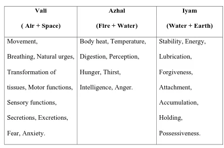
Table-1
Physiological Functions of Humours
Vali
( Air + Space)
Azhal
(Fire + Water)
Iyam
(Water + Earth)
Movement,
Breathing, Natural urges,
Transformation of
tissues, Motor functions,
Sensory functions,
Secretions, Excretions,
Fear, Anxiety.
Body heat, Temperature,
Digestion, Perception, Hunger, Thirst, Intelligence, Anger. Stability, Energy, Lubrication, Forgiveness, Attachment, Accumulation, Holding, Possessiveness.
VEGANGAL – 14 (REFLEXES)
The vegangal represents the natural reflex, excretory, protective
and preventive mechanisms. It involves psycho neuromuscular function of
the body. The 14 vegangal are:
Abaanan, Thummal, Siruneer, Malam, Kottavi, Pasi, Neervetkai,
Erumal, Ellaippu, Thookam, Vaanthi, Kanneer, Sukkilam and Swaasam.
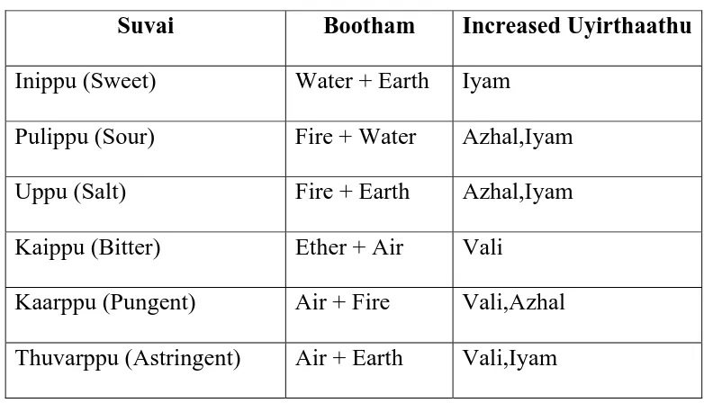
SUVAIGAL – 6 (TASTE)
Suvaigal has related to Panchaboothams and Uyirthaathu.The
stamina and built of the body is mainly based on one’s intake of balanced
[image:15.612.89.532.109.402.2]Table-2
Relation between Suvai, Bootham and Uyir Thaathu
Suvai Bootham Increased Uyirthaathu
Inippu (Sweet) Water + Earth Iyam
Pulippu (Sour) Fire + Water Azhal,Iyam
Uppu (Salt) Fire + Earth Azhal,Iyam
Kaippu (Bitter) Ether + Air Vali
Kaarppu (Pungent) Air + Fire Vali,Azhal
Thuvarppu (Astringent) Air + Earth Vali,Iyam
Any increased or decreased intake of these tastes alter the basic ratio
of Uyirthaathukkal and shows changes in Udalthaathukkal.
UDAL VANMAI- 3 (TYPES OF IMMUNITY)
It is of three types namely Iyarkai vanmai, Seyarkai vanmai and
Kaala vanmai.
UDAL THEE- 3 (BODY FIRES)
It is of four types namely Samaagni, Vishamaagni, Deekshnaagni and
[image:16.612.118.509.114.338.2]SIDDHA PATHOLOGY
‘Neha;ehb Neha;Kjy; ehb mJjzpf;Fk; tha;ehb tha;g;gr; nray;”
- jpUf;Fws;
For the exact diagnosis and to treat the ailment properly, a sound
knowledge about Noi Naadal is essential.
Noi (Pathos) - Disease
Naadal (Logos) - Study
Pathology is thus scientific study of structure and function of the body
in disease. It deals with causes, effects, mechanisms and nature of disease.
DISEASE
clYld; gpize;j capH mDgtpf;Fk; ,d;g czHr;rpf;F
khwhd czHr;rpNa gpzp.
Thus disease is loss of ease to the body.
ETIOLOGY
‘kpfpDk; FiwapDk; Neha;nra;Ak; E}NyhH tspKjyh vz;zpa %d;W”
- jpUf;Fws;
The human body is governed by three cardinal humours namely Vali,
Azhal and Iyam. The relative proportion of these humours are responsible
these basic factors results in disease. It can be altered due to various factors
such as,
1. Dietary Changes
2. Modified life style
3. Immunological status of the individual
4. Environmental changes
5. Seasonal variations
6. Suppression of reflexes
7. Variations in the 7 physical constituents of the body.
8. Kanma Vinnai (congenital causes)
DIETARY CHANGES
Dietary nutrients should be balanced according to the age, sex,
immunological status, seasonal variations and environmental changes.
‘,optwpe; Jz;ghd; fzpd;gk; Nghdpw;Fq; fopNg hpiuahd; fNzha;”
- jpUf;Fws;
Nutritional imbalance is more often a problem, accounting for
increased frequency of so many ailments.
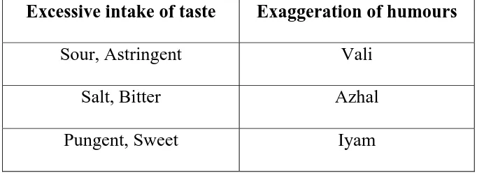
The nutrients in diet mainly based upon six taste (Suvaigal) namely
Sour, Astringent, Salt, Bitter, Pungent and Sweet. Excessive intake of these
‘GspJtH tpQ;Rq;fwp ahw;G+hpf; Fk;thjk;
xsp AtHifg; Ngwpy; gpj;Jr; rPWk; - fpspnkhopNa fhHg;gpdpg;G tpQ;rpw; fgk;tpQ;R Q;rl;bujr;
Nrug; GzH NehaZfhNj”
[image:19.612.144.485.259.383.2]- fz;Zrhkpak;
Table-3
Excessive intake of taste and Exaggeration of humours
Excessive intake of taste Exaggeration of humours
Sour, Astringent Vali
Salt, Bitter Azhal
Pungent, Sweet Iyam
Besides, excessive intake of nutrients, skipping of diets also results in
disease.
MODIFIED LIFE STYLE
Siddhars had taught so many life style manners to lead a healthy life.
They given under the topic Pini Anuga Vithi (Preventive laws to avert the
disease) as,
‘jpz;z kpuz;Ls;Ns rpf;f tlf;fhkw;
ngz;zpd;gh nyhd;iwg; ngUf;fhky; - cz;Zq;fhy; ePHfUf;fp NkhHngUf;fp nea;AUf;fp Az;gtHjk;
NgUiuf;fpw; NghNk gpzp”
Avoiding of constipation, retention of urine and excessive sexual
desire will prevent disease and also one should taking boiled water,
increased intake of skimmed milk, intake of melted ghee also prevent so
many diseases.
Any modifications in this life style manners will cause disease.
UDAL VANMAI (Immunological Status of the Individual)
The immunological status also plays an important role in determining
the disease. It is classified into three types.
1. Iyarkai Vanmai
This is based on Sathuva, Raso and Thamo gunams and it is the
strength which naturally presents in the body.
2. Seyarkai Vanmai
This strength depends upon by protecting the mukkunam based
body by taking proper diet, habits and medicine.
3. Kaala Vanmai
This strength is gained by seasonal variations as well as the age
of a person.
ENVIRONMENTAL CHANGES
‘mz;lj;jpYs;sNj gpz;lk; gpz;lj;jpYs;sNj mz;lk;”
Any interference in the external environment depicts in man’s internal
environment. It alter the basic ratio of three humours and also affects the
human’s defense mechanism and suppresses the immunity resulting in so
many ailments.
The ancient people classified the land into 5 types and the people
dwelling in these lands have more prevalence to certain diseases.
Kurinji - Kaba diseases
Mullai - Pitha diseases
Marutham - Suits for aboding
Neithal - Vatha diseases
Paalai - All kind of diseases.
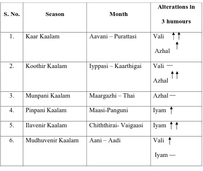
SEASONAL VARIATIONS
Ancient thamizh people classified the annum into six seasons each one
consisting of two months. Naturally, during these seasons the three humours
namely Vali, Azhal and Iyam shows some seasonal variations in their own
Table-4
Seasonal variations and alterations in humours
S. No. Season Month
Alterations in
3 humours
1. Kaar Kaalam Aavani – Purattasi Vali
Azhal
2. Koothir Kaalam Iyppasi – Kaarthigai Vali
Azhal
3. Munpani Kaalam Maargazhi – Thai Azhal
4. Pinpani Kaalam Maasi-Panguni Iyam
5. Ilavenir Kaalam Chiththirai- Vaigaasi Iyam
6. Mudhuvenir Kaalam Aani – Aadi Vali
Iyam
- Exaggerated
- Brisk
- Stable
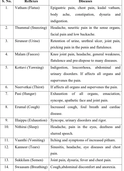
SUPPRESSION OF REFLEXES
There are 14 natural reflexes involved in the normal physiological
Table-5
Suppression of Reflexes and Diseases
S. No. Reflexes Diseases
1. Vatham (Flatus) Epigastric pain, chest pain, kudal vatham,
body ache, constipation, dysuria and
indigestion.
2. Thummal (Sneezing) Headache, neuritic pain in the sense organs,
facial pain and low backache.
3. Siruneer (Urine) Retention of urine, urethral ulcer, joint pain,
pricking pain in the penis and flatulence.
4. Malam (Faeces) Knee joint pain, headache, general weakness,
flatulence and pre-dispose to many diseases.
5. Kottavi (Yawning) Indigestion, leucorrhoea, abdominal and
urinary disorders. If affects all organs and
supervenes the pain.
6. Neervetkai (Thirst) If affects all organs and supervenes the pain.
7. Pasi (Hunger) Exhaustion of all organs, emaciation,
syncope, apathetic face and joint pain.
8. Erumal (Cough) Increased cough, foul breath and cardiac
disease.
9. Illaippu (Exhaustion) Syncope, urinary disorders and rigor.
10. Nithirai (Sleep) Headache, pain in the eyes, deafness and
slurred speech.
11. Vaanthi (Vomiting) Itching and symptoms of increased pitham.
12. Kanneer (Tears) Sinusitis, headache, eye diseases and chest
pain.
13. Sukkilam (Semen) Joint pain, dysuria, fever and chest pain.
THE ALTERED CHARACTERS OF THREE HUMOURS
The inequilibrium state of three humours namely Vali, Azhal and
Iyam is the main reason for the occurrence of disease.
Table-7
Altered characters of three Humours
S. No. Humour Exaggerated character Diminution character
1. Vali Emaciation, Blackish colouration
of the body, Desire to take hot
food, Tremors, Constipation,
Abdominal distension,
Insomnia,Weakness,
Weakness of sense organs,
Giddiness, Sluggishness.
Stiffness, Diminution of
voice, Impaired intellectual
function, Semi consciousness,
Difficulty in doing any kind
of work, Paleness and
coolness of the body,
Excessive salivation,
Heaviness of the body,
Excessive sleep, Abdominal
distension.
2. Azhal Yellowish discolouration of skin,
eye, urine and faeces, polyphagia,
polydypsia, burning sensation all
over the body, insomnia.
Decreased appetite, cold,
pallor, symptoms associated
with defective growth of
Iyam.
3. Iyam Excessive salivation, reduced
appetite, cough, dyspnoea,
heaviness of the body, whiteness
of the body, chillness of the body,
increased sleep, sluggishness.
Giddiness, swelling of the
joints and prominence of
bone. Iyam dissolved in
lungs, Excessive sweating,
VARIATIONS IN THE SEVEN UDAL THAATHUKKAL
When the normal ratio of the three humours are affected, it immediately
[image:25.612.80.553.182.688.2]shows changes in the natural physiological function of Udal thaathukkal.
Table-6
Variations in the seven udal thaathukkal
S.
No.
Udal
Thaathukkal
Increased features Decreased features
1. Saaram
(Chyle)
Features related with decrease in
Iyam and loss of appetite.
Dryness of skin, loss of
weight, tiredness and the
functions of sense organs
are diminished.
2. Senneer
(Blood)
Boils and tumours in different parts
of the body, splenomegaly, pain,
hypertension, redness of the eyes,
leprosy and jaundice.
Desire for cold things,
dryness, discolouration and
paleness of the skin.
3. Oon (Muscle) Tumours or extra growth around the
neck, face, abdomen, thigh and
genitalia.
Lethargy of 5 sensory
organs, pain in the joints,
loss of subcutaneous fat.
4. Kozhuppu
(Fat)
Identical to increased features of Oon,
tiredness, dyspnoea on exertion.
Splenomegaly, loin pain,
emaciation.
5. Enbu (Bone) Excessive ossification and dentition. Weak bone, pain in the
joints, splitting of hair and
nails.
6. Moolai (Bone
marrow)
Heaviness of the body and eyes,
swelling of smaller joints of hand and
feet, oliguria, non- healing ulcers.
Osteoporosis and blurred
vision.
7. Sukkilam/
Suronitham
(Sperm or
Ovum)
Excessive sexual desire, urinary
calculi.
Pain in the genitalia and
KANMA VINNAI (CONGENITAL CAUSES)
The formation of embryo plays a key role in determining the health of
an individual.
‘NgWapsik apd;gk; gpzp%g;G rhf;fhL MWq;fU tpyikg;G”
- fz;Zrhkpak;
According to Siddha concept, the three Kanma Vinnai namely
Aanavam, Maayai and Kanmam are determined during the time of formation
of embryo and also, if there is any derangements of Vali, Azhal and Iyam
humour among the parents during sexual intercourse it also affects the
formation of embryo and results in congenital diseases.
gpzpawp Kiwik
(DIAGNOSTIC METHODS)
The diagnostic tool adopted to evaluate a disease in Siddha system of
medicine is termed as Piniyari Muraimai. It is based on the following
priniciples.
1. Poriyaal Arithal
2. Pulanaal Arithal
3. Vinaathal
Poriyal Arithal means diagnosing through the five organs of
Pulanal Arithal means diagnosing through the five object of senses
namely Skin sensation, Taste, Vision, Smell and Auditory sensation.
Vinaathal is a method of interrogating the complaints of the patient
from his own words or from his thoothuvan (attendars).
The methods including all the above falls under,
ENNVAGAI THERVUGAL
Theraiyar mentioned the 8 types of diagnostic tools as,
‘nka;f;Fwp epwe;njhdp tpopeh tpUkyk; iff;Fwp”
- Njiuah;
In Agathiyar Vaidhya Vallathi – 600, Ennvagai Thervugal mentioned
as,
’njhFf;fYw;w ml;ltpjg; ghPl;ir jd;id Jyf;f KWk; gz;bjNu njspthfg;
gFf;fhpa ehbia eP gpbj;Jg; ghU gfh;fpd;w thh;j;ijg; ghh; ehitg; ghU tFf;fhpa Njfnkdj; njhl;Lg; ghU tskhd rhPuj;jpd; epwj;ijg; ghU rfpf;fhpa kyj;ijg; ghh; ryj;ijg; ghU
1. Naa (Tongue)
2. Niram (Colour)
3. Mozhi (Speech)
4. Vizhi (Eye)
5. Sparisam (Sense of touch)
6. Malam (Stool)
7. Moothiram (Urine)
8. Naadi (Pulse)
On accessing the variations in the above said tools, the physician can
find out the derangements of three humours and come to a proper diagnosis.
1. Naa (Tongue)
By the examination of tongue the following features are noted.
Colour, Size, Shape, Coating, Anomalies, Surface, Movements, Local
lesions, Ulcers, Fissures, Vesicles, Dryness, Moisture, Deviation of
tongue, Pigmentation, Small tongue – Microglossia, Large tongue –
Macroglossia.
2. Niram (Colour)
Pallor, Yellowish, cyanosis, hyperpigmentation,
hypopigmentation, contusions could be noted.
3. Mozhi (Speech)
The volume, clarity and any disturbance in speech are to be
4. Vizhi (Eye)
Here the colour change, lacrimation, visual disturbances are to
be noted. The nature of eye brow and eye lids are also to be noted.
5. Sparisam (Sensation)
Temperature of the Skin, Smoothness, Dryness, Scaling,
Swelling,Tenderness, Sweating, Any abnormal growths, Internal
organ enlargements, Thickening of nerves, Varicosity of veins,
Cutaneous changes, Subcutaneous nodules should be find out.
6. Malam (Stool)
The colour, odour, froth, quantity, consistency of the stool and
presence of any abnormal constituents such as blood, parasites etc.,
are taken as diagnostic criteria.
7. Moothiram (Urine)
The diagnostic method by examining urine is of two types.
1. Neer kuri and
2. Nei kuri
1. Neerkuri
Here the Niram(Colour), Manam(Smell), Nurai(Frothy Nature),
Edai(Specific gravity) and Enjal(Quantity) of the voided urine is
2. Neikuri
According to Theraiyar,
‘mUe;Jkh wpujKk; mtpNuhjkjha; m/fy; myh;jy; mfhyT+d; jtph;e;jow; Fw;wstUe;jp cwq;fp itfiw
Mbf;fyrj; jhtpNa fhJ nga; njhUK$h;j;jf; fiyf;Fl;gL ePhpd; epwf;Fwp nea;f;Fwp epUkpj;jy; flNd”
To see Neikuri, before collecting the urine, the patient is asked to take a
balanced diet and have a good sleep. After waking up from the bed in the
early morning, the first voided urine is collected in a clean glass container
and examined within one hour. A drop of gingely oil is to be dropped in the
urine and seen under direct sunlight. By this method, the character of three
humours is accessed.
The character of Vatha neer
!! ! ‘muntd ePz;bd0Nj thjk;”!
When the drop of oil spreads like a serpent it indicates derangement of
Vali.
The character of Pitha neer
!! ! ‘Mop Nghy; gutpd; m0Nj gpj;jk;”
The character of Kaba neer
‘Kj;njhj;J epw;fpd; nkhoptnjd; fgNk”
If the oil remains like a pearl it denotes the derangement of Iyam.
The character of Thontha neer
‘mutpyhopAk; Mopapy; muTk; mutpy;; Kj;Jk; Mopapy; Kj;Jk; Njhw;wpy; njhe;j Njhlq; fshNk”
If the dropped oil shows a combination of two shapes it indicates
Thontha thodam.
8. Naadi (Pulse)
It is a unique diagnostic method in Siddha system of medicine.
It is responsible for the existence of life. The three humours namely
Vali, Azhal, and Iyam are in the ratio of 1: ½: ¼.
‘toq;fpa thjk;khj;jpiu nahd;whfpy; joq;fpa gpj;je;jd;dpyiu thrp
moq;Fq;fge; jhdlq;fpNa fhNyhby; gpwq;fpa rPth;f;Fg;gpr nfhd;Wkpy;iyNa”
- Fzthflk;
AIM AND OBJECTIVES
Nowadays due to altered dietary habits and modified life style people
suffered by many diseases. It is the mere responsibility of a physician to cure
the disease and to create a healthy society.
The primitive aim of the author to study about Anda vadham is, to
diagnose the disease through Siddha parameters with the help of modern
parameters and to create awareness among the people to prevent the disease.
Anda Vadham is one of the important disease of the abdominal
organs in which the patients are disturbed both functionally and emotionally.
To evaluate the disease is important because if it is not diagnosed and treated
properly it may leads to various complications.
The study mainly made out, to rule out the pathogenesis of this disease
on the basis of etiology, genetic factors, nutritional status, dietary habits, life
style and socio-economic status.
To achieve the aim in a successful manner the specific objectives have
been utilized.
1. To know about the disease in detail it is necessary to collect the
evidences found in Siddha literature regarding Anda Vadham.
2. To evaluate the Siddha basic physiology is needful.
3. To review the altered mukkutram and to establish the disease
Thervugal, Manikkadai Nool and correlating the disease with
Nilam, Kaalam and Sothidam is helpful.
4. For better diagnosis, thorough manual examination of the patient is
needed.
5. To support the study of Anda Vadham modern parameters have
been utilized.
6. The study is concluded by defining the aggravating factors which
leads to complications and finally the study dealt with the
ELUCIDATION ABOUT ANDA VADHAM
According to the literature Thanvanthiri Vaithiyam, Anda Vadham
has been described under Uthara Roga Nithanam as,
‘nfhz;lq;F Flypw;wq;fpf; Fiwe;njhU tpijapw; Nwhd;wp kz;bNa typj;J tPq;fp kWtpiu jdpYe; Njhd;Wk;
gz;bypnyhj;J thq;Fk; gfh;e;jpL kz;lthj kz;lU Kdpth; jhK kwpaNt Aiuf;f Yw;whh;”
.!jd;te;jphp itj;jpak; ,uz;lhk; ghfk;
ghly; vz; 120
Meaning of the words in this poem
mz;lk; :!Scrotum
thjk; :!Energy or power that prevails all over the body
and keeping the tissues in good condition.
nfhz;lq;F :!At that place!
Flypw;wq;fp :!Accumulation of Vayu in the intestines!
Fiwe;njhU :!Atleast one side (unilateral)!
tpijapw; Nwhd;wp :!Occurs in the scrotum!
kz;bNa :!Accumulation
typj;J :!Physical pain!
tPq;fp :!Swelling!
Njhd;Wk; :!Will occur!
gz;b :!Abdomen
xj;J thq;Fk; :!Reducing to an equality
Description
‘nfhz;lq;F Flypw;wq;fpf; Fiwe;njhU tpijapw; Nwhd;wp”
! ! Increased Vayu accumulation in the intestines and descend of
the intestines into the scrotum. Initially it occurs on one side of the scrotum.
‘kz;bNa typj;J tPq;fp kWtpiu jdpYe; Njhd;Wk;”
! !! Pain and swelling of the scrotum. Later the same signs and
symptoms occur on the other side of the scrotum also.
‘gz;bypnyhj;J thq;Fk; gfh;e;jpL kz;lthj kz;lU Kdpth; jhK kwpaNt Aiuf;f Yw;whh;”
! This line indicates that the hernial contents reduce back into the
abdominal cavity.All these represents the clinical features of the disease
Anda Vadham.
The Thanvanthri’s lines are summarized as follows,
Increased Vayu accumulates in the intestines and the increased
intra-abdominal pressure descend the intestine into the scrotum. Initially it occurs
unilaterally. There is pain and swelling in the scrotal region. Later the same
features occurs on the other side of the scrotum.The descended intestine then
reduce back into the abdominal cavity.
DETAILED PATHOLOGICAL VIEW
OF THE DISSERTATION TOPIC
SIDDHA ASPECT
In this dew drop of research work on the topic Anda Vadham the
author try to explain the pathological view through Siddha aspect which
helps in the diagnosis of the disease.
The disease Anda Vadham is mentioned in Thanvanthiri
Vaidhiyam under Uthara Roga Nithanam (Abdominal Diseases).
Uthara Rogam (Abdominal Diseases) are caused by the following
factors:
‘kdKwTz;ifahY kfpo;e;j T+z; FiwjyhYk; fdkyQ; nrwpe;J epd;W fow;Wj ypyhkyhYe; jd;kpfj; NjlYw;Wj; jhDU Nkhfj;jhYQ; rpdkpf TUifahYQ; NrHe;jpL KjuNehNa”
- jd;te;jphp itj;jpak; Kjy; ghfk;
Excessive intake of diet
Reduced intake of diet
Constipation
Excessive sexual desire
Anger
Doing heavy works after full meals
Lying in chill floor
Sleeping in improper position.
All these factors pave a way increased Vayu accumulation in the
abdomen and results in abdominal diseases.
‘gpzpapDw; gj;jpiag; NgRtd; gpzpKjy; thjgpj; jq;fg kd;ke;jphp je;jphp
! !……/!……//!………//!………/!……!…….!
! !……/!……//!………!…………/!……!……”!
! ! ! ! ! - NjiuaH fhg;gpak;
According to Siddha Medicine theory, the cardinal humour Vali is
considered as the chief Administrator and Azhal and Iyam as sub-ordinators.
The derangement of Vali affects the natural characters of the other two
humours Azhal and Iyam respectively.
ALTERED UYIRTHAATHUKKAL IN ANDA VADHAM
‘nfhz;lq;F Flypw;wq;fpf; Fiwe;njhU tpijapw; Nwhd;wp”
In Anda Vadham, the cardinal humour Vali is predominantly affected.
Due to deranged Vali increased vayu accumulates in the intestines, since the
dwelling place of Vali is in the intestines and below the naval region it tends
to accumulate there.
The reason for increased vayu is,
‘ke;jkyhJ thA tuhJ”
Mantham represents indigestion of the food. It occurs as follows,
During derangement of Vali, the digestive fire Samaana Vayu is
affected and it combines with increased Iyam and results in improper
digestion(mantham). The Mantham state is the character of increased Iyam
and decreased Azhal humours. Thus the deranged Vali affects the other two
humours respectively.
Since the Samaana Vayu (Nadukkaal) is deranged in Anda Vadham,
it can not able to maintain the equilibrium state of other vayus like
Praanan,Udhaanan and Kirukaran which plays an important role in
digestion. In turn, the Abaana Vayu(Keelnokkuk Kaal) is also deranged and
hence it’s basic physiological function, expulsion of vayus is affected. All
these, results in increased Vayu accumulation in the intestines.
Thus due to enormous amount of Vayu accumulation in the intestines,
the viscus become very weak and it descends from its original position into
the scrotum.
‘kz;bNa typj;J tPq;fp kWtpiujdpYk; Njhd;Wk;”
The increased Vayu produces localized pain and swelling of the
scrotum which is the deranged character of Vali.
‘gz;bypnyhj;J thq;Fk;”
ALTERED UDAL THAATHUKKAL IN ANDA VADHAM
In Anda Vadham the Udal thaathukkal shows their decreased
character.
Saaram (Chyle)
‘thjkyhJ Nkdp nflhJ”
Generally Vali gives encouragement to mind and gives co-ordination
to seven Udal thaathukkal.In Anda Vadham, Vali is deranged and it depicts
in Saaram and results in weakness of the abdominal muscles.
Senneer (Blood and its fluids)
The blood and its fluids affords strength to the tissues and organs. In
Anda Vadham the senneer attains its decreased character and results in
abdominal muscle weakness.
Oon (Muscle)
The Vali humour helps in keeping the tissues in good condition. When
it is deranged, it depicts in Oon which is responsible for the proper shape of
the organs in accordance to their activity. Thus in Anda Vadham, the normal
anatomical position of the intestines is not maintained and it is descended in
to the scrotum.
Kozhuppu (Fat)
In Anda Vadham, the derangement of Vali decreases the Kozhuppu
thaathu and results in weakening of the abdominal musculature.
Thus the Udal thaathukkal decreased in their basic character and
DETAILED PATHOLOGICAL VIEW OF THE
DISSERTATION TOPIC
MODERN ASPECT
In modern aspect the pathological view of Anda Vadham is described
as follows,
‘nfhz;lq;F Flypw;wq;fpf; Fiwe;njhU tpijapw;Nwhd;wp”
This line indicates increased Vayu accumulation in the intestines
and the descend of the intestines into the scrotum.
The pathology of descend of the intestines is due to,
Generally, the abdominal wall and the internal visceras are covered by
an extensive serous membrane, the peritoneum.
The anterior abdominal wall is musculo-fibrous. It is firm and elastic.
The firmness provides protection to the abdominal viscera and the elasticity
allows expansion of the hollow viscera.
The posterior abdominal wall is osseo-musculo fascial and is rigid. It
provides support and nutrition to the abdomen organs by the attachment of
various peritoneal folds.
The main action of the muscles of the anterior abdominal wall is, it
MUSCLE TONE
The muscle fibers always maintain a state of slight contraction with
certain degree of vigor and tension. This property of muscle is called tone or
tonus.
The smooth muscles themselves control the tone. They are supplied by
both sympathetic and parasympathetic nerves, which antagonize each other
and control the activities of smooth muscles. However, the tone of the
muscle is independent of the nerve fibers.Sometimes, the tonic contraction
occurs due to the action of some hormones.
Stress – Relaxation of Smooth Muscle
One of the important characteristic of smooth muscle, especially the
visceral unitary type of smooth muscle of many hollow organs, is its ability
to return to nearly its original force of contraction, seconds or minutes after it
has been elongated or shortened.
A sudden increased volume due to any cause in the abdominal cavity
stretch the smooth muscle in the abdominal wall which causes and
immediate large increase in pressure in the abdominal cavity. However,
during the next 15 seconds to a minute or so, despite continued stretch of the
abdominal wall, the pressure returns almost exactly back to the original
level. Then, when the volume is increased by another step, the same effect
Conversely, when the volume is suddenly decreased, the pressure falls
very low at first but then rises back in another few seconds or minutes to or
near to the original level. These phenomena are called stress-relaxation and
reverse stress-relaxation.
Their importance is that, except for short periods of time, they allow a
hollow organ to maintain about the same amount of pressure inside its
lumen. In long-term, large changes in volume will occur.
The descend of the intestines is mainly due to
When there is any factor which increases the intra-abdominal pressure
for a prolonged period, the abdominal musculature is highly stretched and it
weakens the muscles and its aponeuroses and the tonicity of the muscle is
weakened. So the position of the visceras are not retained in position.
Besides, the increased intra-abdominal pressure also compresses the
internal visceras (the intestines) and all these reasons are responsible for the
descend of the intestines from its original anatomical position.
The role of inguinal canal in descend of the intestine
Under normal conditions, the inguinal canal plays a defense role.
Since the canal is oblique in direction, when the intra-abdominal pressure is
increased the posterior wall of the canal is pushed forwards and comes in
contact with the anterior wall. Thus the canal is obliterated like a flap-valve.
Opposite the deep ring, the anterior wall of the canal is strengthened
superficial ring the posterior wall is strengthened by the conjoint tendon and
reflected part of the inguinal ligament.
The arched fibres of the internal oblique and the transversus act as
demi-sphincters in increased intra abdominal pressure and obliterate the
canal.
Especially in males, the cremaster muscle contracts during increased
intra-abdominal pressure and pulls the testes towards the superficial ring, so
the outlet of the canal is closed like a plug.
But certain conditions which causes raised intra-abdominal pressure
this shutter mechanism is failed and it paves way for a weak abnormal
opening.
So the already descended intestines pass through this abnormal
opening.
The line also indicates that the intestines descends into the scrotum.
This is because the scrotum is the downward prolongation of the anterior
abdominal wall. The skin of the scrotum is thin and transparent and
elastic.So it easily allows distensions. For this reason the intestines distended
into the scrotum and lodges in it.
The descend of the hernial sac into the scrotum indicates the features
of Complete Inguinal Hernia. The line also mentions the signs and
‘kz;bNa typj;J tPq;fp kWtpiu jdpYe; Njhd;Wk;”
It indicates pain and swelling of the scrotum. Later the above said
signs and symptoms occur on the other side also.
In this disease, the reason for pain is overdistension of the scrotum.
Extreme overfilling of a hollow organ results in pain, presumably because of
overstretch of the tissues themselves. Besides, due to dragging of mesentery
also produces pain.
PHYSIOLOGY OF PAIN
Pain is an unpleasant sensation and emotional experience associated
with or without tissue damage. In general, the viscera have sensory
receptors for no other modalities of sensation besides pain. Any stimulus that
excites pain nerve endings in diffuse areas of the viscera causes visceral
pain.
VISCERAL PAIN
Pain from viscera is unpleasant. It is poorly localized.
CAUSES FOR VISCERAL PAIN
1. Ischaemia
2. Chemical stimuli
3. Spasm of hollow organs and
PATHWAY OF PAIN SENSATION FROM VISCERA
The pain sensation from the scrotum is transmitted by Sympathetic
nerves.
PATHWAY OF PAIN SENSATION
Receptors (Free nerve Endings)
First order Neurons
Second order Neurons
Third order Neurons
Sensory area of Cerebral cortex Hypothalamus
In this mechanism pain is felt in the scrotal region.
CAUSE FOR SWELLING
Swelling is an abnormal enlargement or increase in volume, associated
with accumulation in the tissue of a protein-containing exudate.
In complete inguinal hernia due to laxity of the scrotal skin and its
dependent position the scrotal swelling is common because when the
herniated sac accumulates in the cutaneous bag (scrotum), it is distended and
‘gz;byp nyhj;J thq;Fk;”;
This line indicates the reducible character.It meant that the contents
(intestines) can be returned back into the abdominal cavity, but the sac
remains in its position.It indicates the uncomplicated stage of the disease.
T
he clinical features of Anda Vadam,1. Descend of the intestines into the scrotum-Both unilateral
and bilateral sides.
2. Pain and swelling of the scrotum
3. Reducibility of the intestines into the abdominal character.
All these features clearly depicts the clinical features of Complete
REVIEW OF LITERATURE
The same topic Anda Vadham is mentioned in various literatures.In
the literature Pararasa Sekaram, Anda Vadham is mentioned under –
Vadharoga Nithaanam as,
‘cjuj;jpy; thA kz;b A+jpNa ailrpf; Fj;jpf; fjKw kyr yj;ijf; fl;bNa Athjp ahFk; jpjKwg; gPre; jd;dpw; NruNt apwq;F Nkhh;fhy; ,jkwf; fLf;F ehhp NawpL kz;l thjNk”.
The clinical features are,
Vayu accumulates in the intestines
Abdominal distension
Constipation
Retention of urine
Descend of the intestines into the scrotum
Pain in the scrotum
In the literature Theraiyar Kaapiyam, the same features of Anda
Vadham is mentioned under Vidhai Noi (Scrotal Diseases) under the
heading Kudal Andam. Here, it is mentioned that the increased Vayu is the
cause for Anda Vadham and the increased Vayu is due to the following
factors:
1. The diet which increases the Vali humour
3. Strenous exercises
4. Horse riding
5. Chilled water bath
6. Suppression of the reflexes.
In Yugimuni Vaidhiya Kaviyam – 1000
mz;lthjj;jpd; Fzk;
‘nkj;jNt thjk; Ja;j;J tpiujdpy; Cjpf;nfhz;L gj;jNtkuf;fhy;NghYk; ghpe;jpLk;fphpr;rpfhl;Lk; Fj;JNkFj;jpf;fhl;bf; FlyJgpwl;bf;nfhs;Sk; kw;wpJnrhd;Ndhk; mz;lthjj;jpd;tifap jhNk”
This Poem also depicts the same clinical features of Anda Vadham.
All these literature evidences mentioned that the increased Vayu is the
reason for Anda Vadham and produce the pathological changes in the
abdomen and the scrotum.
To support my study, in Pararasasekaram the derangement of Vali
characters is mentioned as,
thjj;jpdhw; Nwhw;Wtd
‘thjNk fjpj;j NghJ thaT nkOg;gp kPSk;
thjNk apUk yhfpj; njhlh;e;jpLQ; rd;dp thjk;
NgjNk nra;fp uhzp ngUtap Wju Njh\k;
NghjNt Njhd;W nkd;W nghUe;jNt KdptH nrhd;dhh;”
1. Increased Vayu
2. Cough
3. Sanni Vadham
4. Dysentry
5. Ascites and
6. Abdominal diseases.
Hence Saint Thanvanthiri mentioned the disease Anda Vadham under
Uthara Roga Nithanam (Abdominal Diseases) which results due to
exaggeration of the Vali humour.
The altered diet habits and modified life style manners are mainly
THEORETICAL VIEW OF THE
DISSERTATION TOPIC
MODERN ASPECT
ANATOMY
ABDOMEN AND ITS WALLS
The abdomen is a part of the trunk below the diaphragm. It contains a
cavity which is subdivided by the plane of the pelvic inlet into an upper part,
the abdomen proper, and a lower part known as the pelvic cavity.
The abdominal wall and the contained viscera are covered by an
extensive serous membrane, the peritoneum. Histologically, it is composed
of an outer layer of fibrous tissue which gives strength to the membrane and
an inner layer of mesothelial cells which secrete a serous fluid which
lubricates the surface, thus allowing free movements of viscera.
The peritoneum is in the form of a closed sac which is invaginated by
a number of viscera. As a result the peritoneum is divided into,
(1) An outer or parietal layer
(2) An inner or visceral layer, and folds of peritoneum by which the
viscera are suspended.
PARIETAL PERITONEUM
1. It lines the inner surface of the abdominal and pelvic walls and the
lower surface of the diaphragm. It is loosely attached to the walls by
2. Embryologically, it is derived from the somatopleural layer of the
lateral plate mesoderm.
3. Its blood supply and nerve supply are the same as those of the
overlying body wall. Because of the somatic innervation, parietal peritoneum
is pain sensitive.
VISCERAL PERITONEUM
1. It lines the outer surfaces of the viscera, to which it is firmly
adherent and cannot be stripped. In fact it forms a part and parcel of the
viscera.
2. Embryologically, it is derived from the splanchnopleural layer of
the lateral plate mesoderm.
3. Its blood supply and nerve supply are the same as those of the
underlying viscera. Because of the autonomic innervation visceral
peritoneum is pain insensitive.
FOLDS OF PERITONEUM
1. Many organs within the abdomen are suspended by folds of
peritoneum. Such organs are mobile. The degree and direction of mobility
are governed by the size and direction of the peritoneal fold. Other organs
are fixed and immobile. They rest directly on the posterior abdominal wall,
and may be covered by peritoneum on one side. Such organs are said to be
retroperitoneal. Some organs are suspended by peritoneal folds in early
2. Apart from allowing mobility, the peritoneal folds provide
pathways for passage of vessels, nerves and lymphatics.
3. Peritoneal folds are given various names.
i. Large peritoneal folds attached to the stomach are called
omenta .
ii. In many situations double layered folds of peritoneum
connect organs to the abdominal wall or to each other. Such folds are
called ligaments. These may be named after the structures they
connect. Other folds are named according to their shape triangular.
PERITONEAL CAVITY
The viscera which invaginate the peritoneal cavity completely fill it so
that the cavity is reduced to a potential space separating adjacent layers of
peritoneum.
FUNCTIONS OF PERITONEUM
1. Movements of viscera
The chief function of the peritoneum is to provide a slippery surface
for free movements of abdominal viscera.
The efficiency of the intestines is greatly increased as a result of the
wide range of mobility that is possible because the intestines are suspended
by large folds of peritoneum.
2. Protection of viscera
3. Absorption and dialysis
4. Healing power and adhesions
THE GREATER OMENTUM
This is a large fold of peritoneum which hangs down from the greater
curvature of the stomach like an apron and covers the loops of intestines to a
varying extent.
LESSER OMENTUM
This is a fold of peritoneum which extends from the lesser curvature
of the stomach and the first 2cm. of the duodenum to the liver.
THE MESENTERY
The mesentery of the small intestine (or mesentery proper) is a broad,
fan-shaped fold of peritoneum which suspends the coils of jejunum and
ileum from the posterior abdominal wall.
TRANSVERSE MESOCOLON
This is a broad fold of peritoneum which suspends the transverse
colon from the upper part of the posterior abdominal wall.
SIGMOID MESOCOLON
This is a triangular fold of peritoneum which suspends the sigmoid
colon from the pelvic wall.
BOUNDARIES OF ABDOMEN
It consists of Roof, Floor, Anterior and Posterior Walls.
Roof
It is formed by the undersurface of the diaphragm. It moves up and
down with respiration and alters the intra-thoracic and intra-abdominal
Floor
The floor of the pelvic cavity is formed by the pelvic diaphragm in the
posterior part and by the urogenital diaphragm in the anterior part. It
supports the pelvic viscera and regulates the acts of defaecation, micturition
and in female parturition.
Anterior wall
It is musculo - fibrous and is formed by three flat muscles and their
aponeuroses. On each side of linea alba the anterior wall is strengthened by
the recuts abdominis and pyramidalis within the rectus sheath. Anterior
wall is firm and elastic. The firmness provides protection to the abdominal
viscera and the elasticity allows expansion of the hollow viscera.
Posterior wall
It is osseo-musculo fascial and is rigid. Retro-peritoneal organs,
principal vessels and nerves lie in the posterior wall; posterior wall provides
support and nutrition to the abdomen organs by the attachment of various
peritoneal folds.
ANTERIOR ABDOMINAL WALL
The anterior abdominalwall is limited above by the xiphi - sternum,
right and left costal margins, below by the anterior part of the iliac crest, fold
of the groin, pubic tubercle, pubic crest and symphysis pubis and on each
side separated from the posterior abdominal wall by the downward
The anterior wall is firm but elastic and consists of 8 layers from
before backwards.
1.Skin
2.Superficial fascia
3.External oblique muscle and its aponeurosis
4.Internal oblique muscle and its aponeurosis
5.Transversus abdominis muscle and its aponeurosis
6. Fascia transversalis
7.Extra-peritoneal tissue and
8.Parietal peritoneum.
Opposite the linea alba, the layers are reduced to six in number.
Embryologically, the 7th layer of the extra-peritoneal tissue is important because all abdominal visceras virtually lie in this layer; this fact is proved
by the coverings received by the testis during its descent.
DESCRIPTION OF DIFFERENT LAYERS
SKIN
It is thinner and more sensitive than the skin of the posterior
abdominal wall. The skin presents a number of cleavage lines (Langer’s
lines). The skin presents two parts, an outer epidermis which is non- vascular
and an inner dermis or corium which is highly vascular and presents rich
The skin of the anterior abdominal wall consists of longitudinal
groove overlying the linea alba, and a curved groove on each side with the
convexity directed laterally which corresponds with the lateral border of the
rectus abdominis muscle. A surface depression, the umbilicus, affects the
median groove; It is composed of cicatritial tissue and represents remnant of
the foetal end of the umbilical cord.
SUPERIFICIAL FASCIA
Above a line joining the two anterior superior iliac spines it consists of
a single layer. Below that line it splits into superficial fatty layer (Camper’s
fascia) and deep membranous layer (Scarpa’s fascia).
SUPERFICIAL INGUINAL SPACE
It is a potential space in the anterior abdominal wall between the fascia
of Scarpa and the aponeurosis of external oblique.
EXTERNAL OBLIQUE MUSCLE
(OBLIQUUS EXTERNUS ABDOMINIS)
Origin
It arises by eight fleshy slips from the outer surfaces and lower borders
of the lower eight ribs.
Insertion
The most posterior fibres pass almost vertically downwards and are
segment of the iliac crest. Its posterior margin is free and forms the anterior
boundary of the lumbar triangle.
The remaining fibres pass downwards, forwards and medially, and end
in a broad aponeurosis.
INGUINAL LIGAMENT (POUPART’S LIGAMENT)
It is the thickened lower border of the aponeurosis of the external
oblique which is folded backwards presenting a grooved upper surface. It
measures about 12 cm. to 14cm. in length in the adult.
INTERNAL OBLIQUE MUSCLE
(OBLIQUUS INTERNUS ABDOMINIS)
Origin
1. From the lateral two - third of the upper surface of the inguinal
ligament.
2. Anterior 2/3 of intermediate lip of the iliac crest.
3. Posterior layer of thoraco lumbar fascia.
Insertion
1.By forming conjoint tendon it is inserted into pubic crest and
pectineal line.
2.Forms rectus sheath and insert into the linea alba.
3. Lower 3 or 4 ribs.
Rectus abdominis
Actions of the rectus muscles
They provide protection to the abdominal viscera from external injury;
Compress the abdominal cavity and maintain intra-abdominal pressure
and
Produce flexion of the vertebral column.
Pyramidalis
Origin – From the symphysis pubis and the pubic crest.
Insertion – By its apex it is inserted into the linea alba, midway
between the umbilicus and symphysis pubis.
Nerve supply – From the subcostal nerve.
Action – It is a tensor of the linea alba.
Fibres of the deep layer of external oblique aponeurosis pass
downwards and medially as continuation of ipsilateral muscle. On
approaching the linea alba, the fibres decussate and separate into
superficial and deep sets.
FASCIA TRANSVERSALIS
It is an areolar membrane which lines the inner surface of the
transversus muscle and forms an endo-abdominal fascia.
It extends behind the inguinal ligament as the anterior wall of femoral
ACTIONS OF THE MUSCLES OF THE ANTERIOR ABDOMINAL
WALL
Retention- The muscles retain the viscera in position. This is
maintained by the tone of the muscles.
Protection - They protect the viscera from external injury. This is
done mainly by two recti.
Compression – They increase intra-abdominal pressure and
compress the viscera in the act of vomiting, micturition or
defaecation. During compression, the diaphragm is pushed
upwards helping expiration.
Actions on vertebral column
(a) When muscles of both sides act, lumbar vertebrae are
flexed.
(b) In unilateral action of the muscles, the trunk is bent laterally
producing lateral flexion.
(c) When the external oblique of one side contracts
simultaneously with the internal oblique of the other side,
INGUINAL CANAL
It is m usculo-aponeurotic tunnel, about 4 cm. in length, and extends
from the deep inguinal ring to the superficial inguinal ring. The canal is
directed downwards, forwards and medially, above and parallel with the
medial half of the inguinal ligament.
Peculiarities
1.In female the canal is narrow; hence chance of inguinal hernia is
less.
2.In the newborn the canal is directed almost straight forwards,
because muscles of the anterior abdominal wall are not properly
differentiated.
Contents
1.Spermatic cord in males, or round ligament of uterus in females
(entire content).
2. Ilio- Inguinal nerve (partial content). The nerve enters the canal by
piercing the internal oblique muscle about 2.5 cm. below and medial to the
anterior superior iliac spine. It is situated superficial to the spermatic cord,
and leaves the canal through superficial ring. Ilio- Inguinal is a mixed nerve
conveying fibres from the ventral ramus of L1, but after piercing the internal
CONSTITUENTS OF THE SPERMATIC CORD
1. The ductus deferens.
2. The testicular and cremasteric arteries, and the artery of the ductus
deferens.
3. The pampiniform plexus of veins.
4. Lymph vessels from the testis.
5. The genital branch of the genitofemoral nerve, and the plexus of
sympathetic nerves around the artery to the ductus deferens.
6. Remains of the processus vaginalis.
COVERINGS OF THE SPERMATIC CORD
From within outwards these are as follows
1. The internal spermatic fascia, derived from the fascia transversalis,
it covers the cord in its whole extent.
2. The cremasteric fascia is made up of the muscle loops constructing
the cremaster muscle, and the intervening areolar tissue. It is
derived from the internal oblique and transversus abdominis
muscles, and therefore covers the cord below the level of these
muscles.
3. The external spermatic fascia is derived from the external oblique
BOUNDARIES OF THE CANAL
The canal presents anterior and posterior walls,roof and floor, inlet
and outlet.
Anterior wall
It is entirely formed by the skin, superficial fascia and the aponeurosis
of the external oblique. The wall is partially formed in lateral one-third by
the fleshy fibres of the internal oblique.
Posterior wall
It is entirely formed by the fascia transversalis which separates the
canal from the extra-peritoneal tissue and peritoneum. It is partially formed-
(a) In the medial half, by the conjoint tendon in front of the fascia
transversalis.
(b)In the medial one-fourth, by the reflected part of the inguinal ligament
in front of the conjoint tendon.
Roof
The roof is formed by the arched fibres of the internal oblique and
transversus abdominis muscles.
Floor
(a)It is formed by the grooved upper surface of the inguinal ligament,
the posterior margin of which fuses with the fascia transversalis.
(b)Medially, the floor is formed by the upper surface of the lacunar
Inlet
Inlet is formed by the deep inguinal ring which is an oval gap in the
fascia transversalis about 1.25 cm. above the mid-inguinal point. From the
margin of the deep ring a tubular prolongation of internal spermatic fascia
extends over the spermatic cord and testes. In fact the deep ring is not an
opening, but is the neck of the internal spermatic fascia. Contents of the
spermatic cord or round ligament enter the canal through the deep ring.
RELATIONS OF DEEP RING
Above
Arched fibres of the transversus abdominis muscle.
Infront and laterally
Internal oblique muscle.
Medially
Inferior epigastric artery.
Outlet
The outlet is formed by the superficial inguinal ring which is an
oblique triangular gap in the aponeurosis of external oblique, above and
lateral to the pubic crest. From the margin of the ring the external spermatic
fascia balloons over the spermatic cord.
STRUCTURES PASSING THROUGH THE SUPERFICIAL RING
(a)Ilio- inguinal nerve.
(b)Spermatic cord or round ligament.
Measurements of the ring
BOUNDARIES OF SUPERFICIAL RING
Base - formed by the pubic crest.
Apex - Directed above and laterally, formed by the convergence of the
two crura and kept in position by the inter-crural fibres of the external
oblique.
Medially - By the superior crus which is attached to the symphysis
pubis.
Laterally - By the inferior crus which is fixed to the pubic tubercle.
DEFENSIVE MECHANISM OF INGUINAL CANAL
(SHUTTER MECHANISM)
1. The inguinal canal is oblique in direction so that the deep and
superficial rings are situated at different places. In increased intra-
abdominal pressure the posterior wall of the canal is pushed forwards
and comes in contact with the anterior wall obliterating the canal like a
flap-valve.
2. The construction of the canal is such that, opposite the deep ring the
anterior wall of the canal is strengthened by the fleshy fibres of the
internal oblique muscle, and opposite the superficial ring the posterior
wall is strengthened by the conjoint tendon and reflected part of the