PERIPHERAL ULCERATIVE KERATITIS –
A PROSPECTIVE STUDY
Dissertation submitted to The Tamil Nadu Dr. M.G.R. Medical University in partial fulfilment of the requirements for the degree of
MS Ophthalmology
BRANCH - III
OPHTHALMOLOGY
THE TAMIL NADU
DR. M.G.R. MEDICAL UNIVERSITY
CHENNAI –600032
CERTIFICATE
This is to certify that this dissertation entitled “PERIPHERAL
ULCERATIVE KERATITIS – A PROSPECTIVE STUDY” is a
bonafide done by Dr. Naveen keshav.S under the guidance and
supervision in the department of Cornea, Aravind Eye Hospital and Post
Graduate Institute of Ophthalmology in Madurai during his residency
period from June 2015 to May to 2018.
Dr.N.Venkatesh prajna, DO., DNB., FRCOphth.,
Professor and Head of the department,
Aravind Eye Hospital,
Madurai, Tamil Nadu Dr.N.Venkatesh prajna, DO., DNB.,
FRCOphth., Guide,
Professor and Head of the department, Cornea services,
Aravind Eye Hospital, Madurai, Tamil Nadu.
Dr.R.Rathinam, DO,DNB., Ph.D., The Principal,
DECLARATION
I, Dr. Naveen Keshav .S solemnly declare the dissertation titled
“PERIPHERAL ULCERATIVE KERATITIS – A PROSPECTIVE
STUDY” has been prepared by me. I also declare that this bonafide work
or a part of this work was not submitted by me or any other for any
award, degree, diploma to any other university board either in India or
abroad.
This dissertation is submitted to the Tamil Nadu Dr. M. G. R.
Medical University, Chennai in partial fulfilment of the rules and
regulation for the award of M. S. Ophthalmology (BRANCH III) to be
held in May 2018.
Place: Madurai
Date:
Dr. Naveen Keshav. S
Aravind Eye Hospital
Post Graduate Institute of Ophthalmology
ACKNOWLEDGEMENT
I take this opportunity to pay my respect and homage to
Dr. G.Venkataswamy, our founder, whose ideals and philosophy have
guided this institution in all its successful endeavours.
It is a proud privilege and pleasure to express my sincere thanks
towards my mentor and guide Dr. N. Venkatesh Prajna, Prof.& Head of
the Department, Cornea Services, Aravind Eye Hospital, Madurai, for
being a constant source of motivation and encouragement which
ultimately structured my thesis.
I am very grateful to Dr. N. Venkatesh Prajna, Director of
Academics and Head of the Department, Cornea Services, Aravind Eye
Hospital, Madurai, for his constant encouragement, guidance and support
throughout my residency. I shall remain indebted to him.
I offer my sincere thanks to DR. R. D. Ravindran, Chairman,
Aravind Eye Care System for having created an environment enriched
with all the facilities for learning and gaining knowledge. I am privileged
to have on my side Dr. P. Namperumalsamy, Chairman Emeritus,
Director of Research, Dr. G. Natchiar, Director Emeritus (Human
Resource Department), Dr. M. Srinivasan, Director Emeritus and other
I offer my sincere thanks to Dr.Ganesh Gaikwad, Cornea fellow,
Aravind Eye Hospital, Madurai, for helping me structure the study design
and case recruitment, Dr.Hasika Ravula, Cornea Fellow, Aravind Eye
Hospital, Madurai for helping me with case recruitment and discussion
and Dr. Naveen.R, Medical Officer, Aravind Eye Hospital, Madurai for
providing me with slit lamp pictures of the patients in this study.
I am much grateful to Mrs. R. Kumaragurupari, Sr. Librarian,
Mr. R. Govindarajan, Asst. Librarian, for their prompt and efficient
response to my innumerable requests for articles and information.
I extend my sincere thanks to Mrs. Iswarya, Biostatistician, for
her valuable help in the statistical analysis of the study and also the
paramedical staff of the Cornea Department and Medical Records
Department.
I sincerely thank my patients without whom this study would not
have been possible.
Last but not least, I thank my parents and my brother for all their
sacrifices and unfailing love towards me.
CONTENTS
PART I
S. NO. TITLE PAGE NO.
1. INTRODUCTION 1
2. ETIOLOGY 4
3. PATHOGENESIS 27
4. CLASSIFICATION 36
5. TREATMENT 37
PART II
S. NO. TITLE PAGE NO.
1. AIMS AND OBJECTIVE 67
2. MATERIALS AND METHODS 68
3. RESULTS 74
4. DISCUSSION 89
5. CONCLUSION 97
6. ANNEXURES
Bibliography
Abbreviations
Proforma
Consent form
Institutional Review Board approval
Plagiarism Report
1
PERIPHERAL ULCERATIVE KERATITIS –
A PROSPECTIVE STUDY
PART 1
INTRODUCTION
Peripheral ulcerative keratitis is an ulcerative inflammation of the
cornea that is usually associated with a systemic or local autoimmune
disease. It is considered as a potentially life threatening disease as it may
precede or follow an autoimmune disease1. The need for studying the presentation of this peripheral corneal ulceration arises because of its
varied aetiology and lack of specific treatment of choice for PUK.
Though many studies have been conducted, much remains to be done as
the etiopathogenesis is not clearly understood to go about in managing
this disease.
Peripheral ulcerative keratitis is a crescent-shaped destructive
inflammation of the juxta-limbal corneal stroma with an epithelial defect,
stromal degradation and thinning.1, 2About 50 % of the non-infectious PUK have association with collagen vascular disease.3
Mooren’s ulcer being one of the common causes of peripheral
ulcerative keratitis has a rapidly progressive course which initially affects
2
involve the central cornea .Mooren’s was first described by Bowman in
1849 ,then by Mckenzie in 1854 as “a chronic serpignous ulcer or ulcus
rodens”. But the name Mooren’s was given to this condition following
publication of case series reported by Mooren in 1863 and 1867 4The reason why peripheral part of the cornea has affection to systemic
diseases is attributed to its anatomical and physiological pecularities in
comparison with the central cornea as follows:
• Capillaries enter 0.5mm into clear cornea 5
• Subconjunctival lymphatics accompany the capillaries5
• Contains more IgM6, C17, Langerhan’s cell8
Though the mean age of occurrence of PUK is around 65 years, 2
cases have been reported in children10, 11
Early diagnosis and treatment is important because peripheral
ulcerative keratitis can be a window to occult potentially lethal systemic
disease.
It has been hypothesized that PUK could occur following
intra-ocular surgery.1, 13, 14, 15 Studies have also identified a possibility of an infectious disease like blepharitis could trigger the immune system to
3
Many studies have shown that starting immunosuppression therapy not
only treats PUK but also the decreases the mortality due to systemic
vasculitis18. Hence managing the disease with immunosuppression which is tailored to the individual’s response and keeping in mind the adverse
• OC Her • SY arth • NO Pos Exp NON-INF OTHERS CULAR rpes zoste YSTEMIC hritis, Den ON-INFEC st-surgical posure ker FECTIOU
S - Leukae
E
INFECT
er, Herpes
C INFEC
ngue fever
CTIOUS
l, Sicca
ratitis, Car
US SYST
emia, Porp
4
ETIOLO
TIONS -simplex, F CTIONS r, Leishma OCULA syndrom rcinoma. TEMIC phyria
OGY
1, 3- Strepto
Fungal.
- Varic
aniosis
AR – Mo
me, Metah
coccus,
cella-zoste
ooren’s ul
herpetic,
Staphyloc
er, Gono
lcer, Trau
Neuropa
coccus,
ococcal
umatic,
5
RHEUMATOID ARTHRITIS 3, 5
Rheumatoid arthritis affects approximately 0.5 to 1 % of the
worldwide adult population. Incidence of RA is more between 25 and 55
years of age. It occurs more commonly in the female population. The
female: male ratio is 2 to 3:1 probably due to the fact that estrogen
stimulates the production of Tumour Necrosis Factor alpha (TNF-alpha).
The presenting symptom results from inflammation of joints, tendons,
and bursae. Common complaints are early morning small joint stiffness in
a symmetric pattern. The overall mortality rate is twice greater than
general population. Ischemic heart disease is the most common cause of
death followed by infection. There is higher risk for survival with
extra-articular involvement.
Extra-articular manifestation occurs during the course of the
disease or even prior to onset of arthritis. They occur in approximately
25% of patients. Extra-articular disease has more association with
smoking, especially those who are tested positive for serum rheumatoid
factor and anti-cyclic citrullinated proteins. A study showed that women
had 2.5 times more chance of RA on smoking. Subcutaneous nodules,
secondary Sjogren’s syndrome, pulmonary nodules, and anaemia are
most commonly observed. Secondary Śjogren’s is seen in 10 % of
6
RA is caused by the recognition of a self-antigen has come from
the fact that it is an associated disease with accumulation of
HLA-DR4+ individuals. RA has a genetic factor that contributes to its
occurrence. It is 2 to 10 times more common in first-degree relative than
general population. The HLA-DRB 1 allele share an amino acid sequence
at positions70-74 called shared epitope which is associated with
production of anti-CCP antibodies. Among the HLA DR-B1 alleles *0401
has a high risk of disease.
The pathophysiology of RA is complex. Genetic predisposition
with environmental factors acts as trigger for activation of CD4 + T-cell.
The current concept of RA is that inflammation and tissue destruction
result from complex cell-to-cell interactions, including antigen-presenting
cells, CD4+T cells, macrophage activation with resultant secretion of
proinflammatory cytokines, particularly interleukin 1, interleukin 15, and
tumour necrosis factor alpha (TNF-alpha). The pivotal role in
pathobiology of inflammation is by TNF-alpha. It upgrades the adhesion
molecules and influx of leukocytes and activates the fibroblasts. These
fibroblasts secrete matrix metalloproteinases (MMP) as well as other
proteases that are responsible for breakdown of proteoglycans and
7
The diagnosis of rheumatoid arthritis is based on the EULAR
criteria 2010 where it has to satisfy >= 6/10. The diagnosis of RA is
clinical, with arthritis in three or more joints (especially the proximal
interphalangeal, metacarpophalangeal, or wrist joints), morning stiffness,
rheumatoid factor, and autoantibodies to IgG in the serum. Very high
titers of IgM rheumatoid factor are typically present during active
vasculitis in RA. RF was found to be positive in 75-80% of patients with
RA. But Rheumatoid factor may be also found in scleroderma,
polyarteritis nodosa, Wegener’s granulomatosis, systemic lupus
erythematosus, sarcoidosis, and certain infections (e.g. hepatitis B and
C).1-5% of normal population are also positive for RF.
Ocular manifestations of RA, includes keratoconjunctivitis sicca,
episcleritis, anterior scleritis, marginal corneal furrows, and choroidal
lesions and/or retinal vasculitis secondary to posterior scleritis. Corneal
melting, in association with rheumatoid arthritis, appeared as a late
phenomenon. The mean time between diagnosis of RA and the onset of
the corneal melt was 19.6 years. 20
Scleral involvement includes nodular and diffuse anterior scleritis,
as well as potentially severe and blinding conditions, such as necrotizing
scleritis, scleromalacia perforans, and posterior scleritis. Among the
8
abnormalities ranges from 49% to 69%.In patients studied by Tauber et
al, RA accounted for 34% of non-infectious PUK, and 44% of PUK cases
were bilateral3
Peripheral inflammatory cell infiltration and vascularization
followed by permanent scarring-localized to the site adjacent to scleral
inflammation.21
Patients with RA and severe PUK typically progress to corneal
perforation without vigorous immunosuppressive therapy. Patients with
destructive PUK and necrotizing scleritis have a decreased life
expectancy because of systemic vasculitis. In 100% of scleral biopsies
and 83% of conjunctival biopsies of patients with RA-associated
necrotizing scleritis and/or PUK a microangiopathy with fibrinoid
necrosis, neutrophil invasion of the vessels, and/or vascular
immunodeposits with IgA, IgG, IgM, C3, and C4 was identified.
Although the antigen responsible for the immune complex disease
is unknown, one that appears to participate is the patient's own
immunoglobulin. The so-called rheumatoid factor is an immunoglobulin
M (IgM) antibody formed against the patient's IgG immunoglobulin.
Antigen/antibody complexes then form and produce a microvasculitis
within the joint synovium. Capillary and arteriolar vasculitis is routinely
9
Activation of the complement system, through both conventional and
alternative pathways, results in chemotactic attraction of mononuclear
cells and neutrophils. Immune complex phagocytosis and subsequent
release of inflammatory cell enzymes result in tissue destruction. This
microvasculitis is not restricted to the synovium. Rheumatoid arthritis is a
systemic disease in which extra-articular vasculitis is common but is
subtle and frequently overlooked. Microvasculitis lesions occur in nerve,
pleura, pericardium, muscle, subcutaneous tissue, kidney, heart, and eyes
of patients with rheumatoid arthritis. The granulomatous lesions that form
in these areas are a direct consequence of the appearance of a small area
of microvasculitis. PUK is the sensitive indicator to assess a lethal occult
systemic vasculitis. For microvasculitis in RA HLA-DW4 antigen seems
to be involved .Mean duration for arthritis patient to develop PUK is 14
years. Only 1 / 17 patients on immunosuppression died that on
discontinuation over a 10 years follow up in comparison to 9/17 deaths on
conventional therapy for RA with PUK.18
MOOREN’S ULCER
By definition, it is an acute, painful ulceration of the cornea up to
the sclera, but not involving it, in the absence of any on-going ocular
infection or any systemic disease. The presenting complaints of patients
10
typically the outstanding feature. The patient experiences incapacitating
pain and are often well out of proportion to the inflammation.4Decreased visual acuity can also be a presenting feature in certain cases that occurs
secondary to central corneal involvement, or irregular astigmatism due to
peripheral corneal thinning or associated iritis.
Mooren’s ulcer, on examination may seem to begin with patchy,
peripheral stromal infiltrates that then coalesce, more often in the medial
and lateral quadrants than in the superior and inferior ones. In this area
develops an epithelial defect and then a shallow furrow. Limbal
involvement in Mooren’s is contrasting feature in comparison to other
forms of PUK, like that seen with rheumatoid arthritis Wegener’s
granulomatosis or staphylococcal marginal disease. Part of the ulcer may
be quiescent while others are active. The end- stage result is typically a
scarred, vascularized cornea that may be thinned to less than half of its
original thickness. As the end stage of the process approaches, the patient
may experience sudden relief from the excruciating pain that has been
present throughout the course of the disease. Ulcerative process first
spreads circumferentially and then centrally to involve the entire cornea
eventually. The anterior one-third to one-half of the stroma is involved,
11
vascularization then follow, with the disease slowly running its course
over 4 to 18 months 4.
Features of Mooren’s ulcer according to P.G.Watson 21:
• Crescent-shaped peripheral corneal ulcer which commences
slightly central to the corneo-scleral limbus
• Extensive undermining of the central edge of the ulcer
• Stromal yellow/white infiltrates in advance of the ulcer
• Central and circumferential progression of the ulcer leaving a
thin vascularised cornea behind
• No scleral involvement
• No detectable systemic disease
There are two clinical types of Mooren's ulcer according to Wood
and Kaufman22:
Type 1 - Usually unilateral, with mild to moderate symptoms, and
generally responds well to medical and surgical therapy. This type occurs
in older patients and was called as typical or benign Mooren's ulcer.
Type2- Bilateral, with relatively more pain and a generally poor
response to therapy progressing to perforation in more than 1/3rd of cases.
This occurs more commonly in younger patients and came to be known
12
The normal vascular architecture at the limbus gets disrupted prior
to the appearance of the ulcer at that site. This is followed by appearance
of new vessels that sprout from the superficial conjunctival capillaries
and extend to the advancing edge of the ulcer. Vessels leak at the
advancing tips, in the active stage of the disease. Subsequently
vaso-occlusive changes develop. They precede the appearance of destructive
corneal changes which once they have occurred are permanent. Those
areas that are affected due to destructive corneal changes are subject to
new vessel formation.
The limbal capillaries and adjacent episcleral network needs to be
carefully observed either clinically, using red free light, or with anterior
segment fluorescein angiography can indicate whether the disease is
active and progressive or is in a quiescent phase. Disruption of the normal
capillary architecture is the earliest indication of disease. During this
active phase of the disease the new vessel sprouts are straight and leak at
their tips. When the disease becomes quiescent these leaky vessels
anastomose with adjacent normal vessels to form a mature arcade. Hence
leakage from their tips ceases at this stage.23
According to P.G.Watson based on clinical presentation and low –
dose anterior segment fluorescein angiographic findings 3 distinct
13
1. 1.Unilateral Mooren’s ulceration(UM) – in elderly patients,
painful, progressive ulceration with non-perfusion of superficial
vascular plexus of the anterior segment
2. Bilateral aggressive Mooren’s ulceration (BAM) – in young
patients, progress circumferentially and later central cornea.
Angiography shows vascular leakage new vessel into base of ulcer.
3. Bilateral indolent Mooren’s ulceration (BIM) - middle-aged,
progressive peripheral guttering with little inflammatory response.
No angiography changes except for a new vessel extension into the
ulcer Based on the analysis of 287 cases from 20 published series
on Mooren's ulcer in 1990 the aforementioned concepts about the
epidemiology of Mooren's ulcer did not correlate with the available
data. They found that more number of older patients (43%) had
bilateral disease, whereas only one-third of patients younger than
35 years had bilateral disease. Whites were more than twice as
likely to have bilateral disease as blacks. The analysis also revealed
that men had 1.6 times more propensity to develop Mooren's ulcer
than women. This could be attributed to the increased incidence of
ocular trauma in men (an association with Mooren's ulcer) or
cultural practices that discourage female to go to hospital in some
countries. Hence this finding may not reflect a true biological
14
Mooren's ulcer has been associated with many entities hence it is
important to obtain their causal relationship. A possible relationship
between hookworm and Mooren’s ulcer was proposed by Kuriakose ET.
All 6 cases of Mooren’s ulcer seen by him had ancylostomiasis.
Regression of ulcer occurred only after administration of
tetrachlorethylene. He was the first to suggest that hookworm toxins
might have diffused through the perilimbal plexus to cause the disease.25
Another study suggested that the progression of the ulcerative keratitis
was arrested by local therapy in combination with systemic therapy for
the parasitic infection. A causal relationship, suggesting that helminth
toxins or antigens deposited in the cornea may lead to antigen-antibody
reactions or that infection may cause alteration of the host immune
system, allowing the keratitis to occur was proposed. Subsequently,
investigators started looking into the validity of this causal relationship
with helminthiasis. In a prospective observational case control study by
Srinivasan et al involving 15 patients with age and gender matched
controls it was found that there appears to be a significant association
between hookworm infestation and Mooren’s ulcer especially in male
patients of older age group.
In 2 patients with bilateral Mooren's ulcers, chronic hepatitis C
15
presentation .they were also tested positive for serum anti-HCV
antibodies. The keratitis in both patients was observed to improve after
treatment of the hepatitis with interferon alpha2b. They proposed that
molecular mimicry may be involved, with the hepatitis C virus
stimulating an autoimmune response to corneal antigens through
cross-reacting epitopes. Alternately, they also propose that deposition of
immune complexes in the limbal or peripheral corneal tissues may lead to
an immune response and the release of proteolytic enzymes. Chronic
hepatitis C infection is not a rare disorder. So if a causal relationship is
established, then Mooren's ulceration would be expected to be seen more
frequently and their line of management would also differ focusing
towards monoclonal antibodies.26
Other infections that have been associated with Mooren's ulcer are
syphilis and tuberculosis. Mooren's ulcer has also been reported
following local corneal disease. Specific associations have included
physical trauma, foreign bodies, chemical burns, herpes simplex
infection, herpes zoster infection, and surgical procedures like penetrating
keratoplasty and cataract extraction. These cases may not represent true
cases of strictly defined Mooren's ulcer and, if they do, definitive
16
Corneal scrapings for culture will usually establish an infectious
origin for ulcerative keratitis. In those cases, there is also a characteristic
discharge and a response to antibiotics. Mooren's ulcer could be
differentiated from the non-inflammatory corneal degenerations, such as
pellucid marginal degeneration or Terrien's, in which the epithelium
remains intact and pain is absent. The degenerations in Mooren's ulcer
begins generally in the interpalpebral regions, in contrast to the
non-inflammatory corneal degenerations which generally begin in the superior
and inferior quadrants. Staphylococcal marginal keratitis may be
differentiated from Mooren's ulcer by a lack of severe pain, the presence
of blepharitis, a lucid zone between the infiltrate and the limbus, and a
quick response to topical steroid therapy. The presence of a Mooren's-like
ulcer requires an extensive search for occult and potentially lethal
systemic diseases. A thorough medical history and examination are
mandatory, and also a comprehensive laboratory investigation.
Investigations include a complete blood cell count with evaluation of the
differential count, platelet count, erythrocyte sedimentation rate,
rheumatoid factor, antineutrophil cytoplasmic antibody (ANCA),
complement fixation, antinuclear antibodies, circulating immune
complexes, liver functions tests, VDRL and fluorescent treponemal
antibody absorption (FT A-ABS) tests, blood urea nitrogen and
17
Lab testing additional to this may also be required based on the
presentation of the patient. Its mandatory to go about with laboratory
investigations as only when they fail to prove a disease could a diagnosis
of Mooren’s ulcer could be made, as it is a diagnosis of exclusion only.4
GRANULOMATOSIS WITH POLYANGITIS (WEGENER’S)
It is characterised by the presence of granulomatous inflammation
in the upper (92%) or lower (85%) respiratory tract, focal necrotizing
glomerulonephritis (77%), and focal necrotizing vasculitis in small
vessels (involving both arteries and veins). Its prevalence is 3 per
100,000. Renal disease is associated with mean survival of 5 months. 28
A limited form of WG has been recognized that spares the kidneys
and carries a better prognosis than does the classic syndrome. Ocular
involvement does not differ in frequency or severity between classic and
limited WG.
Antineutrophil cytoplasmic antibodies (ANCA) toward proteinase
3 (PR3) with the classic “cytoplasmic” immunofluorescence staining
pattern (cANCA) have a specificity of approximately 90% in
biopsy-proven WG. This c-ANCA is not only a seromarker but also play a role in
18
Conjunctivitis, corneoscleral ulceration, uveitis, episcleritis, and/or
scleritis may be observed in the anterior eye of affected patients.
Corneoscleral involvement begins with perilimbal infiltrates, which may
ulcerate concentrically to form a ring ulcer that may ultimately perforate.
It is important to remember that ulcerative peripheral keratitis is a local
manifestation of a systemic vasculitis. Therefore, control of the disease
requires systemic rather than local treatment.
The incidence of ocular involvement in WG is 52%. Eye
involvement was the presenting feature in 16% of WG patients in one
study18. The eyes may be affected secondary to contiguous granulomatous paranasal sinus disease, which can cause orbital
inflammation, obstruction of the nasolacrimal duct, ocular muscle
involvement, or optic neuropathy.
Focal ocular involvement of the peripheral cornea and sclera is
caused by a small-vessel vasculitis of the intrascleral portions of the
anterior ciliary arteries or perilimbal arteries, or both. Peripheral
ulcerative keratitis and necrotizing scleritis may be the initial
manifestations of the disease and may indicate a generalized vasculitic
process.
Antineutrophil cytoplasmic antibodies appear to also be sensitive
19
patients with WG show serum levels of c-ANCA; however, in limited
disease a perinuclear immunofluorescence pattern (p-ANCA) may be
prominent. Power et al demonstrated that a relapse of scleritis in WG is,
in general, not preceded by a significant rise in ANCA titre, and,
therefore, ANCA levels do not correlate with disease activity, as was
shown for systemic vasculitis. They suggested, however, that a failure of
ANCA titres to return to normal levels may be associated with potential
relapse in patients with limited ophthalmic WG. Furthermore, elevated
levels of soluble IL-2 receptors may be an indicator of future relapses of
WG-associated ocular disease. Peripheral ulcerative keratitis is a
common, often bilateral, manifestation of WG. It usually begins as
paralimbal infiltrates, which lead to epithelial and stromal necrosis with
subsequent furrow-like ulceration. The process may extend concentrically
to form a ring ulcer, or it may progress centrally. Scleral inflammation is
invariably present, ranging from redness to localized necrotic slough.
Several studies from the 1960s and 1970s reported widespread scleral
necrosis and peripheral ulceration in patients with WG, which gradually
deteriorated without therapy. Koyama et al reported a patient with WG
associated with bilateral ocular destruction and replacement of the entire
globes by granulomatous tissue. Bullen et al noticed peripheral corneal
involvement in 11 of 40 patients, of whom five developed corneal
20
have postoperative PUK with necrotizing scleritis. A striking
granulomatous inflammatory reaction was present in patients with
peripheral corneal ulcers and in necrotic sclera, lending support to the
contention that immune reactions play a major role in
WG.Histopathologic examination of eyes with PUK associated with WG
revealed an impressive occlusive necrotizing vasculitis of the anterior
ciliary arteries, supporting the vasculitic nature of the eye
involvement.The immunopathogenesis of WG certainly is complex, as
not only a simple type III immune reaction is involved; a type IV immune
reaction with granulomatous inflammation as its prominent feature also
plays a major role in typical WG lesions.
Peripheral corneal involvement in Wegener’s granulomatosis
usually consists of an ulcerative keratitis. This process is usually bilateral,
is classically associated with an adjacent scleritis, and may be the
presenting sign of the disease. The keratitis usually begins as multiple
peripheral stromal infiltrates, similar to those seen in staphylococcal
hypersensitivity. Eventually, the epithelium and uninvolved stroma
overlying these infiltrates ulcerate.
Progression of the ulcerative keratitis is generally circumferential;
a 360” peripheral ring ulcer may result. In some cases, the ulcer may
21
Mooren’s ulcer. Untreated, the central and peripheral ulcerations may
perforate.
The histopathologic features of peripheral keratitis in Wegener’s
granulomatosis include: necrosis of the epithelium and superficial stroma;
stroma1infiltration by inflammatory cells; and, occasionally, the presence
of epithelioid and giant cells surrounding the ulcer base. This necrotizing
granulomatous inflammation may extend into the adjacent sclera and
ciliary body. The pathogenesis of peripheral ulcerative keratitis in
Wegener’s granulomatosis is believed to involve an occlusive vasculitis
of the adjacent intrascleral blood vessels.
RELAPSING POLYCHONDRITIS
Relapsing polychondritis is a rare autoimmune disorder
characterized by a recurrent inflammation of the cartilaginous tissues
throughout the body, particularly the ears and nose. Incidence of
relapsing polychondritis is 3.5 per million population per year. Average
age of onset is between 32 and 51 years.
Approximately 30% of the cases will have associated
rheumatologic disorder, commonly systemic vasculitis followed by
22
usually antedate the presentation of relapsing polychondritis. About 70%
had respiratory symptoms; 61% had nasal involvement. 30
The clinical diagnosis of relapsing polychondritis is usually
obvious by inflammatory episodes involving the typical sites. McAdam et
al suggested diagnostic criteria, including recurrent chondritis of both
auricles or of the nasal cartilage, polyarthritis, chondritis of the
respiratory tract, cochlear or vestibular damage, and ocular
inflammation.31
The diagnosis of relapsing polychondritis is established when
three of the following criteria are met, when one sign is present along
with histologic confirmation, or when two anatomically separate
locations are involved. There are no specific laboratory tests available to
establish the diagnosis of relapsing polychondritis. A biopsy of nasal or
auricular cartilage is not mandatory.32
Ocular inflammation is common in relapsing polychondritis and
has been estimated to occur in up to 60% of patients. It is evident at the
time of diagnosis in 19% to 32% of cases. In a study of 112 patients,
episcleritis (39%) and scleritis (14%) were the most common ocular
findings, followed by lid edema(9%), iritis (9%), retinopathy (9%),
muscle paresis (5%), optic neuritis (5%), and peripheral ulcerative
23
associated with inflammation at other sites, commonly the nose and the
joints. 33
A study reported sclerokeratitis or marginal corneal ulceration in
11% of their patients. A severe form of peripheral ring ulcer was
associated with scleromalacia and erythema nodosum. 34 A case of bilateral destructive PUK leading to perforation, endophthalmitis, and
ultimately enucleation of both eyes was reported. 35
SYSTEMIC LUPUS ERYTHEMATOSIS
Systemic lupus erythematosis (SLE) is a multisystem disorder
which may affect the articular, cutaneous, renal, hematologic, pulmonary,
neurologic, cardiovascular, and ocular systems.
Ocular complications of SLE include scleritis, keratitis, cornea1
furrowing, and retinal vasculitis.
Corneal stromal involvement in SLE is distinctly rare. Bilateral
deep interstitial keratitis in a bandshaped pattern has been reported in two
SLE patients. Discoid lupus ervthematosis (DLE) is a related collagen
vascular disorder that affects only the skin and mucous membrane. As
with SLE, superficial punctate keratitis is the major cornea1 complication
in DLE. Diagnosis can be made by skin biopsy, and the keratitis resolves
24
POLYARTERITIS NODOSA
It is a necrotizing vasculitis of small and medium-sized vessels
throughout the body. It occurs more frequently in males in the middle
decades of life. It is a diagnosis of exclusion. It is a nongranulomatous
vasculitis of arteries. The annual incidence of PAN is 2.4/million.37
More than 50 % of cases have only weight loss, fever,
mononeuritis multiplex (51%), livedo reticularis, fatigue,
myalgias/arthralgias (64%) as presentation. 60% have renal involvement
which manifest as hypertension, renal insufficiency or haemorrhage. It
can occur either as a primary vasculitis or in association with hepatitis B
or C or other viral infections. Leukocytes are elevated with predominant
neutrophils.
Ocular involvement occurs in approximately 20% of polyarteritis
nodosa patients and includes scleritis, choroidal vasculitis, retinal
vasculitis, optic atrophy, exudative retinal detachment, papilledema, and
keratitis. The keratitis in polyarteritis nodosa is usually peripheral,
bilateral, ulcerative, and progressive. It may begin with marginal stromal
infiltrates, and eventually the overlying epithelium and anterior stroma
ulcerate. This process generally spreads circumferentially, and may also
extend Mooren’s-like into the central cornea. Frequently, the keratitis is
25
granulomatosis, peripheral ulcerative keratitis may be the presenting sign.
Orbital pseudotumor, papilledema or papillitis, and extraocular muscle
dysfunction have been reported. Involvement of the posterior segment
occurs both as a result of the arteritis and secondary to the associated
systemic hypertension. Involvement of the anterior ciliary arteries may
result in conjunctival, episcleral, and corneal lesions. The histological
features are scleral with surrounding granulomatous reaction. 38
IgG, IgM, and complements were demonstrated in the arteriole
wall of a conjunctival lesion in a patient with PAN, indicating an immune
complex–mediated vasculitis. 39
A furrow –like ulceration in paralimbal region with infiltrate and
vascularisation morphologically similar to Mooren’s ulcer has been
observed as the presenting manifestation of PAN.40
MICROSCOPIC POLYANGITIS AND CHURG-STRAUSS SYNDROME3
Churg-Strauss syndrome is defined by the combination of asthma,
peripheral eosinophilia(>1000 cells/microL), and a systemic necrotizing
vasculitis. Elevated ESR, fibrinogen seen in 81% cases. Pulmonary
infiltrates, mononeuritis multiplex (72%), allergic rhinitis sinusitis
(61%),purpura (51%) and cardiac involvement are more common than in
26
Microscopic polyangiitis is characterized by a small-vessel
vasculitis, usually associated with necrotizing glomerulonephritis,
respiratory tract lesions, and the absence of granulomatous inflammation.
It is rare, with an annual incidence of 3.6/million. 41
The presence of antineutrophil cytoplasmic antibodies against
myeloperoxidase (p-ANCA) has become an important diagnostic tool to
establish a diagnosis of MPA or CSS. 75% of patients with MPA are
ANCA-positive, with ANCAs directed toward myeloperoxidase in 70%
of cases.39
Ocular manifestations-
Bilateral PUK associated with MPA that progressed both centrally
and circumferentially with undermining of the central edge of the ulcer
and corneal perforation similar to Mooren’s ulcer. However, involvement
of the adjacent sclera clearly distinguishes PUK in PAN, MPA, or CSS
from Mooren’s ulcer. In CSS, multiple ocular lesions including
conjunctival granuloma, episcleritis, peripheral ulcerative keratitis,
uveitis, multifocal choroidal ischemia, ischemic optic neuropathy, and
cranial nerve palsies have been reported. However, ocular involvement is
27
PATHOGENESIS
The exact pathophysiologic mechanism of PUK remains unclear,
but the same pathogenic mechanism is thought to occur in all forms of
PUK. Research suggest that both humoral-mediated and cell-mediated
autoimmune processes are involved. Reactions to corneal antigens,
circulating immune complex deposition, and hypersensitivity reactions to
exogenous antigens are other mechanisms implicated in the pathogenesis
of PUK42
The peripheral cornea has distinct morphologic and immunologic
characteristics that predispose it to immune inflammation. Unlike the
avascular central cornea, the limbus and the peripheral cornea receive a
portion of their nutrient supply from the capillary arcades, which extend
only approximately 0.5 mm into the clear cornea5. The vascular architecture of the limbus is suitable for accumulation of IgM6, the first component of complement cascade C17, and other high molecular weight molecules and immune complexes in the limbus and corneal periphery. It
was found that IgM was present only in the peripheral cornea and absent
28
The pathogenesis of most vasculitic syndromes is not completely
understood, one common feature of vasculitis appears to be the
involvement of the immune system, as indicated by the presence of
circulating autoantibodies. However, the antigen specificity of these
antibodies and the pathogenesis of the vessel damage differ among the
various vasculitides. Antibodies binding to cell-surface antigens and a
subsequent complement-mediated attack have been proposed as one
possible mechanism for vascular damage (immune complex–mediated
vasculitis). Vasculitis may also be initiated by a process involving
leukocyte-mediated cytotoxicity caused by antineutrophil cytoplasmic
antibodies (ANCA) directed against neutrophil granule enzymes, mainly
antiprotease 3 and antimyeloperoxidase (ANCA-positive, pauci-immune
vasculitis).In vitro studies showed that ANCA promotes neutrophil
activation and endothelial injury 5
Two types of pathological change have been noted in ulceration of
the cornea in rheumatoid arthritis in patients with marginal guttering
(Iwamoto et al., 1972).43
1. Inflammatory group- marked vascularization in and around the
corneal lesion and dilatation of the adjacent conjunctival blood
vessels. Often these patients are treated with local steroid eye drops
29
away. Electron microscopy of the cornea shows changes
resembling hypersensitivity reactions with accumulation of
lymphocytes, neutrophils, and oedema, and fibrinoid necrosis of
small blood vessels.
2. The quiescent group - less vascularization and no evidence of
inflammation. The electron microscopic appearances show fatty
degenerative changes in the stroma.
Vasculitis patients have antibodies to two 66-kDa corneal antigens
and that autoantibodies to these antigens are mutually exclusive. It has
also been shown that antibodies to BCEA-B are associated with CSS,
whereas BCEA-A antibodies are associated with WG and RA 44
There is a high level of association between anti-BCEA-A
antibodies and PUK complications in WG patients(50%). Because PUK
is often a presenting problem in WG.
But patients with RA develop PUK much later, 10 to 15 years into
the on-going systemic disease. At such a late stage, the anti-BCEA-A
antibodies are among a wide spectrum of circulating autoantibodies. They
may contribute to the autoimmune reaction in the eye secondary to an
advanced systemic disease. The different characteristics of PUK in RA
and WG may account for the difference in the frequency of anti-BCEA-A
30
Anti-BCEA-B antibodies have a 60% association with CSS, but are
present in only 13% of patients with WG. This is the first time that a
significant association between an antigen and CSS has been shown. It
helps to distinguish CSS from WG in the early stages of the disease. To
fully understand the relevance of these two new antigens, both in the
disease process and as diagnostic tools, longitudinal studies are needed.45
Indirect immunofluorescence on bovine corneal sections
demonstrated that antibodies bound to epithelial antigens in two distinct
patterns: a lattice-like pattern, probably staining intercellular membrane
antigens, and a diffuse pattern covering the entire surface of the
epithelium. Both patterns were associated with PUK rather than systemic
disease whilst the presence of the lattice pattern was more associated
with the onset of the PUK. 46
The detection of a lattice pattern by immunofluorescence was
related to patients with PUK and particularly with WG (100%). The
lattice pattern was present at the onset of disease in all the patients with
WG and PUK and in 50% of cases the staining was lost upon remission
of the PUK.
Among a number of corneal antigens targeted that were being
31
proteins two antigens were found to be of particular importance. They are
54 k-Da and 70kDa antigens. 46
Antibodies to the 54 kDa antigen, the major corneal-specific
antigen, were also detected by enzyme-linked immunosorbent assay
(ELISA). The protein has also been identified biochemically as an
aldehyde dehydrogenase in bovine and human cornea with both structural
and enzymatic roles. Longitudinal studies showed that these antibodies
often first occurred after an episode of PUK. This information supports
the hypothesis that antibodies to the 54 kDa protein are produced as a
response to tissue damage in the cornea brought about by the disease
process, and that they do not play any role in the initiation of the
condition.
Antibodies to the 70 kDa antigen were related to the Wegener's
granulomatosis rather than the PUK .Antibodies directed against the
corneal epithelium were found in 91.3% of patients with PUK compared
with 36% of patients without PUK. This suggests that the presence of
antibodies alone is insufficient to cause corneal disease. Although
autoantibodies have been demonstrated in the serum of the patient
studied,no in vivo fixation of the antibodies was observed. It is therefore
32
However, it is suggestive that there exists a relation between the
occurrence of the antibodies and the disease under study.
As for Mooren’s ulcer,14 CD4/CD8 ratio is significantly higher than normal in the adjacent bulbar conjunctiva. In the inflammatory
lesion of Mooren’s ulcer, it is thought that cyclosporin A inhibits the
function of helper T lymphocytes and stimulates suppressor/cytotoxic T
lymphocytes. Therefore, the helper T lymphocytes are more likely to
participate in Mooren’s ulcer. Infiltration of macrophages was also
observed in the conjunctival submucosa. Since infiltration of T
lymphocytes and macrophages was observed in the Mooren’s ulcer lesion
site, it seems that some abnormalities of the immune system are involved
in the pathogenesis of the disorder. a recent study has shown that
paramyosin that is present in the hookworm that is capable of binding to c
(CaGC) inciting a reaction and resulting in Mooren’s ulcer. CaGC is a
protein released by activated neutrophils and involved in host defence
against filarial infections. The identification of binding protein of the
helminth Brugia malayi to CaGC and the ability of binding complexes to
induce keratitis explains the pathophysiology of development of
33
ROLE OF MATRIX METALLOPROTEINASE IN PUK 49
Activated matrix metalloproteinase I (MMP-Ior collagenase I), the
enzyme that exhibits specificity for type I collagen, the major component
of the corneal extracellular matrix, has long been implicated as a
causative agent of PUK. The results of an early investigation indicated
that this enzyme is secreted by limbal inflammatory cells and/or
conjunctival epithelial cells lying adjacent to the corneal ulcers induced
by rheumatoid arthritis. More recently it has been suggested that MMP-I
is produced within the stroma of affected corneas, either by the corneal
keratocytes themselves or by infiltrating macrophages. There exists
uncertainty with respect to the source of this enzyme, its activation
mechanism, and whether other proteolytic enzymes are also involved in
the initiation or maintenance of the diseased state.
The role of proteolytic enzymes in PUK is primarily on the
stimulated production of interstitial collagenase (MMP-1) and/or an
apparent reduction in the tissue concentration of its inactivating protein
ligand,TIMP-1. Although it has been reported that MMP-1 is an
endogenous corneal enzyme, secreted by stromal keratocytes readily
hydrolyse type IV and type I collagen denatured by heating at 60°C for
34
Expression of MMP-8 (a neutrophil collagenase) has been
associated with inflammatory conditions and plays a crucial role in
wound healing and tissue remodelling. Overexpression of MMP-8 may be
involved in the pathogenesis of non-healing chronic ulcerations
systemically. Therefore MMP-8 may play a role in triggering or
exacerbating a corneal ulceration in cases associated with the use of
NSAIDs.
The PUK disease progression correlates with the abnormal
production of MMP-2 in corneal stromal tissue and with the appearance
of MMP-9 in tear secretions. Both these enzymes exhibit specificity for
type IV basement membrane collagen. The MMP-2 is secreted by the
corneal keratocytes, the MMP-9 secretory cells have not been identified
yet.
Production or overproduction of gelatinase (MMP-2 an MMP-9)
correlated with clinical manifestations of PUK progression, we
hypothesised that these enzymes, once activated, may initiate perforation
by breaching the corneal basement membranes (epithelial cell and
Descemet’s). By hydrolysing newly synthesised, non-cross-linked
interstitial collagens, they could also limit tissue repair and facilitate
infiltration of inflammatory cells and their proteolytic enzymes (including
35
Corneal perforation is usually a localised event and mostly occurs
at a site inflammatory cells could produce adjacent to invasive
inflammatory cells in cases of PUK. These enzymes or growth factors
that locally stimulate the production of activated corneal gelatinases,
including the MMP-9 produced by corneal epithelial cells. Following
action of these enzymes on type IV basement membrane collagen and
other susceptible components of the corneal matrix, the penetration and
diffusion of macrophages and inflammatory cell proteases, including the
collagenase that hydrolyses the interstitial (types I and III) collagens,
36
CLASSIFICATION
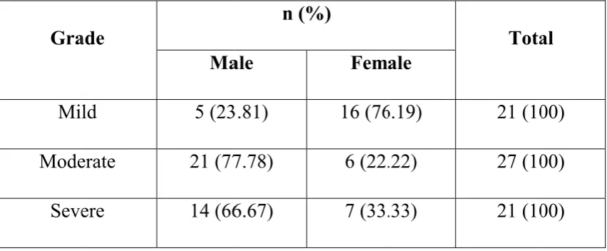
Tauber et al graded corneal ulceration in PUK based on the depth3 as
<25% – grade 1
25-50% - grade 2
50- 75%- grade 3
75 – 100% - grade 4
According to Sharma et al, PUK classified as follows1:
SEVERITY DESCRIPTION OF ULCER
MILD 2’ clock hours, superficially upto anterior stroma
MODERATE 2’ - 4’ clock hours, with anterior stromal involvement/
any clock hours with mid stromal involvement
37
TREATMENT OF PUK
50The management of PUK depends on the severity of findings
within the cornea and the extent of extra-ocular disease. The treatment
initiated for the systemic autoimmune disease has been found to have
beneficial effects on ocular manifestations. Regardless of the
management of corneal disease, results will be disappointing unless
aggressive treatment of the systemic disease is considered. The current
treatment strategy for PUK with underlying systemic disease is a
combination of systemic corticosteroids with a cytotoxic agent during the
acute phase of the disease. The exact cytotoxic agent may differ
according to the underlying systemic disease.
The primary goal of medical treatment for ocular disease is to
reduce inflammation, promote epithelial healing, and to minimize the
stromal loss. Though there have been advancements in
immune-modulatory and biologic agents, the outcome of PUK depends primarily
on the accompanying disease, and timely diagnosis and treatment. This
re-emphasis the need for identifying the underlying cause of PUK. Hence
a stepladder approach should be considered for the management of PUK
38
AIM OF MANAGEMENT OF PUK:
1. Identifying the etiology
2. Facilitating epithelial wound repair
3. Check ulceration and support repair
ESTABLISHMENT OF ETIOLOGY AND PRIMARY THERAPY
• Examine eyelids, corneal sensation and tears-Schirmer’s test
• Obtain cultures and scrapings- gram stain, KOH mount
• Debride devitalized tissue
• Initiate antimicrobial therapy
• Initiate anti-inflammatory therapy
• Systemic evaluation (e.g., for collagen-vascular disease)
Total count, Differential count, Erythrocyte sedimentation rate,
C-reactive protein, Random blood sugar, Rheumatoid factor, Urine routine,
Chest X-Ray, Mantoux test, Anti-nuclear antibody, anti-HCV antibodies,
Anticyclic citrullinated peptide antibodies, Complete metabolic panel
Sacroiliac joint radiographs, IgE levels, rapid plasma regain,
gastrointestinal evaluation fluorescent treponemal antibody
39
Facilitate epithelial wound repair
Administer tear substitutes, lubricants; punctal occlusion can be done in severely dry eyes
Eyelid closure (pressure patching, taping, or tarsorrhaphy) Therapeutic soft contact lens
Surgical therapy
Conjunctival transplantation Keratoepithelioplasty
Check ulceration and support repair
Tissue adhesive
Glued-on hard contact lens Anti-inflammatory agents Progestational steroids Systemic corticosteroids Immunosuppression Surgical therapy Conjunctival flap Conjunctival resection Corneoscleral lamellar graft Lamellar keratoplasty
40
LOCAL TREATMENT
Local bacterial and viral infections that cause PUK are usually
relieved with local targeted treatment. Lubricate the ocular surface with
preservative-free lubricating agents. This is done as most of the patients
with PUK also suffer from tear film abnormalities. These agents are also
helpful for removing or diluting harmful inflammatory proteins and
mediators on the ocular surface. In patients with marginal ulcerative
keratitis without an accompanying systemic disease, eyelid hygiene and
topical corticosteroids can be used and tapered according to the clinical
response. A topical antibiotic is recommended before corticosteroid use.
Collagenase inhibitors or collagenase synthetase inhibitors, such as
topical l% medroxyprogesterone and topical 20% acetylcysteine, may be
of limited benefit in reducing additional stromal ulceration. Topical
corticosteroids are not appropriate in patients with related systemic
disease, because these drugs inhibit new collagen production and thereby
increase the risk of perforation. Oral tetracycline derivatives may provide
additional benefit in preventing further stromal loss by decreasing
protease activity.
41
followed by tapering doses) may be effective in promoting epithelial closure and may suppress stromal inflammation and collagenolysis.
In patients with abnormalities of eyelid-globe relationships, correction of the anatomical disorder will facilitate epithelial recovery. Correction may include epilation or cryoablation of aberrant lashes in
trichiasis and appropriate repair of eyelid ectropion or entropion. The
liberal use of artificial tears and ointments may promote epithelial
migration in many patients with tear deficiency, exposure, or irregular
epithelium. Preparations conatining sensitizing preservatives, like
thimerosal must be avoided. Prefer an unpreserved eye drop or ointment.
In patients with severe dry-eye conditions, punctal occlusion by thermal
or electrical cauterization may augment the effect of tear substitutes.
Eyelid closure can encourage resolution of persistent epithelial
defects. In some cases, pressure patching or the use of tape may be
sufficient to encourage reepithelialisation.
Fibronectin has been shown to be an integral part of corneal
epithelial wound healing. Uncontrolled clinical trials have suggested it
may be effective in the treatment of persistent epithelial defects. In
alkali-burned rabbit corneas, fibronectin has been shown to improve epithelial
cell adhesion. Still, the use of this compound is investigational and will
42
In patients with persistent epithelial defects following chemical
burns in whom healthy donor tissue is not available in the fellow eye,
lenticules of donor cornea covered by epithelium may be placed at the
corneoscleral limbus. The epithelium spreads from the donor lenticule
and resurfaces the recipient cornea with functionally normal epithelium.
CHECK ULCERATION AND SUPPORT REPAIR
Management should be aimed at suppressing or at least delay stromal ulceration or perforation until neovascularization can provide serum antiproteases, macroglobulins, and other inhibitors of collagenolytic enzymes or until management strategies such as immunosuppression can reduce the inflammatory response, and healing provides adequate structural reinforcement. Depending on the situation, any one or a combination of the following approaches may be employed during this phase of therapy.
Tissue Adhesives 51
43
awaiting perforation and its attendant complications, this proactive measure of early use of tissue adhesive in nearly any case of persistent epithelial defect with progressive stromal ulceration is a boon. The cyanoacrylate glue is easily and quickly applied to the nonperforated cornea at the slit lamp with minimal preparation and complication. The mechanism of action is believed to be through the exclusion of inflammatory cells from the ulcerating stroma. When the glue is applied, a durable bandage contact lens is usually applied to provide comfort and to reduce the risk of glue dislodgment. Prophylactic antibiotics are mandatory. Cycloplegics, lubricants, and steroids can help reduce inflammation and discomfort.
Anti-inflammatory Agents52
Topical corticosteroids are advocated in the early therapy. In eyes
in which severe inflammation is present but ulceration is progressive,
their use can become dangerous. . As corticosteroids can interfere with
collagen synthesis, alternatively progestational steroids reduce
inflammation, curb collagenase activity with less delay in collagen
synthesis.
In severe cases of herpetic stromal keratitis or uveitis, the use of
systemic steroids is justified as a means of halting inflammatory process
44
can also be effective in peripheral ulcerative keratitis associated with
relapsing polychondritis, rheumatoid arthritis, and systemic lupus
erythematosis. Dosage selection depends on both systemic manifestations
and ocular response. Most patients are begun on 60 to 300 mg prednisone
per day, followed by tapering of the dose and switching to an
alternate-day regimen if possible. In many cases, the combination of systemic
steroids with immunosuppression is usually more effective than either
modality alone.
Corticosteroids, whether administered topically or systemically, are
usually ineffective in peripheral ulcerative keratitis associated with
Wegener's granulomatosis or Mooren's ulcer. They may be effective,
however, in reducing inflammation while the patient is being
immunosuppressed.
Systemic immune modulation Glucocorticoids
Systemic corticosteroids are the traditional first-line therapy for
acute phases of PUK, but alone are often unable to inhibit disease
progression or overcome the autoimmune disease. The usual starting dose
is 1 mg/kg/day maximum 60 mg/day), followed by a tapering schedule
based on clinical response. Pulsed methylprednisolone 1 g/day for 3
45
with imminent danger of vision loss. Immunosuppressive drugs or
biologic agents are administered with or without glucocorticoids in cases
refractory to glucocorticoids and when glucocorticoid-associated adverse
effects become an issue. Common complications of systemic
corticosteroids, such as osteoporosis, exacerbation of hypertension and
diabetes, electrolyte imbalance, and gastrointestinal bleeding, may be
avoided with initiation of immunosuppressive drugs. Although steroids
have a profoundly positive effect on ocular and systemic symptoms, they
fail to reduce the high mortality rate in patients with rheumatoid
vasculitis18. Therefore, the addition of cytotoxic chemotherapy comes up to successfully treat these patients.
Immunosuppressives/immunomodulators
Immunosuppressives used in cases of peripheral ulcerative keratitis
with auto-immune etiology include antimetabolites, alkylating agents, T
cell inhibitors, and biologic agents. Cyclophosphamide is the most
commonly used drug.
Methotrexate, azathioprine, mycophenolate mofetil, and
leflunomide are suitable antimetabolite agents. Methotrexate and
azathioprine are the two most commonly used antimetabolites in cases
unresponsive to oral corticosteroids and with recalcitrant rheumatoid
46
azathioprine 1.0–2.5 mg/kg/day have been reported to be effective.
Recent studies indicated better inflammatory control and fewer side
effects with mycophenolate mofetil (1.0 g twice daily) than with
methotrexate or azathioprine. Clinical reports now suggest that
leflunomide might be efficacious in the treatment of ocular inflammation.
The alkylating agents, cyclophosphamide and chlorambucil, are
suggested for use in severe progressive cases and in cases unresponsive to
methotrexate or other antimetabolites.
In a retrospective case series, Messmer and Foster reported that
cytotoxic immunosuppressive agents are highly effective in patients
resistant to systemic corticosteroids52. Cyclophosphamide was reported to be the most effective agent in their series; however, methotrexate was
reported to be very effective with less potential toxicity and was
suggested as a potential first choice for immunosuppression.
Cyclophosphamide may be administered orally at doses of 1–2 m/kg/day
or as pulsed intravenous therapy every 3–4 weeks under rheumatologic or
internal medicine guidance. Data on the use of cyclosporine A in patients
with rheumatoid arthritis and severe inflammatory eye disease suggest
that cyclosporine could be the initial immunosuppressant treatment of
choice in idiopathic cases or in those not associated with systemic
47
nephrotoxicity. Hence, cases associated with systemic vasculitis more
potent immunosuppressives like cytotoxic (eg, cyclophosphamide) or
antimetabolite (eg, methotrexate) therapy is instituted.
Biologic agents
Infliximab is a specific, chimeric monoclonal antibody against
pro-inflammatory cytokine tumor necrosis factor alpha (TNF-α). It has been
approved for use by the US Food and Drug Administration in 1999. Use
of infliximab for ocular inflammation was first reported in 2001 for
patients with panuveitis and rheumatoid arthritis-associated scleritis. It is
currently indicated for treatment of connective tissue or vasculitic
autoimmune diseases, and accompanying PUK, as well as other ocular
inflammatory states, such as necrotizing scleritis and uveitis. It is a
specific, chimeric monoclonal antibody against proinflammatory cytokine
tumor necrosis factor alpha (TNF-α), which stimulates production of the
matrix metalloproteinases responsible for corneal stromal lysis in PUK. It
binds both soluble and transmembrane TNF-α by blocking its receptor.
Cells expressing transmembrane TNF-α bound to infliximab may also be
susceptible to complement-mediated lysis, potentially increasing its
anti-inflammatory effect. Dosing of infliximab varies from 3 mg/kg
intravenously for rheumatoid arthritis to 5 mg/kg intravenously for
48
8 weeks for up to 18 months. Improvement usually occurs 1–2 weeks
after the first infusion. Although the optimal frequency and dosing of
infliximab for PUK and/or corneal perforation have not yet been
established, a dosing regimen similar to that used for rheumatoid arthritis
seems reasonable. The maintenance of remission must be weighed against
potential adverse events, because the long-term efficacy and safety of
biologics for use in ocular inflammation is unknown. Before
administering infliximab, opportunistic infections such as tuberculosis
must be ruled out, in addition to absolute contraindications such as
congestive heart failure. The reported side effects of long-term use
include increased risk of opportunistic infections, anaphylaxis, diarrhea,
cardiac failure, and resistance. More serious side effects, such as
lymphoproliferative disorders, malignancy, hepatotoxicity, and
endogenous endophthalmitis, have been reported. Moreover, increased
risk of thrombosis was reported, ranging from branch retinal vein
occlusion to myocardial infarction and pulmonary embolus; therefore,
special care must be taken when using infliximab and other biologic
agents. Other biologics, including etanercept (Enbrel) and rituximab
(Rituxan), have been used for the treatment of PUK. Etanercept is a
human recombinant dimeric fusion protein that mimics the effects of
naturally occurring soluble TNF-α receptors. It has been used for the
49
less efficacious than infliximab for the treatment of ocular inflammation.
This may be due to the ability of infliximab to bind to membrane-bound
TNF-α, in addition to free-floating cytokines.
In a study by Thomas et al, 3 patients with progressive rheumatoid
arthritis associated keratolysis refractory to steroids and
immunosuppressives were given infliximab infusion. There was marked
reduction in conjunctival injection and epithelial defects healed. Clinical
improvement occurred in first week of infusion.