“HEMODYNAMIC CHANGES ON LMA INSERTION WITH SAME
DOSE OF ETOMIDATE AND VARYING DOSES OF PROPOFOL”
Dissertation Submitted in
Partial fulfilment of the University regulations for
MD DEGREE IN
ANAESTHESIOLOGY
(BRANCH X)
MAY 2019
GOVERNMENT THENI MEDICAL COLLEGE
THE TAMIL NADU Dr. M.G.R. MEDICAL UNIVERSITY
CERTIFICATE
This is to certify that the dissertation titled
“Hemodynamic changes on LMA
insertion with same dose of Etomidate and varying doses of Propofol”
is a
Bonafide
original work done by
DR.V.ANANDH
during May 2016 -
May 2019
in partial fulfilment of the requirements for
M.D. (Anaesthesiology) Branch X-
Examination of The Tamilnadu Dr.M.G.R. Medical University to be held in May
2019.
Prof. DR.S.VIJAYA, Prof. DR.S.VIJAYARAGAVAN,
M.D., M.D., D.A.,
Professor and Guide, Professor and HOD,
Department of Anaesthesiology, Department of Anaesthesiology,
Govt. Theni Medical College, Govt. Theni Medical College,
Theni. Theni.
Prof. DR.K.RAJENDRAN, M.S., D.Ortho.,
Dean,
DECLARATION
I
DR.V.ANANDH
solemnly declare that this dissertation, titled
“Hemodynamic changes on LMA insertion with same dose of Etomidate and
varying doses of Propofol”
is a Bonafide record of work done by me in the
Department of Anaesthesiology,
Govt. Theni Medical College and Hospital, Theni
under the guidance of
Prof. DR.S.VIJAYA, M.D.,
Professor of Anaesthesiology,
Govt. Theni Medical College & Hospital, Theni.
This dissertation is submitted to The Tamilnadu Dr.M.G.R. Medical
University, Chennai in partial fulfilment of the University regulations for the award
of degree of M.D. (Anaesthesiology), Branch X- examination to be held in
MAY-2019.
Place: Theni
Date:
DR.V.ANANDH
ACKNOWLEDGEMENT
With deep sense of gratitude I thank God almighty for his grace and close presence, which strengthened and sustained me through this endeavour.
Perfection of work is possible only by the union of master brains, expertise hands and dedicated hearts of enthusiastic people at the right time. There by it gives me an immense pleasure to thank all the contributors who added oil to the glowing lamp of my study from the time of its ignition. Their valuable contributions reflect in the perfection of this study
I wish to express my sincere thanks to Prof. DR. K.RAJENDRAN, M.S.,
D.Ortho, Dean, Govt. Theni Medical College, Theni and the former Dean Prof. DR. T.
THIRUNAVUKKARASU, M.D., D.A., for granting me permission to do my study in
this esteemed institution.
I lend this opportunity to express my sincere heart full thanks and gratitude to Prof.
DR.S.VIJAYARAGAVAN, M.D., D.A., Professor and Head of the Department of
Anaesthesiology, Govt. Theni Medical College, Theni for his motivation, constant supervision and for providing all necessary arrangements for the conduct of the study, without which this dissertation would not have materialized.
I would like to place on record my indebtedness to my guide Prof. DR.S.VIJAYA,
M.D., Professor of Anaesthesiology, Govt. Theni Medical College, Theni for her constant
encouragement, constructivecriticism and suggestions throughout the period of the study. I express my profound thanks to Prof. DR.KANNAN BOJARAAJ, M.D., D.A.,
Govt. Theni Medical College, Theni for their whole hearted help and support in doing this study.
I am extremely thankful to DR.S.LOGANATHAN, M.D., Assistant Professor of Anaesthesiology, Govt. Theni Medical College, Theni for his sagacious advice and appropriate guidance to complete this study.
I thank all the Assistant Professors and Senior Residents of Department of Anaesthesiology for their keen interest and encouragement during this study.
I thank all the Professors in the Department of Surgery, Orthopaedics, Obstetrics and Gynaecology, Govt. Theni Medical College, Theni for their able help and support during the course of the study.
I also wish to thank all my colleagues for their constant help during this study. My thanks are due to all the theatre personnel for their willing cooperation and assistance.
I am deeply grateful to all the patients included in the study, for their whole hearted co-operation in spite of their illness made this study possible.
CONTENTS
SERIAL NO CHAPTERS PAGE NO
1 INTRODUCTION 1
2 OBJECTIVES 5
3 REVIEW OF LITERATURE 6
4 MATERIALS AND METHODS 36
5 RESULTS 48
6 DISCUSSION 76
7 SUMMARY 85
8 CONCLUSION 88
9 LIMITATIONS 89
ANNEXURES
ANNEXURE I: BIBLIOGRAPHY
ANNEXURE II: MASTER CHART
ANNEXURE III: KEY FOR MASTER CHART
ANNEXURE IV: PROFORMA
ANNEXURE V: ETHICAL APPROVAL
CERTIFICATE
ANNEXURE VI: PLAGIARISM REPORT
i
x
xxii
xxiii
xxv
INTRODUCTION
Airway management is of prime importance with regards to patient safety and is also the most important responsibility of anaesthesiologist to maintain a clear patent airway to facilitate administration and maintenance of anaesthetic agents besides sustaining the oxygen delivery through lungs. Hence various devices and methods were tried and used both by ancient and modern physicians for maintaining the airway and the ever-lasting search for the quick, safe and convenient method goes on. (1)
The cornerstone in the field of anaesthesia is the discovery of endotracheal intubation (2) and is usually done by direct laryngoscopy under direct vision. In the late 80s, only face mask and endotracheal tube (ETT) were the only means available to maintain the airway patency (3). Though
endotracheal intubation is safe in healthy patients, it may trigger hemodynamic changes and induce reflex cardio-vascular responses such as hypertension, tachycardia and arrhythmias which may in-turn lead to life-threatening complications like myocardial ischemia, left ventricular failure and cerebral haemorrhage (4, 5).
To avert these reflex hemodynamic changes, alternate devices such as fibre-optic scope, light wand and laryngeal mask airway (LMA) were used (6).
many variants of LMA were introduced. The laryngeal mask airway (LMA) is a potential tool in the management of difficult airway and today it is widely used as a part of routine airway management in an elective setting (8,9). The hemodynamic response to the insertion of laryngeal mask Airway (LMA) is considerably less than that is observed after laryngoscopy and endotracheal intubation (10).
The smooth insertion of laryngeal mask airway (LMA) entails adequate mouth opening with suppressed Airway reflex (11). Various
intravenous agents like thiopentone, etomidate, propofol and midazolam have been used for induction of anaesthesia with the purpose of attaining hemodynamic stability, attenuation of stress responses and maintenance of balance between myocardial oxygen supply and demand (12).
Etomidate is an IV anaesthetic agent which is used either alone or in combination with other agents for induction of anaesthesia and also for maintenance of anaesthesia in different contexts. It is a rapid onset short acting agent and it produces significantly less cardiovascular depression than the other induction agents. Etomidate does not depress the airway reflex and is also not associated with histamine release. Administration of etomidate may cause nausea, vomiting, pain on injection, myoclonus and adrenocortical suppression (15).
When propofol alone is administered for insertion of laryngeal mask airway (LMA) it can be associated with complications like coughing, gag reflex and laryngospasm. To avoid these complications, numerous agents were tried along with propofol and also to decrease the hemodynamic instability.
The combination of propofol and etomidate decreases the dose requirement for both the drugs, provides advantages of both the drugs such as better airway quality and muscle relaxation for LMA insertion and also has fewer effects on respiration and circulation as it provides hemodynamic stability and is much safer and more effective than propofol or etomidate alone. (13, 16, 17). Etomidate has shown that it is superior in comparison to other
with propofol. (18). Studies have shown that reduction in sympathetic tone and
direct venodilator effect of propofol is dose-dependent and in turn affects the hypotension produced by propofol which offsets the pressor responses of LMA insertion (19, 20, 21).
OBJECTIVES
1) To compare the Haemodynamic parameters during LMA Insertion with same dose of Etomidate and varying doses of Propofol.
2) To compare the ease of LMA insertion by number of attempts, total duration and efficiency of established airway.
3) To compare the duration of apnea.
4) To find out the incidence of side effects associated with the drugs and complications associated with LMA Insertion.
REVIEW OF LITERATURE
Airway management and Patient Safety is the most important aspect of management during general anaesthesia. Various devices that tried by ancient and modern physicians for maintenance of airway and delivery of anaesthetic agents. The most important milestone in the field of anaesthesia was the discovery of endotracheal intubation and it is usually done by using direct laryngoscopy under direct vision (22, 23). The endotracheal intubation is
the gold standard in safe and healthy patients. It has disadvantages of triggering hemodynamic changes and reflexes which can be dangerous to some groups of patients with other comorbidities (24, 25, 26, and 27).
Endotracheal intubation is known to induce reflex cardiovascular responses such as hypertension, tachycardia, arrhythmias, myocardial ischemia, infarction besides hypoxia, hypercapnia, laryngospasm, bronchospasm and sometimes rarely increased intracranial pressure and increased intraocular pressure (28, 29, and 30).
To avoid this reflex hemodynamic changes, alternate devices in the form of Supraglottic Airway devices (SADs) which includes laryngeal mask Airway (LMA) and its variants are used (31, 32, 33). The first of the modern
classic LMA for gynaecological laparoscopic surgeries with controlled ventilation(34, 35, 36).
These supraglottic devices which have their own advantages and disadvantages when compared to traditional and conventional face mask and endotracheal tube intubation (37, 38) as mentioned below:
Indications for the use of supraglottic airway devices
1. For elective ventilation: the supraglottic Airway devices are an effective alternative for mask anaesthesia in the operating theatres and also an alternative for the endotracheal tube intubation for short and elective surgical procedures under general anaesthesia (37). Sometimes to avoid the damage to
vocal cords and to avoid the complications of endotracheal intubation such as in professional singers Supraglottic Airway devices are preferred.
2. In difficult Airway: supraglottic device is an integral component of both anticipated and unanticipated difficult Airway management. They are used as a rescue device in situations of failed intubation or ventilation. LMA insertion can be tried while preparing for cricothyroidotomy in a patient who cannot be intubated or ventilated (38).
4. As a conduit for intubation: supraglottic airway devices can be used for this purpose when intubation is unsuccessful. The endotracheal tube can be passed through the supraglottic airway device or using a special intubating LMA either directly or by assistance with the bougie or a fibre optic scope
5. Can also be used as a bridge to extubation
Advantages of supraglottic airway device
1. Hands-free maintenance of the airway which removes the fatigue component to the operator (39).
2. These are more secure and reliable means of maintenance of airway
3. Also there are lesser chances of gastric insufflations
4. Lesser hemodynamic response and reflex than intubation.
Disadvantages of supraglottic airway devices
The incidence of sore throat is higher roughly about 17% for supraglottic airway devices as against 3 % for facemasks.
Contraindications for supraglottic airway devices
(40, 41, 42, 43, 44)1. Obstruction in the upper Airway in the form of tumour, abscess, hematoma, edema etc...
3. All patients at risk of aspiration such as no adequate fasting, intestinal obstructionand patients undergoing emergency surgeries.
4. In patients with disrupted upper Airway, facial trauma, burns following caustic ingestion
5. Among morbidly obese patients, pregnancy more than 14 weeks, patients who have received prior opioids medication
6. Conditions compromising lung compliance or increased resistance to breathing such as pulmonary Fibrosis, status asthmaticus etc.
However newer and safer supraglottic Airway devices provide high sealing pressures so that they can be used in conditions like obesity and stiff lungs (45,
46)
Miller’s Classification of supraglottic Airway devices
(47)A. Cuffed Peri-laryngeal sealers
1. Without directional sealing: Laryngeal Mask Airway (LMA), Intubating Laryngeal Mask Airway (ILMA), Soft-seal LM, Ambu LM
2. With directional sealing: Proseal- Laryngeal Mask Airway (PLMA) B. Cuffed Pharyngeal sealers
1. Without esophageal sealing cuff: Cobra-PLA
C. Cuffless Pre-shaped sealers: Stream-lined liner of the pharynx airway (SLIPA), I-gel, Baska Mask.
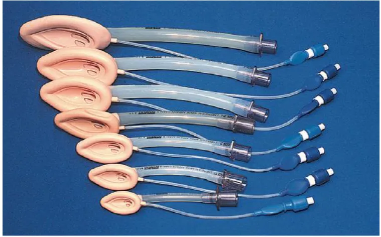
Laryngeal Mask Airway (LMA)
The laryngeal Mask Airway was conceived and designed by Dr. Archie Brain in 1981 and after years of research it was finally released for use in 1988 (48). The laryngeal Mask Airway clearly fills a niche between the
face mask and the endotracheal tube in terms of both Anatomical position and the degree of invasiveness. It is also reusable as set is made from Medical grade silicone rubber.
Figure showing Classic LMA
Table showing
Laryngeal Mask Airway (LMA) size and cuff
inflation volume
Weight in
Kg
LMA size
Cuff
inflation
volume
<5
1
4
5-10
1.5
7
10-20
2
10
20 – 30
2.5
14
30 – 50
3
20
50 – 70
4
30
70 – 100
5
40
[image:20.595.120.500.534.770.2]>100
6
50
The following is the standard method of insertion of laryngeal mask Airway (LMA) (49):
1. First of all anaesthesia should be deep enough to permit Insertion of LMA and we should not try to insert the LMA immediately following barbiturate induction unless a relaxant drug is given
2. Next position the head and neck as per normal tracheal intubation by keeping the neck flexed and head extended by pushing the head from behind with one hand while inserting LMA into the mouth with the other hand
3. While inserting, hold the LMA like a pen with the index finger placed anteriorly on the junction of the cuff and the airway tube. Then press it against the hard palate and verify whether it lies flat against the palate and that the tip is not folded over before further pushing into the pharynx
4. Now use the index finger to push the LMA backwards with continuously maintaining pressure against the palate
pressure seal. During inflation of the cuff we should not hold the tube as this will prevent the device from settling in the correct location.
7. Then connect to the circuit by holding the tube to prevent any displacement. Now gently inflate the lungs to check for the correct placement and confirm it. Insert a roll of gauze as a bite block and tape the device into place so that the proximal end of the tube is pointing caudally. The tube would be pressed back against the palate and posterior pharyngeal wall if it is correctly placed.
The following are the signs of correct placement of LMA:
1. Upon inflation of the LMA there will be a slight outward movement of the tube
2. A small oval swelling will be present in the neck around the thyroid and cricoid area
3. There will be no cuff visible in the oral cavity
4. There will be expansion of the chest wall on ventilation
There are other methods of inserting the laryngeal mask airway (LMA) like (a) Reverse technique (180o rotation)
(b) Fully or partially inflated cuff
(c) Techniques based on different head positions (d) Laryngoscope guided LMA insertion
(f) Using an introducer (g)Insertion in prone position.
Manoeuvres which can aid in LMA insertion: Jaw thrust, Chin-lift, anterior traction of tongue, superior laryngeal nerve block, partial removal or insertion.
Complications of LMA
(50)• The most common complication of laryngeal mask Airway is the post-operative sore throat. Excessive pressure or malposition off the cuff can be exacerbated by incorrect mask size use of nitrous oxide and prolonged surgery. To avoid this the most important factor to be monitored is the intraoperative cuff pressure which if maintained constantly drastically reduces the incidence of sore throat in the postoperative period
• Trauma to the pharyngeal cavity, airway edema and arytenoid dislocation is not uncommon. Rarely hypoglossal nerve injury, numbness of the tongue secondary to lingual nerve injury, recurrent laryngeal nerve injury and vocal cord paralysis may occur. Usually the nerve injuries are because of the wrong positioning of the LMA or excessive cuff pressure.
Induction techniques for LMA insertion
Intravenous induction
: Many drugs are used either alone or in combination for induction in the intravenous route which is by far the preferred route in most of the conditions. The following drugs have been used for IV induction for LMA insertion (51, 52, and 53):• Propofol
• Thiopentone
• Etomidate
• Etomidate + propofol
• Fentanyl + propofol
• Ketamine+ propofol
Inhalational induction
: Sometimes in infants and children where IV access is not possible, inhalational induction is the preferred route. The following drugs have been used for inhalational induction for LMA insertion(54, 55, and 56):
• Sevoflurane
Propofol
Propofol is a 2, 6 diisopropyl phenol and it has become one of the most frequently used intravenous anaesthetics today. It has a rapid onset predictable half-time and provides rapid emergence from anaesthesia (57, 58, 59).
Also it has a favourable side effect profile and antiemetic property which aids in many conditions including induction and maintenance of general anaesthesia, Intensive Care units (ICU) sedation and as a sedative-hypnotic in the context of outpatient procedures.
Drug structure of propofol
Commercial preparation
levels when prolonged IV infusions are used and also it supports bacterial growth (60, 61, 62, 63).
A generic formulation of propofol incorporates sodium metabisulphite as the preservative and has a lower PH between 4.5 to 6.4 whereas Diprivan uses the disodium edetate as a preservative with sodium hydroxide to bring the pH between 7 to 8.5.
Like thiopental, etomidate and ketamine, propofol is also not a clinical compound. Propofol cannot be mixed with any other drugs as it may result in coalescence of oil droplets and may pose a risk of pulmonary embolism which can rarely occur when mixed with lidocaine to prevent the pain on IV injection (64).
Propofol should be administered within 6 hours of opening the ampoule as sepsis and death have been linked to contaminated propofol preparations.
Pharmacokinetics
A. Absorption
: Propofol is available only for intravenous administration for the induction of anaesthesia and for moderate to Deep sedationC. Metabolism
: Propofol undergoes conjugation to an inactive glucuronide in the liver or it may get metabolized to quinol which is excreted as sulphate and glucuronide conjugates of the hydroxylated metabolite via cytochrome P450 and extra hepatic metabolism also probably contributes as the drug clearance exceeds the hepatic blood flow. Fortunately renal and hepatic diseases do not have much significant effect on the metabolism of propofol(66)
D. Excretion
: The metabolites of propofol are excreted in the urine unchanged and the elimination half-life is 9.3 to 69.3 minutes. The clearance is much higher in children and it is decreased in the presence of renal failure. The terminal elimination half-life maybe prolonged following extended administration although under normal conditions propofol is non-cumulative(66).
Mechanism of action
The interaction of the propofol with specific components of GABA-A receptor appears to reduce the rate of dissociation of neurotransmitter which is inhibitory in its prime action hence propofol increases the duration of GABA activated opening of the chloride channels resulting in the hyperpolarization of the cell membranes. In contrast to the other volatile anaesthetics propofol does not alter the spinal motor neuron excitability which suggests that immobility during propofol anaesthesia is not because of drug-induced spinal cord depression (68).
Effects of propofol on major systems
A. Effects on cardiovascular system
The most important effect on the cardiovascular system is the reduction in arterial blood pressure because of the drop in systemic vascular resistance (because of the inhibition of sympathetic vasoconstriction), preload and cardiac contractility. Moreover propofol causes hypotension following induction which is usually compensated by the stimulation accompanying the laryngoscopy and intubation. The factors which are associated with the propofol induced hypotension include rapid injection of the drug, old age and using large doses (69). In the usual doses propofol impairs the normal arterial
function. The coronary sinus lactate production increases in some patients though the myocardial oxygen consumption and coronary blood flow usually decreases which demonstrates that there is some mismatch between the myocardial oxygen supply and demand (70).
B. Effects on the respiratory system
There is a profound respiratory depression which usually causes apnea following an induction dose of propofol. There is inhibition of hypoxic ventilation drive and also depresses the normal response to hypercarbia even when used in sub-anaesthetic doses for conscious sedation (71). When
compared to thiopental there is greater depression of upper Airway reflexes with propofol and hence alloying intubation, endoscopy or laryngeal mask airway placement in the absence of neuromuscular blockade. Induction with propofol is associated with lower incidence of wheezing in asthmatic patients and non-asthmatic patients when compared with barbiturates or etomidate even though propofol is known to cause release of histamine (72).
C. Effects on the central nervous system
manifested if blood propofol concentration is above 200 ng / ml and hence propofol remains the preferred drug for outpatient anaesthesia. Sometimes Induction with propofol call can be accompanied with excitatory phenomena such as spontaneous movement, opisthotonus, muscle twitching or hiccups. Though this excitatory phenomenon can mimic tonic-clonic seizures, propofol also has anticonvulsant property which is used successfully to terminate status epilepticus and hence propofol can be safely administered even to epileptic patients. The other advantage of propofol is that it decreases intraocular pressure. Tolerance does not develop after long term propofol infusions and hence propofol is an uncommon agent of physical dependence or addiction.
Side effect profile
Propofol related infusion syndrome (PRIS) is a potential deadly side effect of propofol which is extremely rare and it was first described in children in the late 90s and subsequently in the adults after its use for sedation in the ICU. The clinical features of PRIS include metabolic acidosis, hyperkalaemia, hyperlipidaemia, rhabdomyolysis, hepatomegaly, renal failure, arrhythmias, major ECG changes and progression to cardiac failure.
The pathophysiology of PRIS is unclear even today and it is proposed that it may involve mitochondrial toxicity which uncouples the intracellular respiratory chain. Some researchers hypothesize that propofol related infusion syndrome is because of the indication of fatty acid oxidation (74). The clinical manifestation of PRIS is always dose dependent and duration dependent. Short duration infusion of high doses has been associated with cardiac failure. The case fatality rate in PRIS cases is above 50 %. In 2006 the FDA recommended the maximum propofol infusion dose to be 4 mg / kg / hr.
Sometimes there is production of green urine which is reported especially in ICU settings following prolonged infusions of propofol. It is stipulated that increased extra hepatic metabolism of propofol and excretion of the metabolites in urine causes greenish discoloration (75).
described in patients with history of atopy/allergy and in patients with history of other drug allergies especially to neuromuscular blocking drugs (75).
Propofol does not trigger malignant hyperthermia and can be administered to patients with hereditary coproporphyria. Also secretion of cortisol is not affected by group of all even when administered for prolonged period or duration there is temporary abolition of tremors in patients with Parkinsonism after administration of propofol and hence it may not be suited for patients undergoing stereotactic neurosurgery during which the symptom is required to correctly identify the anatomic location (75).
Etomidate
It is a carboxylated imidazole and it is chemically unrelated to any other drug used for intravenous induction of anaesthesia. The imidazole nucleus makes the etomidate to be water soluble like midazolam at an acidic pH and lipid soluble at physiologic pH (76).
Commercial preparations
Drug Structure of Etomidate
and occasional venous irritation. Recently it has been changed to fat emulsion which has virtually abolished the pain on injection and venous irritation. But the incidence of myoclonus remains unchanged even in the new formulation. Oral formulation of etomidate for administration through trans-mucosal route has shown to provide dose-dependent sedation. The administration in this route results in direct systemic absorption while bypassing the hepatic first pass metabolism which results in higher blood concentrations achieved at much rapid rate compared to the drug being swallowed (76).
Pharmacokinetics
A. Absorption:
Etomidate is available only for intravenous administration for the induction of anaesthesia and it is sometimes used for production of unconscious sedation in circumstances such as prior to the placement of retro bulbar blocksnon-ionized fraction at physiological pH. The decrease in plasma concentration to awakening levels is mainly because of the redistribution of the drug. Because of the redistribution, the pharmacokinetics of the drug can be explained only by a two Compartment model (77)
C. Biotransformation
: The hepatic microsomal enzymes and plasma hepatic esterases rapidly hydrolyze etomidate to an inactive metaboliteD. Excretion
: The metabolites of etomidate are excreted in the urine unchanged.Mechanism of action
nervous system that control extrapyramidal motor activity and these results in 30 to 60 % incidence of myoclonus with etomidate induction of anaesthesia
(78).
Effects of etomidate on major systems
(79)A.
Effects on cardiovascular system
Etomidate has very minimal effects on the cardiovascular system which is manifested as and mild reduction in peripheral vascular resistance. This results in slight decrease in arterial pressure whereas the myocardial contractility and cardiac output usually remains unchanged. Etomidate is not known to release histamine. Analgesia is not produced by etomidate. For this reason, opioid administration before induction with etomidate may be useful to blunt hemodynamic responses by laryngoscopy.
B.
Effects on the respiratory system
When compared with barbiturates or benzodiazepines, etomidate is relatively safe as ventilation is least affected even in doses used for induction do not usually result in apnea unless opiates are pre-administered
C.
Effects on the central nervous system
nausea and vomiting is very common with etomidate administration when compared to propofol or Barbiturates.
D.
Effects on the endocrine system
Even with usual doses of etomidate used for induction, there is a transient inhibition of enzymes involved in cortisol and aldosterone synthesis because of the adrenocortical suppression exhibited by etomidate. Long term infusion of etomidate is associated with increased mortality among the critically ill patients mainly because of the adrenocortical suppression
Side effects
The most important adverse effect of etomidate is the adrenocortical suppression. Etomidate exhibits inhibitory action against the enzyme 11 Beta hydroxylase and prevents the conversion of cholesterol to cortisol. Even one dose of etomidate is sufficient enough to transiently suppress the adrenocortical axis. Some studies suggest curtailing this effect by pre-treatment with dexamethasone. However some researchers concluded that there is no direct effect on outcomes following bolus administration of etomidate even in high-risk septic population (80).
The following table shows the comparison between propofol and etomidate regarding the hemodynamic changes in percentage (%) after
induction of anaesthesia
Parameter Etomidate Propofol
Heart rate -5 ± 10 -10 ± 10
Mean arterial blood
pressure 0 to 17 -10 – 40
Systemic vascular
resistance -10 ± 14 -15 – 25
Pulmonary artery
pressure -9 ± 8 0 – 10
Pulmonary vascular
resistance -18 ± 6 0 – 10
Pulmonary artery
occluded pressure Unchanged Unchanged
Right atrial pressure Unchanged 0 – 10
Cardiac Index -20 ± 14 -10-30
Stroke volume 0 - 20 -10-25
Left ventricular stroke work index
The following are the studies related to hemodynamic changes
during LMA insertion
Balasubramaniam S et al (81) compared the hemodynamic responses
between laryngeal mask airway (LMA) and endotracheal tube (ETT) intubation in a single blinded randomised controlled trial with 60 normotensive individuals who were posted for elective surgeries under general anaesthesia. They divided the samples into two groups with 30 in each group and studied the heart rate, blood pressure and rate pressure product. These hemodynamic parameters were studied at baseline, pre- intubation, 1, 2, 3, 5 and 10 minutes after extubation. They concluded that there was a steady increase in hemodynamic responses in both the groups but the mean increase in heart rate, blood pressure and rate pressure product was significantly lower (p<0.05) for the LMA group in comparison to the endotracheal tube group.
Renu Bala et al (82) assessed the hemodynamic changes with insertion
time intervals. They concluded that all the three supraglottic devices do not differ in hemodynamic changes but I-gel has a higher success rate in comparison to classic LMA and proseal LMA.
Jarineshin H et al (83) compared the immediate hemodynamic effects
of insertion of supreme LMA and classic LMA with laryngoscopy and endotracheal intubation by studying a sample of 150 patients and observing the hemodynamic parameters such as heart rate and blood pressure before and after induction of anaesthesia. They concluded that the systolic blood pressure and diastolic blood pressure was significantly lower in the endotracheal tube group in comparison to the LMA group 5 minutes after the insertion of device. However there was no significant difference between classic LMA and supreme LMA in terms of hemodynamic parameters
Singh D et al (84) compared the hemodynamic changes during
coronary artery disease and cerebrovascular disease where minimal changes in hemodynamics are desirable.
The following are the studies related to the use of drugs etomidate
and propofol for induction
Liu et al (85) explored the preventive effect of pre-treatment with
propofol on etomidate related myoclonus in a prospective double-blind randomised controlled study with the sample of 363 patients scheduled for painless GI endoscopy. The subjects were divided into 5 groups with one group receiving etomidate alone and 3 groups receiving incremental doses of propofol pre-treatment before etomidate anaesthesia and fifth group receiving 1 to 2 mg / kg of propofol for anaesthesia. They are observed that incidence of myoclonus in the propofol group was less than the etomidate alone group and the incidence of hypoxemia in the propofol group was higher than the etomidate alone group whereas the incidence of adverse effects in the proposal + etomidate group was lower than the etomidate alone group.
Meng Q et al (86) Compare the safety and efficacy of etomidate and
in the etomidate alone group was significantly higher in comparison to other groups and hence propofol in combination with etomidate is a safe and effective alternate for elderly patients.
Price et al (87) studied the changes in cardiac index and systemic
vascular resistance during induction of anaesthesia with thiopentone, propofol, methohexitone and etomidate in a sample of 90 subjects randomly allocated to four groups to receive alfentanil followed by thiopentone, methohexitone, propofol or etomidate to obtund the eyelash reflex. They concluded that the cardiac stability of etomidate is not so complete in all groups of patients as there was significant decrease in cardiac index of 16 % among patients who received etomidate alone.
Kumar et al (88) compared the effects of propofol and etomidate on
Agarwal et al (89) compared propofol and etomidate for its hemodynamic effects and adverse effects on 100 patients posted for surgeries under general anaesthesia. The samples were divided into two groups with one group receiving propofol 2 mg / kg and another group receiving etomidate 0.3 mg / kg. They observed that patients in etomidate group had little change in mean arterial pressure and heart rate compared to propofol group and myoclonus activity was higher in etomidate group whereas pain on injection was more in propofol group. They concluded that etomidate is a better alternate agent for induction than propofol.
Meena et al (22) compared to the effectiveness of three different
Aghdaii et al (90) evaluated the hemodynamic effects of midazolam with etomidate by comparing it with propofol and ketamine combination for the induction of anaesthesia in 100 patients with left ventricular dysfunction undergoing coronary artery bypass graft surgery. The patients were randomly allocated into two groups to receive either propofol and ketamine or etomidate with midazolam. They found that the hemodynamic response was comparable in both the groups except for cardiac index after one minute and 3 minute of intubation.
Yagan et al (91) measured the hemodynamic responses between use of
etomidate and propofol in combination and the use of both the drugs separately by dividing the subjects into three groups. They observed a significant reduction in mean arterial pressure values after intubation in the propofol group when compared to etomidate alone group and the combination group. They concluded that propofol-etomidate combination is a very good alternative when extremes of hypotensive and hypertensive responses are expected.
Kaushal et al (92) studied the hemodynamic instability and attenuation
Kaur et al (93) studied the induction and recovery characteristics along with hemodynamic parameters of etomidate-lipuro and propofol-lipuro in a prospective randomized double-blinded study with 60 cardiac patients of either sex. They observed the incidence of Apnea and pain on injection and induction time was less in etomidate group than propofol group but myoclonus and postoperative nausea and vomiting was more common in etomidate group. The mean systolic blood pressure after 15 minutes and mean diastolic blood pressure after 5 minutes was lower in propofol group when compared to etomidate group which led them to conclude that etomidate is a better alternative to propofol in cardiac patients to attain hemodynamic stability and faster induction.
Hosseinzadeh H et al (16) evaluated the hemodynamic effects of
ketamine and propofol (group A) in comparison to etomidate and propofol (group B) for anaesthesia induction. They studied a total of 60 patients above 50 years of age posted for elective surgeries with Group A receiving ketamine 0.75 mg / kg and propofol 1 mg / kg and Group B receiving etomidate 0.2 mg /kg and propofol 1 mg / kg. They observed that there was a significant reduction in systolic, diastolic and mean arterial pressure after induction and after 6 minutes of intubation in both the groups and hence they concluded that both the methods of induction are effective to maintaining hemodynamic stability.
Hosseinzadeh H et al (94) compare the three methods of induction of
etomidate alone with regards to hemodynamic stability after insertion of laryngeal mask airway in elective surgeries. About 90 patients were divided into three groups with 30 subjects in each group. Group A received propofol 2.5 mg / kg, group B received etomidate 0.3 mg / kg and Group C received propofol 1 mg / kg plus etomidate 0.2 mg / kg. They observed heart rate, systolic, diastolic blood pressure and mean arterial pressure before induction and 30 minutes after induction. They also noted the number of attempts for laryngeal mask Airway insertion and the ease of placement of LMA in all the three groups. They observe the significant difference in hemodynamic changes between group A in comparison to group B and Group C. They also noted that heart rate was lower in group A and group B besides the number of attempts and ease of insertion was significantly different in group B when compared to group A and Group C they concluded that etomidate Plus propofol is effective for facilitating LMA insertion with an advantage of cardiovascular stability .
Hashaam B et al (95) compared the hemodynamic effects of etomidate
MATERIALS AND METHODS
STUDY AREA
The study was conducted among the patients posted for elective surgeries in Department of Anaesthesiology, Government Theni Medical College & Hospital, Theni.
STUDY POPULATION
The study population included 90 patients posted for elective surgeries like
• Fibroadenoma Breast - Excision
• Gynaecomastia – Webster’s procedure
TIMELINE
The study work is described in detail in the following Henry Gantt chart from its inception to the completion.
Activity
2016 2017 2018
8 9 10 11 12 1 2 3 4 5 6 7 8 9 10 11 12 1 2 3 4 5 6 7 8 9 10
Protocol preparation,
Submission Questionnaire
Preparation, Ethical clearance Data Collection
Data Entry & Analysis
STUDY PERIOD
The data collection for the study was done between May 2017 to September 2018.
SAMPLE SIZE
Based on the literature (22), to discover a 10 unit change in BP/PR (α=0.05, β=0.2 and power=0.08) sample size of 90 was considered.
GROUP A- Etomidate 0.2 mg/kg + Propofol 0.5 mg/kg (30)
GROUP B- Etomidate 0.2 mg/kg + Propofol 1 mg/kg (30)
GROUP C- Etomidate 0.2 mg/kg + Propofol 1.5 mg/kg (30)
So the total sample size was considered as 90.
INCLUSION CRITERIA
1. Either gender
2. Age between 20-60 years 3. ASA I and II
4. Elective surgeries like
➢ Fibroadenoma Breast - Excision ➢ Gynaecomastia – Webster’s procedure
EXCLUSION CRITERIA
1. Individuals who did not give consent. 2. Pregnancy
3. Alcohol and drug abusers
4. Patients allergic to drugs involved in the study 5. Cardiovascular diseases
6. Respiratory diseases
7. Significant Neurological illnesses like Epilepsy 8. Hepatic and Renal diseases
9. Emergency surgeries 10. BMI > 30
11. ASA III and IV
12. Anticipated difficult intubation such as MPG III, IV and Thyromental distance < 6.5cm, Mouth opening < 2.5 cm.
13. Existence of a considerable pathology in the larynx or pharynx
STUDY DESIGN
It was a double blinded randomized clinical trial.
SAMPLING DESIGN
STUDY TECHNIQUE
METHODOLOGY
1. Patients were kept overnight fasting and were premedicated with T.Alprazolam 0.5mg and T.Ranitidine 150mg on the night day before surgery.
2. All patients were preloaded with Ringer Lactate (6ml/kg) 3. 18 gauge IV cannula was secured.
4. Standard anaesthesia monitors including Electrocardiogram (ECG), Non Invasive Blood Pressure (NIBP) and Pulse Oximeter were attached and Baseline Hemodynamic parameters were recorded.
5. Inj. Glycopyrrolate (0.01 mg/kg) iv was given.
6. Preoxygenation done for 3minutes with 100% oxygen.
7. Midazolam 0.025mg/kg iv, Fentanyl 2microgram/kg iv, and Lidocaine 1mg/kg IV was given.
8. Patients were randomly assigned to one of three groups.
9. GROUP A patients received - Etomidate 0.2 mg/kg + Propofol 0.5 mg/kg. GROUP B patients received - Etomidate 0.2 mg/kg + Propofol 1 mg/kg GROUP C patients received - Etomidate 0.2 mg/kg + Propofol 1.5 mg/kg
10.All study drugs were prepared by Anaesthesiologist who had been blinded to the details of the study.
11.The volume of the medications (20 ml) and the speed of the injection (30 seconds) were equal in all three groups.
13.60 seconds after loss of consciousness (which was confirmed by inability to reply to verbal commands and loss of eyelash reflex), appropriate size of Classical LMA insertion was done with partially inflated technique by Second Anaesthesiologist. 14.Proper placement of LMA was assessed by chest expansion and capnography. The
success rate of LMA insertion was evaluated based on the ventilation status.
15.Ease of ventilation was defined as the presence of normal capnogram wave and lack of air leak after LMA insertion.
16.In case of poor ventilation, LMA was removed and reinserted. A rescue Propofol dose of 1mg/kg was used whenever there was resistance to LMA insertion or the patient moved.
17.Patient was excluded from the study in case of failure in LMA insertion after second attempt.
18.Duration of LMA insertion [from the period of insertion of LMA to the checking of bilateral air entry] was recorded.
19.Following successful LMA insertion, connected to close circuit and Duration of apnea was noted.
20.Anaesthesia was maintained by equal mixture of Oxygen – Nitrous Oxide (4 litre per minute), sevoflurane 0.8% and along with intermittent bolus of Non-Depolarizing Muscle Relaxant (Atracurium) as required throughout the surgery.
21.Hemodynamic parameters like Blood pressure (Systolic, Diastolic and Mean Arterial Pressure), Heart rate and oxygen saturation were recorded at 1min, 3min, 5min, 7min and 10min after induction.
23.LMA was removed after adequate recovery of muscle power.
24.Side effects like Pain on injection, Hypotension (>20% reduction), Bradycardia (HR<60), Post-operative nausea, vomiting (PONV) and Myoclonus was noted.
STUDY PARAMETERS
1.
Blood pressure
(Systolic, Diastolic and Mean Arterial Pressure) - [prior to induction, 30 seconds, 60 seconds, 1min, 3min, 5min, 7min and 10min after induction].2.
Heart rate
[prior to induction, 30 seconds, 60 seconds, 1min, 3min, 5min, 7min and 10min after induction].3.
Spo2
[prior to induction, 30 seconds, 60 seconds, 1min, 3min, 5min, 7min and 10min after induction].4.
Ease of LMA Insertion
[number of attempts (1/ 2/ >2), duration (in seconds) of LMA Insertion].5.
Duration of Apnea
.
6.
Side effects
[Pain on injection, Hypotension (>20% reduction), Bradycardia (HR<60), Post-operative nausea vomiting (PONV) and Myoclonus].STATISTICAL ANALYSIS
All the data was initially entered to Microsoft Excel 2010 and later these spreadsheets were used for analysis. Statistical analysis was done using SPSS version 22.0.
1.
DESCRIPTIVE STATISTICS
Descriptive statistics were calculated as frequency, percentage, mean and standard deviation, median and inter-quartile range. Descriptive data were represented using various tables, graphs, diagrams etc.
2.
INFERENTIAL STATISTICS
For inferential statistics, various tests of significance were used according to the type of variables dealt with. For all the statistical tests of significance, p value of <0.05 was considered to reject the null hypothesis. Chi square test was used to compare the various categorical variables. To reduce the error in approximation, Yates’s correction for continuity is used. Factorial Repeated Measures ANOVA was used to compare between the 3 groups. Post hoc analysis with Bonferroni correction for multiple comparison was performed when statistically significant differences were observed in the Repeated Measures Analysis.
3.
TESTS FOR NORMALITY
(p=0.679 i.e. p>0.05), the data follow a normal distribution, and Parametric tests were used.
4.
SIGNIFICANCE LEVEL
RESULTS
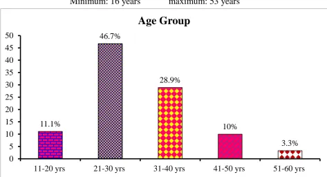
Table 1: Age distribution of the study population (n=90)
Age group Frequency Percent
11 - 20 years 10 11.1
21 - 30 years 42 46.7
31 - 40 years 26 28.9
41 - 50 years 9 10
51 - 60 years 3 3.3
Total 90 100.0
Mean Age (± S.D): 30.07 (± 8.95) years
Minimum: 16 years maximum: 53 years
11.1%
46.7%
28.9%
10%
3.3% 0
5 10 15 20 25 30 35 40 45 50
11-20 yrs 21-30 yrs 31-40 yrs 41-50 yrs 51-60 yrs
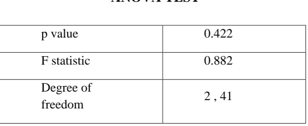
Table 2: Comparison of age among the three study groups (n=90)
Group N
Mean age (years)
Std. Deviation
95% Confidence Interval for Mean Lower Bound Upper Bound
A 30 32.82 9.250 26.60 39.03
B 30 28.32 6.111 25.37 31.26
C 30 30.29 11.698 23.53 37.04
Total 90 30.07 8.956 27.35 32.79
ANOVA test was applied to test the difference in mean age between the groups.
ANOVA TEST
p value 0.422
F statistic 0.882
Degree of
freedom 2 , 41
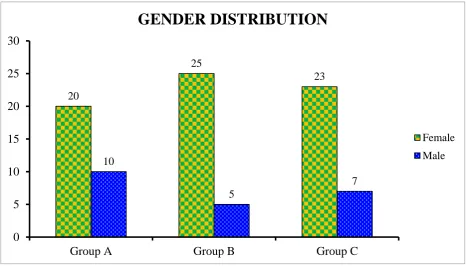
Table 3: Comparison of gender distribution among the study groups (n=90)
Group Female N (%) Male N (%) Total N (%)A 20
(29.4)
10
(45.5) 30 (33.3)
B 25
(36.7) 5 (22.7) 30 (33.3)
C 23
(33.9) 7 (31.8) 30 (33.3)
Total 68
(100)
22
(100) 100 (100)
Chi-square value: 1.646 p value:0.439
Gender distributions of subjects in the three groups were comparable and the minimal difference observed was not statistically significant.
20 25 23 10 5 7 0 5 10 15 20 25 30
Group A Group B Group C
GENDER DISTRIBUTION
Female
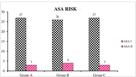
Table 4: Comparison of weight among the three study groups (n=90)
Group N
Mean weight
(kg)
Std. Deviation
95% Confidence Interval for Mean Lower Bound Upper Bound
A 30 55.00 7.457 49.99 60.01
B 30 54.26 5.733 51.50 57.03
C 30 55.14 7.263 50.95 59.34
Total 90 54.73 6.543 52.74 56.72
ANOVA test was applied to test the difference in mean weight between the groups.
ANOVA TEST
p value 0.671
F statistic 0.082
Degree of
freedom 2 , 41
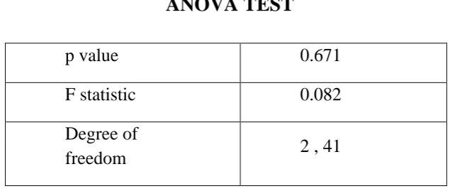
Table 5: Comparison of diagnosis among the study groups (n=90)
Diagnosis A N (%) B N (%) C N (%) Total N (%)Fibroadenoma Breast 18 (60) 24 (80) 20 (66.7) 62 (68.9)
Gynaecomastia 5 (16.7) 3 (10) 4 (13.3) 12 (13.3)
Others 7 (23.3) 3 (10) 6 (20) 16 (17.8)
Total 30 (100) 30 (100) 30 (100) 90 (100)
Chi-square value: 8.372 p value:0.212
The difference in diagnosis observed between three groups was not statistically significant. 18 24 20 5 3 4 7 3 6 0 5 10 15 20 25 30
Group A Group B Group C
DIAGNOSIS
Fibroadenoma
Gynecomastia
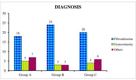
Table 6: Comparison of ASA risk among the study groups (n=90)
Group ASA I
N (%)
ASA II N (%)
Total N (%)
A 27
(33.7) 3 (30)
30 (33.3)
B 26
(32.6) 4 (40)
30 (33.3)
C 27
(33.7) 3 (30)
30 (33.3)
Total 80
(100) 10 (100)
90 (100)
Chi-square value: 2.757 p value:0.252
Distribution of subjects according to ASA risk was comparable in the three groups and the minimal difference observed was not statistically
significant.
27
26 27
3 4 3
0 5 10 15 20 25 30
Group A Group B Group C
ASA RISK
ASA I
ASA II
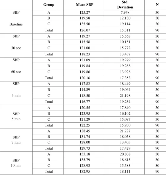
Table 7: Group-wise comparison of systolic blood pressure at various time
periods (n=90)
Group Mean SBP Std.
Deviation N
SBP
Baseline
A 125.27 7.938 30
B 119.58 12.130 30
C 135.50 19.114 30
Total 126.07 15.311 90
SBP
30 sec
A 119.27 15.563 30
B 115.58 10.151 30
C 121.00 15.772 30
Total 118.23 13.437 90
SBP
60 sec
A 121.09 19.279 30
B 119.84 19.288 30
C 119.86 13.928 30
Total 120.16 17.353 90
SBP
3 min
A 117.82 18.449 30
B 114.89 19.064 30
C 118.50 21.198 30
Total 116.77 19.234 90
SBP 5 min
A 120.55 17.840 30
B 123.95 16.102 30
C 121.29 15.097 30
Total 122.25 15.930 90
SBP 7 min
A 128.45 21.727 30
B 131.74 18.058 30
C 128.00 13.405 30
Total 129.73 17.429 90
SBP 10 min
A 133.18 20.808 30
B 135.79 18.615 30
C 128.93 15.583 30
Factorial- Repeated measures ANOVA was applied to test the difference in mean systolic blood pressure at various time intervals between the three groups.
Model Systolic blood
pressure
Systolic blood pressure * Group
Wilks’s
Lambda 0.414 0.602
df 6,36 12,72
p value <0.001 0.041
Between 3 groups - Systolic blood pressure
F statistic 1.156
df 2,41
p value 0.0473
Post-Hoc Test for SBP (n=90)
Time interval Group A vs Group B Group A vs Group C Group B vs Group C
Baseline vs 30 sec 0.781 0.048 0.027
Baseline vs 3 min 0.652 0.038 0.033
Table 8: Group-wise comparison of diastolic blood pressure at various time
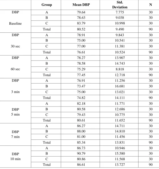
periods (n=90)
Group Mean DBP Std.
Deviation N
DBP
Baseline
A 79.64 7.775 30
B 78.63 9.038 30
C 83.79 10.998 30
Total 80.52 9.490 90
DBP
30 sec
A 78.91 9.843 30
B 75.00 10.541 30
C 77.00 11.381 30
Total 76.61 10.524 90
DBP
60 sec
A 78.27 13.907 30
B 78.58 14.743 30
C 75.29 8.818 30
Total 77.45 12.718 90
DBP
3 min
A 76.91 11.256 30
B 73.47 16.681 30
C 75.00 13.021 30
Total 74.82 14.111 90
DBP 5 min
A 82.18 11.771 30
B 80.58 12.686 30
C 79.43 10.775 30
Total 80.61 11.652 90
DBP 7 min
A 86.27 14.711 30
B 88.00 14.810 30
C 81.00 11.456 30
Total 85.34 13.831 90
DBP 10 min
A 86.73 10.946 30
B 90.79 15.580 30
C 80.86 11.568 30
Factorial- Repeated measures ANOVA was applied to test the difference in mean diastolic blood pressure at various time intervals between the three groups.
Model Diastolic blood
pressure
Diastolic blood pressure *
Group
Wilks’s Lambda 0.570 0.735
df 6,36 12,72
p value 0.002 0.043
Between 3 groups - Diastolic blood pressure
F statistic 1.252
df 2,41
p value 0.045
Post-Hoc Test for DBP (n=90)
Time interval Group A vs Group B Group A vs Group C Group B vs Group C
Baseline vs 30 sec 0.048 0.035 0.019
Baseline vs 7 min 0.912 0.044 0.027
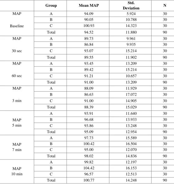
Table 9: Group-wise comparison of mean arterial pressure at various time
periods (n=90)
Group Mean MAP Std.
Deviation N
MAP
Baseline
A 94.09 5.924 30
B 90.05 10.788 30
C 100.93 14.323 30
Total 94.52 11.880 90
MAP
30 sec
A 89.73 9.961 30
B 86.84 9.935 30
C 93.07 15.214 30
Total 89.55 11.902 90
MAP
60 sec
A 93.45 13.209 30
B 89.42 15.214 30
C 91.21 10.657 30
Total 91.00 13.209 90
MAP
3 min
A 88.09 11.929 30
B 86.63 17.072 30
C 91.00 14.905 30
Total 88.39 15.029 90
MAP 5 min
A 93.91 11.640 30
B 96.68 13.933 30
C 93.86 13.248 30
Total 95.09 12.954 90
MAP 7 min
A 97.73 15.589 30
B 100.42 16.504 30
C 95.00 12.070 30
Total 98.02 14.836 90
MAP 10 min
A 99.82 12.197 30
B 104.42 16.153 30
C 96.57 12.513 30
Factorial- Repeated measures ANOVA was applied to test the difference in mean arterial pressure at various time intervals between the three groups.
Model Mean arterial
pressure
Mean arterial pressure *
Group
Wilks’s Lambda 0.520 0.640
df 6,36 12,72
p value <0.001 0.024
Between 3 groups - Mean arterial pressure
F statistic 1.044
df 2,41
p value 0.014
Post-Hoc Test for MAP (n=90)
Time interval Group A vs Group B Group A vs Group C Group B vs Group C
Baseline vs 30 sec 0.718 0.024 0.016
Baseline vs 7 min 0.582 0.039 0.512
Table 10: Group-wise comparison of pulse rate at various time periods
(n=90)
Group Mean PR Std. Deviation N
Pulse rate
Baseline
A 86.55 15.162 30
B 85.11 15.797 30
C 81.57 10.530 30
Total 84.34 13.980 90
Pulse rate
30 sec
A 89.09 14.515 30
B 90.47 16.290 30
C 85.29 10.395 30
Total 88.48 14.069 90
Pulse rate
60 sec
A 86.73 12.410 30
B 94.16 18.836 30
C 84.93 9.311 30
Total 89.36 15.129 90
Pulse rate
3 min
A 89.00 14.262 30
B 88.79 18.201 30
C 82.71 11.684 30
Total 86.91 15.351 90
Pulse rate
5 min
A 86.82 13.482 30
B 85.37 21.474 30
C 78.86 9.984 30
Total 83.66 16.637 90
Pulse rate
7 min
A 83.00 12.050 30
B 84.47 20.359 30
C 80.57 10.449 30
Total 82.86 15.593 90
Pulse rate
10 min
A 86.55 11.970 30
B 91.37 18.643 30
C 78.50 9.485 30
[image:75.595.15.580.121.700.2]Factorial-Repeated measures ANOVA was applied to test the difference in mean pulse rate at various time intervals between the three groups.
Model Mean pulse rate Mean pulse rate
* Group
Wilks’s Lambda 0.670 0.646
df 6,36 12,72
p value 0.019 0.012
Between 3 groups - Mean pulse rate
F statistic 1.197
df 2,41
p value 0.008
Post-Hoc Test for pulse rate (n=90)
Time interval Group A vs Group B Group A vs Group C Group B vs Group C
Baseline vs 60 sec 0.981 0.920 0.009
Baseline vs 5 min 0.487 0.024 0.021
Table 11: Group-wise comparison of oxygen saturation at various time
periods (n=90)
Group
Mean SpO2 Std.
Deviation N
SpO2
Baseline
A 99.64 .809 30
B 99.89 .315 30
C 99.86 .535 30
Total 99.82 .540 90
SpO2
30 sec
A 99.82 .603 30
B 99.95 .229 30
C 100.00 .000 30
Total 99.93 .334 90
SpO2
60 sec
A 99.91 .302 30
B 99.89 .459 30
C 100.00 .000 30
Total 99.93 .334 90
SpO2
3 min
A 99.82 .405 30
B 100.00 .000 30
C 99.93 .267 30
Total 99.93 .255 90
SpO2
5 min
A 99.73 .467 30
B 100.00 .000 30
C 99.14 3.207 30
Total 99.66 1.817 90
SpO2
7 min
A 99.64 .674 30
B 100.00 .000 30
C 100.00 .000 30
Total 99.91 .362 90
SpO2
10 min
A 99.45 .688 30
B 100.00 .000 30
C 100.00 .000 30
[image:78.595.20.577.111.713.2]Factorial- Repeated measures ANOVA was applied to test the difference in mean oxygen saturation at various time intervals between the three groups.
Model Mean SpO2
Mean SpO2*
Group
Wilks’s Lambda 0.726 0.591
df 6,36 12,72
p value 0.059 0.064
Between 3 groups – Mean SpO2
F statistic 2.015
df 2,41
Table 12: Comparison of number of attempts for LMA insertion among the
study groups (n=90)
Group
1 attempt N (%)
2 attempts N (%)
Total N (%)
A 26 (31.7) 4 (50) 30
(33.3)
B 28 (34.1) 2 (25) 30
(33.3)
C 28 (34.1) 2 (25) 30
(33.3)
Total 82 (100) 8 (100) 100
(100) Chi-square value: 0.764 p value:0.682
Table 13: Comparison of time taken for LMA insertion among the three
study groups (n=90)
Group N
Mean Time for LMA
(seconds)
Std. Deviation
95% Confidence Interval for Mean
Lower
Bound Upper Bound
A 30 14.36 8.34 8.76 19.96
B 30 13.75 6.87 7.63 17.56
C 30 12.43 4.95 6.58 16.11
Total 90 13.51 6.72 7.66 17.87
ANOVA test was applied to test the difference in mean time taken for LMA insertion between the groups.
ANOVA TEST
p value 0.258
F statistic 1.399
Degree of
[image:82.595.141.456.541.682.2]Table 14: Comparison of duration of apnea among the three study groups
(n=90)
Group N
Mean Duration of apnea
(seconds)
Std. Deviation
95% Confidence Interval for Mean
Lower
Bound Upper Bound
A 30 99.09 103.000 29.89 168.29
B 30 112.16 102.418 46.85 180.99
C 30 134.36 147.198 49.37 219.35
Total 90 115.20 117.538 42.03 189.54
ANOVA test was applied to test the difference in mean duration of apnea between the groups.
ANOVA TEST
p value 0.595
F statistic 0.526
Degree of
[image:84.595.130.457.537.679.2]Table 15: Comparison of side effects among the study groups (n=90)
Group Pain on
injection N (%) Hypotension N (%) Myoclonus N (%) Bradycardia N (%) PONV N (%) Total N (%)
A 0 (0) 1 (25) 2 (100) 0 (0) 0 (0) 30 (33.3)
B 0 (0) 1 (25) 0 (0) 0 (0) 0 (0) 30 (33.3)
C 0 (0) 2 (50) 0 (0) 0 (0) 0 (0) 30 (33.3)
Total 0 (0) 4 (100) 2 (100) 0 (0) 0 (0) 100 (100)
0 0 0 1 1 1 1 1 2 2 2
A B C
1 1
2 2
0 0
Pain on injection
Hypotension
Myoclonus
Bradycardia
PONV
Table 16: Comparison of complications in LMA insertion among the study
groups (n=90)
Group Cough N (%) Gag reflex N (%) Laryngospasm N (%) Total N (%)A 1 (100) 0 (0) 0 (0) 30 (33.3)
B 0 (0) 0 (0) 0 (0) 30 (33.3)
C 0 (0) 0 (0) 0 (0) 30 (33.3)
Total 1 (100) 0 (0) 0 (0) 100 (100)
0 0 0 0 0 1 1 1 1 1 1
A B C
1
0 0
0 0 0
Cough
Gag Reflex
Laryngospasm
DISCUSSION
GENERAL CONSIDERATIONS
The result of the present study demonstrated that there is improved hemodynamic stability and minimal respiratory depression when propofol is administered along with etomidate, instead of either of this agents used alone for anaesthesia during elective surgeries. Co-administration of propofol and etomidate has a favorable side effects profile, allowed rapid recovery to full activity and provides high levels of patient satisfaction. (86)
The laryngeal mask has been shown to be an effective means of securing clean airway during surgeries. Its insertion does not require penetration of larynx, thereby making the placement less stimulating than tracheal tube insertion or extubation. (81)
Hemodynamic changes associated with LMA insertion are similar to those of guedel’s airway and less than those with SLIPA. (82)
DISTRIBUTION OF DEMOGRAPHIC VARIABLES
Highest percentage 42 (46.7%) of study population were in the age group of 21-30 years, 26 (28.9%) were in the age between 31-40 years, 10 (11.1%) in the age group of 11-20 ye