“ TO STUDY THE ROLE OF OCTREOTIDE IN THE MANAGEMENT OF POST MASTECTOMY LYMPHORRHEA ”
Dissertation submitted
in partial fulfilment of the requirements for the degree of
M.S.DEGREE -BRANCH-I GENERAL SURGERY
STANLEY MEDICAL COLLEGE
THE TAMILNADU Dr.MGR MEDICAL UNIVERSITY CHENNAI-TAMILNADU
MAY 2018
OCTREOTIDE IN THE MANAGEMENT OF POSTMASTECTOMY
LYMPHORRHEA” is the bonafide work done by Dr.SRILAXMI MURUGAN Postgraduatestudent (2015-2018) in the Department of General
Surgery,Government Stanley Medical Collegeand Hospital, Chennai under my guidance and supervision ,in partialfulfillment of the requirementsof The
Tamilnadu Dr.MGR Medical University,Chennai for the M.S. Degree Branch I General SurgeryExamination to be held in May 2018.
Prof.Dr.A.K.RAJENDRAN,M.S,D.ORTHO Professor & HOD
Department of General Surgery Stanley Medical College
Chennai-600001
Prof.Dr.S.PONNAMBALA NAMASHIVAYAM M.D,D.A,DNB THE DEAN
STANLEY MEDICAL COLLEGE CHENNAI 600001
I, DR.SRILAXMI MURUGAN [REG NO: 221511071]solemnly declare that this dissertation titled “TO STUDY THE ROLE OF OCTREOTIDE IN THE MANAGEMENT OF POST MASTECTOMY LYMPHORRHEA” is a bonafide work done by myselfin the Department of General Surgery, Government StanleyMedical CollegeHospital, Chennai under the guidance and supervision of our Unit CHIEF andHEAD OF THE DEPARTMENT Prof. Dr.A.K.RAJENDRAN,M.S,D.ORTHO.
I also affirm this work was not submitted by myself or any others for any award, degree to any other University either in India or elsewhere.This is submitted to The Tamilnadu Dr.M.G.R Medical University,Chennai in Partial fulfilment of the rules and regulations for the award Master of Surgery Degree Branch I ( General Surgery).
Place:
Date: Dr.SRILAXMI MURUGAN
I thank Prof.Dr.S.PONNAMBALA NAMASHIVAYAM,M.D.D.A,
THE DEAN of Government Stanley Medical College and Hospital,Chennai for
permitting me to use all resources at his diposal for my dissertation work.
I thank Prof.Dr.A.K.RAJENDRAN,M.S,D.ORTHO HEAD OF THE DEPARTMENT, Department of General Surgery, Government Stanley
Medical College and Hospital,Chennai for having given me the opportunity to conduct this Study and for his initation and guidance throughout this study and my course.
I would like to Acknowledge the help Rendered by our
unit Assistant Proffessors Dr.Arun Babu M.S, Dr.Vijayalakshmi,M.S, DGO
and Dr.Jayalakshmi,M.S,DGO for their help during the study.
I would like to express my Gratitude to our Co-Postgraduates
in all units of our Department and our junior postgraduates Dr.Monika.S,
Dr.lakshamanamurthy, Dr.Vinoth Jayaram, Dr.Arun
DR.SOWMIYA.
I express my gratitude to my patients for their willing co-operation in this study.
Finally I thank my partner in life and science
study and also for my house surgeons Dr.S.Solai, Dr.B.Skandy,
Dr.R.Sowmya for the timely help.
1. Introduction 1
2. CA Breast 4
3. Anatomy of Axilla 9 a. Boundaries
b. Contents
c. Axillary Lymph Nodes
4. Lymph and Lymphatic System 20
5. Modified Radical Mastectomy 25
6. Lymphorrhoea 38
a. Pathophysiology b. Complication c. Treatment
7. Octreotide 49
8. Clinical Trial 56
9. Analysis 60
10. Discussion 76
11. Similar Studies 83
12. Conclusion 84
13. Reference 85
14. Proforma 87
INTRODUCTION
Carcinoma of the breast is the most common non-skin malignancy in women. 1.3 million women/year are diagnosed to have carcinoma breast and 77% of incidence is seen in women >50 years. It is second only to lung cancer as a cause of cancer death:555,000/year death due to carcinoma breast. Its incidence increases with age, though all women are at risk. It occurs in 1 of 8 women (12.2%). Breast cancer has ranked number one cancer among Indian females with age adjusted rate as high as 25.8 per 100,000 women and mortality 12.7
per 100,000 women. The age adjusted incidence rate of carcinoma of the breast
was found as high as 37.9 per 100,000 women for Chennai1.One third of women with breast cancer die of breast cancer. Now the mortality rate is decreasing owing to early detection.
Surgery is considered as the primary treatment of breast cancer.
Operative resection is most effective in the treatment of breast cancer when the cancer is limited to a localized anatomic area. The tumour and an adequate margin of surrounding breast tissue must be removed, to ensure complete removal. The modified radical mastectomy is an effective operation for breast cancer, provides staging information through removal of axillary lymph nodes, and provides a cosmetic result superior to that of the standard radical
Lymphorrhoea is the most frequent postoperative complication seen after mastectomy and axillary surgery with an incidence of 3% to 85%2. It is so common that it is now believed to be a side effect of surgery rather than a complication. . It can be associated with other more serious complications like skin flap necrosis, delayed wound healing, infection, and lymphedema.
Associated morbidity in the form of prolonged drainage is not only troublesome to the patient but can also significantly impact treatment by delaying adjuvant therapy and increasing the risk for infection. A reoperation may be necessary for cases of longstanding persistent lymphorhoea.This may lead to
lymphedema which may become a permanent and progressive condition.If allowed to progress it may become extremely treatment resistant.Left untreated it leads to chronic inflammation and infection and hardening of skin that in turn leads to further lymph vessel damage and distorsion of the shape of the affected body part.Approximately 30% to50% of post mastectomy patients develop some form of LYMPHEDEMA3
.
The fluid is a natural "food-source" for bacteria. The open draining wound becomes what is referred to as an entry foci for bacteria. This leads
can very quickly become large gaping areas that may eventually lead to the need for skin grafts
Post mastectomy lymphorrhoea remains an unresolved quandary
The risk factors for its formation are still elusive. several techniques and their alternatives have been practiced and published in English literature, but there seems to be no consensus
4Somatostatin is a tetra-decapeptide isolated in the early 1970s from sheep
hypothalami. It has been found in virtually every organ of the body, and it is particularly abundant in the gastrointestinal tract.
Somatostatin exerts an inhibitory action on numerous physiological functions, acting as a classical endocrine hormone, a local (paracrine) regulator, or a neurotransmitter. Somatostatin analogues can be used to treat a range of disorders mostly relating to hormone hypersecretion.
Analogues of somatostatin have been developed with greater specificity and duration of action than endogenous somatostatin
The aim of the study is to find out if octreotide plays a role in controlling post mastectomy lymphorrhoea.
5CARCINOMA BREAST
Breast cancer is the most common invasive cancer in women, and the second main cause of cancer death in women, after lung cancer.
Advances in screening and treatment have improved survival rates dramatically since 1989. There are around 3.1 million breast cancer survivors in the United States (U.S.). The chance of any woman dying from breast cancer is around 1 in 37, or 2.7 percent.
In 2017, around 252, 710 new diagnoses of breast cancer are expected in women, and around 40,610 women are likely to die from the disease.
Awareness of the symptoms and the need for screening are important ways of reducing the risk.
Symptoms
The first symptoms of breast cancer are usually an area of thickened tissue in the breast, or a lump in the breast or in an armpit.
a pain in the armpits or breast that does not change with the monthly cycle
pitting or redness of the skin of the breast, like the skin of an orange
a rash around or on one of the nipples
a discharge from a nipple, possibly containing blood
a sunken or inverted nipple
a change in the size or shape of the breast
peeling, flaking, or scaling of the skin on the breast or nipple
Most lumps are not cancerous, but women should have them checked by a health care professional.
Diagnosis
A diagnosis often occurs as the result of routine screening, or when a woman approaches her doctor after detecting symptoms.
Breast examination
Selfbreast examination has a very important role in early detection of carcinoma breast. when the patient presents to the physician,the breasts must be examined with the patients in different positions like with the arms above the head,with the arms in the side of the body.
Imaging tests
A mammogram is a type of x-ray commonly used for routine breast cancer screening. It produces images that can help detect any suspicious mass
A suspicious result can be followed up by further tests. However,
mammography has low sensitivity. which can lead to unnecessary stress and sometimes interventions.
An ultrasound scan can help differentiate between a solid or cystic leision
An MRI scan gives more accurate information regarding the local infiltration of the disease and the presence of lymphnodes
Biopsy
Diagnosis also involves staging the cancer, to establish:
the size of the tumor
extent
invasive or non-invasive
Distant metastasis
Staging gives valuable information regarding the best modality of treatment
Treatment
Treatment will depend on:
The type of breast cancer
The stage of the cancer
Sensitivity to hormones
The patient's performance status and preferences The main options include:
Radiation therapy
Surgery
Hormone therapy
Chemotherapy
Factors affecting the choice will include the stage of the cancer, other medical conditions, and individual preference.
SURGERY
Various options are available. It depends on the size of the lesion,multifocality and individual preference
Lumpectomy: Removing the tumour and a small margin of healthy tissue around it can help prevent the spread of the cancer. This is preferred when the tumour is small and confined to one quadrant
Mastectomy: Simple mastectomy involves removing the lobules, ducts, fatty tissue, nipple, areola, and some skin. Radical mastectomy removes muscle from the chest wall and the lymph nodes in the armpit as well.
Axillary lymph node dissection: If the sentinel node is positive for
malignancy, then all the axillary nodes are dissected out.Currently only level 1 and level 2 nodes are removed.
Reconstruction: Following breast surgery, breast reconstruction can be done. This can be done at the same sitting, or at a later date. The options include silicone breast implants, muscle flaps like tram flap etc.
ANATOMY OF AXILLA AND AXILLARY LYMPHATICS
The overall 3d shape of the axilla looks slightly like a pyramid. The borders consist of four sides and a base with an opening at the apex.
Apex – Also known as the axillary inlet, this is formed by lateral border of the first rib, superior border of scapula, and the posterior border of
the clavicle.
Medial wall – Consists of the serratus anterior and the thoracic wall (ribs and intercostal muscles).
Anterior wall – Contains the pectoralis major and the underlying pectoralis minor and the subclavius muscles.
Posterior wall – Formed by the subscapularis, teres major and latissimus dorsi.
CONTENTS OF THE AXILLA
Axillary artery – It is the main artery supplying the upper limb. It is commonly referred as having three parts, one medial to the pectoralis minor, one posterior to pectoralis minor, and one lateral to pectoralis minor. The medial and
posterior parts travel in the axilla.
Axillary vein – The main vein draining the upper limb, its two largest tributaries are the cephalic and basilic veins.
Brachial plexus – A collection of spinal nerves that form the peripheral nerves of the upper limb.
Biceps brachii and coracobrachialis – These muscle tendons move through the axilla, where they attach to the coracoid process of the scapula.
There are five axillary lymph node groups, namely the lateral (humeral), anterior (pectoral), posterior (subscapular), central and apical nodes. The apical nodes are the final common pathway for all of the axillary lymph nodes.
LATERAL(HUMERAL) LYMPH NODES OR AXILLARY VEIN GROUP
Identified at the anatomical confluence of lateral vein with the lattismus dorsi
Location: Posteromedial to axillary vein Receives: Lymph from most of the upper limb Drains into: Central, apical and deep cervical nodes
ANTERIOR (PECTORAL) LYMPH NODE OR EXTRA MAMMARY GROUP
These nodes receive the principle volume of lymph drainage from the breast
Location: Inferior border of pectoralis minor, near lateral thoracic vessels Receives: Lymph from skin and muscles of the supraumbilical anterolateral body wall, breast
POSTERIOR(SUBSCAPULAR LYMPH NODES)
Location: Along subscapular vessels on inferior margin of posterior axillary wall
Receives: Lymph from skin and muscles of posterior and inferior body wall Drains into: Central and apical nodes
CENTRAL LYMPH NODES
This nodal group is the most palpable and numerous of axillary lymphatics, and because of its superficial position may be accurate clinical assessment of metastatic disease.
Location: Fat of the axilla
Receives: The above mentioned lateral, anterior and posterior lymph node groups
Drains into: Apical nodes
APICAL(TERMINAL) LYMPH NODES
LYMPHATIC PATHWAYS
Connections may lead across the median plane and hence to the contralateral breast. Lymphatics may reach the sheath of the rectus abdominis and the sub-peritoneal and sub-hepatic plexuses.
There is free communication between nodes below and above the clavicle and between the axillary and cervical nodes.
Surgical levels
There are three surgical levels of axillary lymph nodes:
Level i: below the lower edge of the pectoralis minor muscle
Level ii: underneath/posterior the pectoralis minor muscle
Level iii: above/medial the pectoralis minor muscle
DEFINITION OF LYMPH
7The word lymph is derived from the name of the ancient roman deity of
freshwater, “Lympha”
Lymph is the fluid that circulates throughout the lymphatic system. The lymph is formed when the interstitial fluid (the fluid which lies in
the interstices of all body tissues) is collected through lymph capillaries. Interstitial fluid forms at the arterial (coming from the heart) end of capillaries because of the higher pressure of blood compared to veins, and most of it returns to its venous ends and venules; the rest (up to 10%) enters the lymph capillaries as lymph which is then transported through larger lymphatic vessels to lymph nodes, where it is cleaned by lymphocytes, before emptying ultimately into the right or the left subclavian vein, where it mixes back with the blood.
Since the lymph is derived from the interstitial fluid, its composition
continually changes as the blood and the surrounding cells continually exchange substances with the interstitial fluid. It is generally similar to blood plasma, which is the fluid extracellular matrix (ECM) of whole blood. Lymph
pick up bacteria and bring them to lymph nodes, where they are
destroyed. Metastatic cancer cells can also be transported via lymph. Lymph also transports fats from the digestive system (beginning in the lacteals) to the blood via chylomicrons.
Lymph has a composition comparable to that of blood plasma, but it may differ slightly. Lymph contains white blood cells. In particular, the lymph that leaves a lymph node is richer in lymphocytes. Thus lymph when formed is a watery clear liquid with the same composition as the interstitial fluid. However, as it flows through the lymph nodes it comes in contact with blood, and tends to accumulate more cells (particularly, lymphocytes) and proteins. Likewise, the lymph formed in the human digestive system called chyle is rich
in triglycerides (fat), and looks milky white because of its lipid content.
LYMPHATIC SYSTEM (Formation & recovery)
If interstitial fluid wasn't recovered, the tissues would soon drown in the excess, and the blood within arteries and veins would become increasingly concentrated due to continual loss of fluid. Neither of these events is desirable, so the body has devised a system to return interstitial fluid to the bloodstream.
Scattered within the networks of blood capillaries throughout the body are lymphatic capillaries, which are porous and leaky, tiny tubules. The pressure in the lymphatic capillaries is lower than in the blood capillaries and the surrounding tissues, so the fluid leaking out of the blood capillaries tends to move into the lymphatic capillaries, much like the water in a river runs
downhill.
Mirroring the blood capillaries, which join to become venules and then veins before returning to the heart, the lymphatic capillaries join into
All lymph eventually returns to one of two ducts in the upper central part of the chest. The thoracic duct originates in the abdomen, where it collects lymph from the legs, intestine, and other internal organs. As it proceeds upward into the chest, the thoracic duct collects lymph from the thoracic organs, left arm, and the left side of the head and neck.
LYMPH COMPOSITION
Lymph contains a variety of substances, including proteins, salts, glucose, fats, water, and white blood cells. Unlike your blood, lymph does not normally contain any red blood cells.
transparent, and its chemical composition is similar to blood plasma (the liquid portion of blood). However, lymph contains less protein than plasma.
MODIFIED RADICAL MASTECTOMY
8A modified radical mastectomy is a procedure in which the entire breast is
removed, including the skin, areola, nipple, and most axillary lymph nodes; the pectoralis major muscle is spared. Historically, a modified radical mastectomy was the primary method of treatment of breast cancer. As the treatment of breast cancer evolved, breast conservation has become more widely used.However, mastectomy still remains a viable option for women with breast cancer.
Indications
Prior radiation therapy to the breast or chest wall
Radiation therapy contraindicated by pregnancy (except patients in the third trimester who can receive radiation postpartum)
Inflammatory breast cancer
Widespread disease that is multi - centric, located in more than one quadrant, and cannot be removed through a single incision with negative margins
A positive pathologic margin after repeat re-excision and suboptimal cosmetic outcome
Relative indications for mastectomy include the following:
Active connective tissue disease involving skin (eg, scleroderma, lupus)
Tumours greater that 5 cm in diameter
Focally positive margin
Patients who are younger than 35 years of age or premenopausal with known brca1/2 mutations have an increased risk of local recurrence.
Prophylactic bilateral mastectomy may be considered for risk reduction.
Contraindications
remains systemic therapy. Mastectomy is currently not the standard of care for patients with metastatic disease. Additional contraindications involve patients who are unable to receive general anaesthesia.
Technical considerations
Best practices
There has been recent national debate over the indication for axillary lymph node dissection. Current indications for a level i or ii axillary dissection in patient undergoing a mastectomy include the following:
Preoperative diagnosis of positive axillary lymph node metastasis on fine-needle aspiration or core biopsy
Prior inadequate axillary lymph node dissection
Positive intraoperative sentinel lymph node biopsy
Failed mapping for a sentinel lymph node biopsy
Clinically suspicious nodes at the time of surgery
Neoadjuvant chemotherapy (outside clinical trials)
Axillary local recurrence
patients for whom a full axillary dissection will not influence the recommendation regarding systemic treatment.
Complication prevention
Complications associated with a modified radical mastectomy include issues associated with wound healing, such as hematoma, infection, dehiscence, chronic seroma, and skin necrosis. The risk of skin necrosis often involves the superior flap and the wound edges. It is often treated with only local
debridement and wound care. (a retrospective study by van bastelaar et al indicated that following either a mastectomy and sentinel node biopsy or a modified radical mastectomy, flap fixation using sutures or artiss fibrin sealant lessened the need for postoperative seroma aspiration compared with a drain-only approach.
Patient preparation
Anaesthesia
Dissection
A thoracic paravertebral block may also be used to provide both procedural and postprocedural analgesic effects, leading to a reduction in postoperative pain both immediately and over the following 24 hours. [12] Positioning
Patients are placed in the operating room table in the supine position, with the arm at a 90-degree angle from the body.
Postoperative care
A study by ferreira laso et al indicated that continuous infusion of local anaesthetic following modified radical mastectomy results in decreased pain and reduced analgesic use but has no impact on rates of nausea and vomiting. The randomized, double-blind, placebo-controlled trial involved 73 women who underwent modified radical mastectomy, including 34 who received
Technique
These markings are checked to confirm that there is adequate skin for closure with minimal tension. The skin is then incised.The next step is to make viable skin flaps that leave subcutaneous tissue and superficial vasculature but do not compromise the need to remove the entire mammary gland. These flaps are approximately 5 mm in thickness. The plane is identified by careful retraction with skin hooks and adequate countertraction, allowing the surgeon to identify the avascular plane (superficial breast fascia) between the breast and
subcutaneous tissue. Either a knife, scissors, harmonic scalp, or electrocautery can be used, depending on the surgeon’s preference.
infused into the avascular plane to facilitate dissection and minimize blood loss during the surgery.
The flaps are raised to the borders of the breast as previously defined. The pectoralis fascia is divided both superiorly and medially. The pectoralis fascia is removed with the breast; muscle should only be removed when there is gross involvement. The dissection proceeds to the lateral edge of the pectoralis. See the images below.
SUPERIOR FLAP DISSECTION UP TO THE PECTORALIS MUSCLE FACIA.
Depending on surgeon preference, the breast may now be completely removed or axillary dissection may continue, allowing the breast to give gravity traction and assist with exposure. See the image below.
The axillary lymph node dissection follows the borders of the axilla and includes level i and ii lymph nodes. The axilla is bordered by the axillary vein superiorly, the latissimus dorsi laterally, pectoralis muscle medially, and the serratus muscle anteriorly.
When performing an axillary dissection with a simple mastectomy, a separate incision is not required. However, if a skin-sparing mastectomy is performed, a separate incision may be needed.
Depending on surgeon preference, the breast may now be completely removed or axillary dissection may continue, allowing the breast to give gravity traction and assist with exposure. See the image below.
LIFTING THE BREAST OF THE PECTORALIS MUSCLE WITH THE FASCIA IN THE SPECIMEN
When performing an axillary dissection with a simple mastectomy, a separate incision is not required. However, if a skin-sparing mastectomy is performed, a separate incision may be needed.
The axilla is first entered by opening the clavipectoral fascia. See the image below.
Entering the axilla at the lateral border of pectoralis major muscle.
The axillary vein is identified by locating the lateral border of the pectoralis major; the vein is identified as it runs posterior to the pectoralis muscle with careful blunt dissection and retraction inferiorly of the axillary contents. Once identified, lymphatics can be tied, clipped, or cauterized, depending on
surgeon preference.
After the vein is identified, careful steps are taken to preserve its
and then enters the latissimus dorsi. The long thoracic nerve should be
preserved; it runs medial to the thoracodorsal bundle and is identified close to the chest wall posteriorly. See the image below.
Preserving the axillary vein and the thoracodorsal nerve (along with the thoracodorsal vascular bundle) to the latissimus dorsi and serratus anterior in the axillary dissection.
Once the axillary dissection is completed, two drains are placed: one in the axilla and one anterior to the pectoralis muscle. Drains should be shortened to allow for placement of the drain within a pocket for patient comfort and to avoid clotting in the tubing. The skin is then closed in an interrupted or running fashion according to the surgeon’s preference. See the image below.
drain placement and skin closing after mastectomy.
PATHOPHYSIOLOGY OF LYMPHORRHOEA
9The lymph system is a one-way drainage route designed to rid the "tissues" of
unwanted material and excess fluid. It therefore represents a garbage route and overflow pipe, with its essential function being to return to the blood vascular compartment protein, colloids, and particulate matter too large to re-enter the blood compartment directly.
Two types of lymphatic vessel exist: first, the smaller initial lymphatic, which includes the smallest lymphatic capillary and the larger pre-collector vessel and second, the collecting lymphatic vessel into which the pre-collectors drain. The collecting lymphatics are the main limb lymphatic vessels that provide the afferent flow to the lymph nodes. They behave like a series of smooth muscle hearts that are responsible mainly for the propulsion of lymph
centripetally. intrinsic pumping of collecting vessels is the essential motor for lymph propulsion, but flow can be only as good as the supply of lymph to these collectors. In other words, the physiology is identical to starling's law of the heart.
Causes of lymph drainage failure
Lymphorrhoea is an abnormal collection of protein-rich fluid in the interstitium resulting from obstruction of lymphatic drainage. Lymphatic
obstruction causes an increase in the protein content of the extravascular tissue, with subsequent retention of water and swelling of the soft tissue. The increase in the extravascular protein stimulates proliferation of fibroblasts, organization of the fluid, and the development of a nonpitting swelling of the affected
10Complications of lymphorrhoea
Lymphoedema
Lymphedema can occur anywhere in the body at any point where the drainage of lymph is compromised by any of the above referenced contributing factors. In its early stages, lymphedema cannot easily be distinguished from edema.
A patient’s initial presentation is usually dismissed as simple swelling or edema until elevation of the extremity, or diuretic therapy prove to be
inadequate measures, and don’t resolve the swelling. During the beginning stages, pitting is apparent, the skin is soft, and limb elevation assists in resolving the edema but as the disease progresses, pitting is no longer evident, the skin becomes hard, and elevation does not relieve the swelling. (king, 2006).
Lymphedema can be a stigmatizing condition which can result in the patient developing open, weeping blisters, usually on the affected lower extremities. The following prominent clinical features have been identified (king, 2006, narzarko 2009):
Positive stemmers sign: Inability to pinch a fold of skin at the root of the second toe
Fibrosis: The skin becomes hard and tight; it no longer pits because of fibrous tissue formation in the interstitial spaces.
Papillomatosis: affected skin resembles cobblestones as a result of dilation of the lymphatic system and formation of fibrous tissue. Hyperkeratosis: skin becomes scaly and thickens.
2)Complications resulting from bacterial or fungal infections
The lymphatic system plays an important role in the body’s immune defense; any disruption in lymph flow may result in reduced immune surveillance. Lymphedema is defined as a high-protein swelling; the accumulation of water and protein molecules creates an ideal breeding environment for pathogens in the affected tissues. This often results in a high susceptibility to infections in lymphedemateous tissues.
Cellulitis, an acute infection of the skin and subcutis, is a common complication associated with lymphedema. Cellulitis is characterized by fever, diffuse
redness, tenderness, pain and edema, and is treated with antibiotics.
Lymphedema patients suffering from recurring episodes of cellulitis may be placed on prophylactic antibacterial therapy and be required to use
antibiotics as a preventative.
3) Complications on the skin
When lymphedema remains untreated, protein continues to accumulate and contributes to a progressive hardening of the soft tissues, known
as lymphostatic fibrosis.
This hardening is caused by a proliferation of connective tissue cells and can be compared to scar tissue. This hardening of the soft tissue can be successfully reversed with appropriate lymphedema treatment.
Other skin complications include lymphatic cysts, blister-like formations on the surface of the skin, which develop due to lymphatic blockage. Lymphatic cysts contain lymph fluid and can easily break open, thus presenting an
entryway for pathogens. It is recommended to cover lymphatic cysts with sterile gauze to avoid damage and possible secondary infections.
4) Lymphedema and the risk of malignancies
Angiosarcoma is a rare form of a malignant tumor that may develop as a result of long-standing lymphedema. It may involve the upper or lower extremities and has a tendency to metastasize quickly.
Stewart-treves syndrome typically appears as bluish-reddish macular lesions or nodules on the skin of the ipsilateral upper extremity. This disease generally develops approximately 10 years after treatment for breast cancer, and it is usually (but not always) seen in patients whose risk of lymphedema has been ampli- fied by regional irradiation in addition to alnd. Treatment strategies have included wide local excision, amputation, chemotherapy, and/or radiation, with disappointing results. Most patients succumb to hematogenously disseminated metastases to lung and visceral organs, with a median survival of approximately 2 years.
5) Psycho-social implications
of the patient’s life. Most individuals affected by lymphedema are able to continue a normal life; however, some patients experience depression, anxiety, and impairments related to their intimate, work, and social relationships.While lymphedema may be caused by developmental abnormalities of the lymphatic system (primary lymphedema), the highest incidence of lymphedema in the united states is observed following cancer surgery including the removal and/or radiation of lymph nodes (secondary lymphedema). The fact of being diagnosed with cancer and the often sterile and impersonal nature of cancer care can be devastating and exact an additional toll on the patient’s emotional well-being.
DIFFERENT MODALITIES OF TREATMENT FOR LYMPHORREA
Prevention and reduction of seroma formation
There are several techniques in practice that have been reported to prevent or reduce seroma formation, but no single method has been shown to be
consistently and reliably effective. They can be discussed as
(1) surgical techniques,
Tissue dissection techniques:
Wound closure to reduce or obliterate dead space
(2) the use of sealants and sclerotherapy,
(4) the use of drains,
-Suction versus passive drainage -Single versus multiple drains
(5) shoulder exercise (delayed vs early) and
THE ROLE OF OCTREOTIDE
OCTREOTIDE PHARMACOLOGY11Description
Octreotide is the acetate salt of a cyclic octapeptide. It is a long-acting octapeptide with pharmacologic properties mimicking those of the natural hormone somatostatin.
Indication
For treatment of acromegaly and reduction of side effects from cancer chemotherapy
Structured indications
acromegaly
metastatic carcinoid tumour
profuse watery diarrhea
Gastro-intestinal fistula Pharmacodynamics
splanchnic blood flow, and inhibits release of serotonin, gastrin, vasoactive intestinal peptide, secretin, motilin, and pancreatic
polypeptide. Octreotide has been used to treat the symptoms associated with metastatic carcinoid tumors (flushing and diarrhoea), and vasoactive intestinal peptide (VIP) secreting adenomas (watery diarrhoea).
Octreotide substantially reduces and in many cases can normalize growth hormone and/or IGF-1 (Somatomedin c) levels in patients with
acromegaly.
Mechanism of action
Octreotide binds to somatostatin receptors. These receptors are coupled via pertussis toxin sensitive g proteins which lead to inhibition of
adenylyl cyclase. Octreotide binding to these receptors also stimulates phosphotyrosine phosphatase and activation of the Na(+)/H(+) exchanger via pertussis toxin insensitive g proteins.
Absorption
micrograms. Peak concentrations are reached within 20 to 30 minutes and are 20 to 40% of corresponding values after intravenous injection. Peak concentrations and values for areas under the plasma concentration-time curve linearly correlate with the dosage.
Volume of distribution
13.6 l [healthy volunteers]
21.6 ± 8.5 l [patients with acromegaly]
Protein binding
in blood, octreotide is mainly distributed in the plasma, 65% being bound to lipoproteins.
Metabolism
hepatic metabolism of octreotide is extensive (30 to 40%) and about 11 to 20% of the dose is excreted unchanged in the urine
Route of elimination
1.7 to 1.9 hours
Clearance
7 – 10 l/hr [healthy after 100-mcg sc injection]
8.8 l/hr [mild renal impairment after 100-mcg sc injection]
7.3 l/hr [moderate renal impairment after 100-mcg sc injection]
7.6 l/hr [severe renal impairment after 100-mcg sc injection]
4.5 l/hr [severe renal failure requiring dialysis after 100-mcg sc injection]
5.9 l/hr [patients with liver cirrhosis after 100-mcg sc injection]
8.2 l/hr [patients with fatty liver disease after 100-mcg sc injection]
Common side effects
Gallbladder problems,
Slow heart rate (bradycardia),
Irregular heartbeat (arrhythmias),
diarrhoea,
nausea,
stomach pain,
gas (flatulence),
Abnormal stools,
Stomach distention,
constipation,
Low and high blood glucose,
Low thyroid levels,
headache,
Injection site pain
Pancreatitis
Toxicity
Symptoms of overdose
Abdominal or stomach discomfort
Decreased appetite
Diarrhoea
Dizziness, faintness, or lightheadedness when getting up suddenly from a lying or sitting position
Fast, shallow breathing
General feeling of discomfort
Light-coloured stools
Muscle pain or cramping
No blood pressure or pulse
Redness of the face, neck, arms, and occasionally, upper chest
Shortness of breath
Sleepiness
Stopping of heart
Unusual drowsiness, dullness, tiredness, weakness, or feeling of sluggishness
Upper right abdominal or stomach pain
Weakness
Weight loss
INTERACTIONS
Beta blockers
Tenormin (atenolol), normodyne or trandate (labetalol), lopressoror toprol xl (metoprolol), corgard (nadolol), or inderal (propranolol)
Calcium channel blockers, such as amvaz
and norvasc(amlodipine); cardizem, dilacor, or tiazac (diltiazem); plendil (felodipine); adalat or procardia (nifedipine); sular (nisoldipine); or calan, isoptin, or verelan (verapamil)
Gengraf, neoral, or sandimmune (cyclosporine)
Insulin and oral medications for diabetes
Quinidine
CLINICAL TRAIL
ROLE OF OCTREOTIDE IN THE
MANAGEMENT OF POST
MASTECTOMY LYMPHORRHEA-
A PROSPECTIVE RANDOMISED
BACKGROUND
• Axillary lymph node dissection is an integral part of modified radical mastectomy which is the prime modality of treatment for curable breast cancer
• Post mastectomy lymphorrhoea is a disabling morbidity with no definite cure in the horizon
• Octreotide is a synthetic somatostatin analogue with general antisecretory effects that is used to control lymphorrhea in thoracic duct injury and after radical neck dissection
AIM
• The aim of the study is to find if octreotide has a role in the treatment of post axillary lymphadenectomy lymphorrhea.
PLACE OF STUDY: government stanley medical college hospital
DURATION: november 2016 to september 2017
STUDY DESIGN:
• This is a prospective randomized controlled trial.
• Randomly allotted to
Treatment group 25
Control group 25
INCLUSION CRITERIA:
All female patients diagnosed to have carcinoma breast within the age group of 30yrs to 80yrs who underwent modified radicle mastectomy were included
EXCLUSION CRITERIA:
Patients with the following comorbidities were excluded:
Diabetes
Hepatobiliary disease
Thyroid disorders
Cardiac diseases
Pancreatitis
METHOLOGY:
• The treatment group received0.1 mg octreotide subcutaneously thrice daily for 5 consecutive days, starting on the first postoperative day, • The control group did not receive any treatment.
• The fluid in the axillary suction drain(secured in the intra op period) is measured in milliliters from post operative day 1, for 10 consecutive days for both the treatment and control groups. If there was > 30ml collection in the drain for three successive days then the drain was removed before the tenth day
• The day of removal of the drain was also taken in to consideration
• The fluid was confirmed to be lymph through biochemical tests
ANALYSIS:
Mean quantity and duration of lymphorrhea in control and treatment group are compared
STATISTICAL METHODS:
Amount of lymph drained and day of removal of drain was considered as primary outcome variables. Treatment and controlwas considered as primary explanatory variables. Neo adjuvant chemo/radio, stage of disease and age was considered as others explanatory variable.
DESCRIPTIVE ANALYSIS:
Descriptive analysis was carried out by mean and standard deviation for
quantitative variables, frequency and proportion for categorical variables. Data was also represented using appropriate diagrams like bar diagram, pie diagram and box plots.
The association between categorical explanatory variables and quantitative outcome was assessed by comparing the mean values. The mean differences along with their 95% Class interval were presented. Independent sample t-test was used to assess statistical significance. Association between quantitative explanatory and outcome variables was assessed by calculating person
The association between explanatory variables and categorical outcomes was assessed by cross tabulation and comparison of percentages. Chi square test was used to test statistical significance.
P value < 0.05 was considered statistically significant. IBM SPSS version 22
was used for statistical analysis.(1)
[image:69.595.63.532.415.517.2]RESULTS:
Table 1: Descriptive analysis of group in study population (N=50)
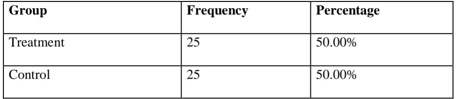
Group Frequency Percentage
Treatment 25 50.00%
Control 25 50.00%
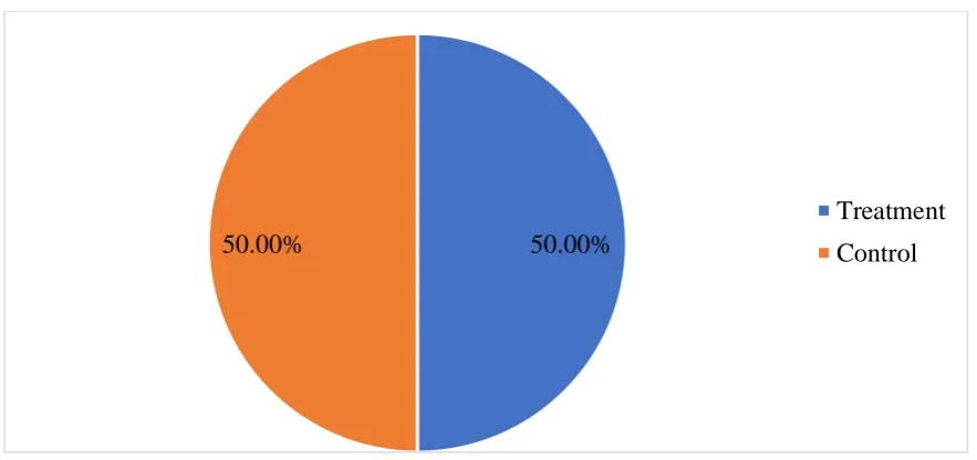
Among the study population, 50% people were in treatment group and 50% people were in control group. (table 1 & figure 1)
Figure :Bar chart of group distribution in study population (N=50) 50.00%
50.00%
Treatment
Control
50.00% 50.00%
0.00% 10.00% 20.00% 30.00% 40.00% 50.00% 60.00%
Treatment Control
P
er
cent
ag
e
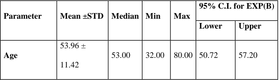
Table 2: Descriptive analysis for age in study population (N= 50)
Parameter Mean ±STD Median Min Max
95% C.I. for EXP(B) Lower Upper
Age
53.96 ± 11.42
53.00 32.00 80.00 50.72 57.20
[image:71.595.67.531.567.736.2]The mean of age was 53.96 years with a standard deviation of 11.42. The youngest person was 32 years old and the oldest person was 80-years-old.(table2)
Table 3: Descriptive analysis of age group in study population (N=50)
Age group Frequency Percentage
Up to 39 years 3 6.00%
40 to 49 years 15 30.00%
50 to 59 years 17 34.00%
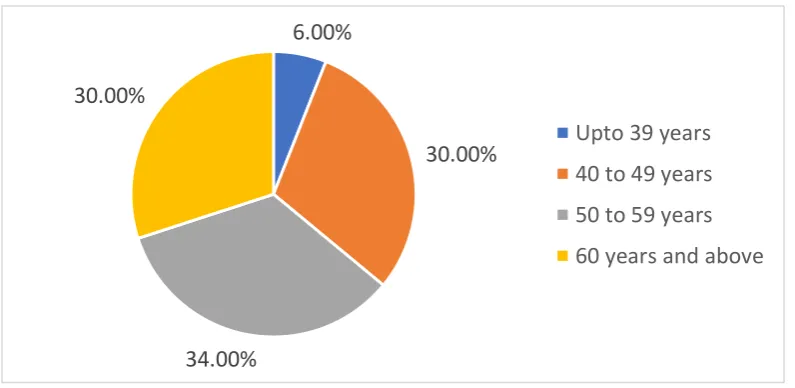
Majority (34%) subjects belonged to 50 to 59-years age group, followed by up to 40 to 49 and 60 and above-years age group (30%),up to 39 years age group (6%).(table 3 & figure 2)
Figure 2: Bar chart of Age group distribution in study population (N=50)
Figure:Pie chart of Age group distribution in study population (N=50) 6.00%
30.00% 34.00% 30.00%
0.00% 10.00% 20.00% 30.00% 40.00%
Upto 39 years 40 to 49 years 50 to 59 years 60 years and above P er cent ag e Age group 6.00% 30.00% 34.00% 30.00%
Upto 39 years
40 to 49 years
50 to 59 years
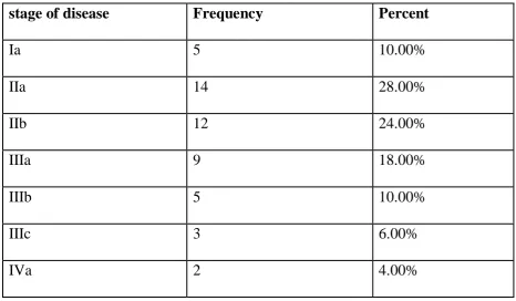
[image:72.595.71.469.527.721.2]Table 4: Descriptive analysis of stage of disease in study population (N=50)
stage of disease Frequency Percent
Ia 5 10.00%
IIa 14 28.00%
IIb 12 24.00%
IIIa 9 18.00%
IIIb 5 10.00%
IIIc 3 6.00%
IVa 2 4.00%
Figure 3: Pie chart of stage of disease distribution in study population (N=50)
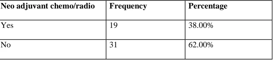
Table 5: Descriptive analysis of Neo adjuvant chemo/radio in study population (N=50)
Neo adjuvant chemo/radio Frequency Percentage
Yes 19 38.00%
No 31 62.00%
10.00%
28.00%
24.00% 18.00%
10.00% 6.00%
4.00%
ia
iia
iib
iiia
iiib
iiic
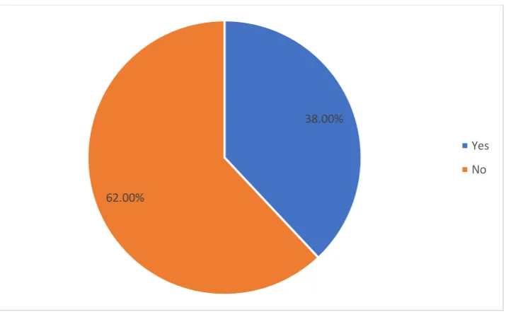
[image:74.595.71.532.604.706.2]Among the study population, 19(38%) were given neo adjuvant chemo/radio and 31(62%) no neo adjuvant chemo/radio was given.(table 5 & figure 4).
Figure 4: Pie chart of Neo adjuvant chemo/radio distribution in study population (N=50)
38.00%
62.00%
Yes
Figure: Bar chart of Neo adjuvant chemo/radio distribution in study population (N=50)
Table 6: Association of group with Neo adjuvant chemo/radio of study population (N=50)
Neo adjuvant chemo/radio
Group
Chi square P-value Treatment
(N=25)
Control (N=25)
Yes 9 (36%) 10 (40%)
0.085 0.771
No 16 (64%) 15 (60%)
38.00% 62.00% 0.00% 10.00% 20.00% 30.00% 40.00% 50.00% 60.00% 70.00% Yes No P er cent ag e
[image:76.595.67.531.543.706.2]Among the treatment group, 9 (36%) were given neo adjuvant chemo/radio and 16 (64%) were not given neo adjuvant chemo/radio. The proportion of neo adjuvant chemo/radio and no neo adjuvant chemo/radio was 10 (40%) and 15 (60%) in control group. The % of people who received neo adjuvant
chemo/radio was same in the treatment and control group.Thedifference
[image:77.595.70.530.412.741.2]between two study groups was statically not significant. (P-value 0.771).
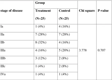
Table 7: Association of group with stage of disease of study population (N=50)
stage of disease
Group
Chi square P-value Treatment
(N=25)
Control (N=25)
Ia 1 (4%) 4 (16%)
3.778 0.707
IIa 7 (28%) 7 (28%)
IIb 8 (32%) 4 (16%)
IIIa 4 (16%) 5 (20%)
IIIb 3 (12%) 2 (8%)
IIIc 1 (4%) 2 (8%)
Among the treatment group, 1 (4%) were Ia stage of disease. The number of IIa, IIb, IIIa, IIIb, IIIc and IVa was 7 (28%), 8 (32%),4 (16%),3 (12%),1 (4%) and 1 (4%) respectively. The proportion of stage of disease Ia, IIa, IIb, IIIa, IIIb, IIIc and IVa was 4 (16%), 7 (28%),4 (16%), 5 (20%), 2 (8%), 2 (8%) and 1 (4%) in control group. The difference in the stage of disease between two study
[image:78.595.68.531.450.745.2]groups was statically not significant. (P-value 0.707). (table 7)
Table 8: Comparison of mean of volume of fluid drained in ml two study groups (N=50)
volume of fluid Drained in ml
Mean ±SD
P value Treatment
(N=25)
Control (N=25)
Day1 69.6 ± 25.65 77 ± 23.89 0.297
Day2 52 ± 24.79 65.2 ± 18.34 0.037
Day3 36.4 ± 20.64 55.4 ± 17.97 <0.001
Day4 28.2 ± 23.45 50.2 ± 19.34 <0.001
Day5 17.2 ± 15.82 37.4 ± 18.21 <0.001
Day7 4.8 ± 11.22 20 ± 18.26 <0.001
Day8 3.6 ± 9.95 12.6 ± 18.09 0.034
Day9 2.4 ± 8.31 7.6 ± 15.28 0.044
Day10 0.4 ± 2 3.8 ± 13.17 0.048
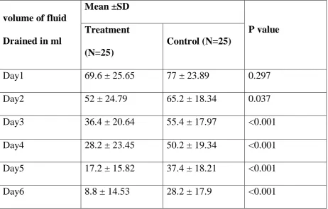
The mean volume of fluid drained in ml day1 of treatment group was 69.6 ± 25.65 and in control group 77 ± 23.89 it was, the difference in volume of fluid drained in ml day1 between the two groups was statistically not significant (p value 0.297). The mean volume of fluid drained in ml day 2 of treatment group was 52 ± 24.79 and in control group 65.2 ± 18.34 it was, the difference in volume of fluid drained in ml day 2 between the two groups was statistically significant (p value 0.037). The mean volume of fluid drained in ml day 3 of treatment group was 36.4 ± 20.64 and in control group 55.4 ± 17.97 it was, the difference in volume of fluid drained in ml day 3 between the two groups was statistically significant (p value <0.001). The mean volume of fluid drained in ml day 4 of treatment group was 28.2 ± 23.45 and in control group 50.2 ± 19.34 it was, the difference in volume of fluid drained in ml day 4 between the two groups was statistically significant (p value <0.001). The mean volume of fluid drained in ml day 5 of treatment group was 17.2 ± 15.82 and in control group 37.4 ± 18.21 it was, the difference in volume of fluid drained in ml day 5
control group 28.2 ± 17.9 it was, the difference in volume of fluid drained in ml day 6 between the two groups was statistically significant (p value <0.001). The mean volume of fluid drained in ml day 7 of treatment group was 4.8 ± 11.22 and in control group 20 ± 18.26 it was, the difference in volume of fluid drained in ml day 7 between the two groups was statistically significant (p value
Figure 5: Comparison of volume of fluid Drained in ml across study groups (N=50) 69.6 52 36.4 28.2 17.2 8.8
4.8 3.6 2.4
0.4 77 65.2 55.4 50.2 37.4 28.2 20 12.6 7.6 3.8 0 10 20 30 40 50 60 70 80 90
Day1 Day2 Day3 Day4 Day5 Day6 Day7 Day8 Day9 Day10
Mea
n
stage of disease
Treatment Control 69.6 52 36.4 28.2 17.2 8.8
4.8 3.6 2.4
0.4 77 65.2 55.4 50.2 37.4 28.2 20 12.6 7.6 3.8 0 10 20 30 40 50 60 70 80 90
Day1 Day2 Day3 Day4 Day5 Day6 Day7 Day8 Day9 Day10
Me
an
volume of fluid Drained in ml
Table 9: Comparison of Day of removal of drainage tub and study group (N=50)
Group Day of removal of
drainage tub Median (IQR)
P value (Mann-Whitney U-Test)
Treatment 5(5 to 6)
<0.001
Control 8 ( 6 to 9)
DISCUSSION
A randomized control trial was undertaken in government Stanley medical college hospital from November 2016 to October 2017 among 50 female carcinoma breast patients who have undergone modified radicle mastectomy between the age group of 30 years to 80 years to determine the role of
octreotide in post mastectomy lymphorrhoea.the patients were randomly
allotted to treatment and control group by computer generated random number AGE DISTRIBUTION OF STUDY POPULATION
The mean of age was53.96 years with a standard deviation of 11.42. The youngest person was 32 years old and the oldest person was 80-years-old. Majority (34%) subjects belonged to 50 to 59-years age group, followed by up to 40 to 49 and 60 and above-years age group (30%),up to 39 years age group (6%).THERE WAS NO STATISTICAL DIFFERENCE IN THE AGE
DISTRIBUTION BETWEEN THE THE TREATMENT GROUP AND CONTROL
GROUP.
Age group Frequency Percentage
Up to 39 years 3 6.00%
40 to 49 years 15 30.00%
50 to 59 years 17 34.00%
DISTRIBUTION OF THE STAGE OF THE DISEASE AMONG THE STUDY POPULATION
[image:85.595.70.529.406.736.2]Among the treatment group, 1 (4%) were I a stage of disease. The number of Ia , IIb , IIIa, IIIb, IIIc and IVa was 7 (28%), 8 (32%),4 (16%),3 (12%),1 (4%) and 1 (4%) respectively. The proportion of stage of disease Ia , IIa, II b, III a, III b, III c and IV a was 4 (16%), 7 (28%),4 (16%), 5 (20%), 2 (8%), 2 (8%) and 1 (4%) in control group. The difference in the stage of disease between two study groups was statically not significant. (P-value 0.707). (table 7)
Table 7: Association of group with stage of disease of study population
stage of disease
Group
Chi square P-value Treatment
(N=25)
Control (N=25)
Ia 1 (4%) 4 (16%)
3.778 0.707
IIa 7 (28%) 7 (28%)
IIb 8 (32%) 4 (16%)
IIIa 4 (16%) 5 (20%)
IIIb 3 (12%) 2 (8%)
IIIc 1 (4%) 2 (8%)
NEOADJUVANT CHEMO/RADIO THERAPY IN TREATMENT AND CONTROL GROUP
[image:86.595.70.533.510.678.2]Among the treatment group, 9 (36%) were given neo adjuvant chemo/radio and 16 (64%) were not given neo adjuvant chemo/radio. The proportion of neo adjuvant chemo/radio and no neo adjuvant chemo/radio was 10 (40%) and 15 (60%) in control group. The % of people who received neo adjuvant chemo/radio was same in the treatment and control group. The difference between two study groups was statically not significant. (P-value 0.771).
Table 6: Association of group with Neo adjuvant chemo/radio of study population (N=50)
Neo adjuvant chemo/radio
Group
Chi square P-value Treatment
(N=25)
Control (N=25)
Yes 9 (36%) 10 (40%)
0.085 0.771
AMOUNT OF FLUID DRAINED IN THE TREATMENT AND CONTROL GROUP FROM POD 1 TO POD 10
DAY 1 : The mean volume of fluid drained in ml day1 of treatment group was 69.6 ± 25.65 and in control group 77 ± 23.89 it was, the difference in volume of fluid drained in ml day1 between the two groups was statistically not
significant (p value 0.297).
DAY 2 : The mean volume of fluid drained in ml day 2 of treatment group was 52 ± 24.79 and in control group 65.2 ± 18.34 it was, the difference in volume of fluid drained in ml day 2 between the two groups was statistically
significant.(p valu0.037). DAY 3 : The mean volume of fluid drained in ml day 3 of treatment group was
36.4 ± 20.64 and in control group 55.4 ± 17.97 it was, the difference in volume of fluid drained in ml day 3 between the two groups was statistically significant (p value <0.001) DAY 4 The mean volume of fluid drained in ml day 4 of treatment group was 28.2 ± 23.45 and in control group 50.2 ± 19.34 it was, the difference in volume of fluid drained in ml day 4
DAY 5 : The mean volume of fluid drained in ml day 5 of treatment group was 17.2 ± 15.82 and in control between the two groups was statistically significant
group 37.4 ± 18.21 it was, the difference in volume of fluid drained in ml day 5
between the two groups was statistically significant (p value <0.001). DAY 6 : The mean volume of fluid drained in ml day 6 of treatment group was
8.8 ± 14.53 and in control group 28.2 ± 17.9 it was, the difference in volume of fluid drained in ml day 6 between the two groups was statistically significant (p
value <0.001). DAY 7 : The mean volume of fluid drained in ml day 7 of treatment group was
4.8 ± 11.22 and in control group 20 ± 18.26 it was, the difference in volume of fluid drained in ml day 7 between the two groups was statistically significant (p value <0.001). DAY 8 : The mean volume of fluid drained in ml day 8 of treatment group was 3.6 ± 9.95 and in control group 12.6 ± 18.09 it was, the difference in volume of fluid drained in ml day 8 between the two groups was statistically significant (p
value 0.034). DAY 9 : The mean volume of fluid drained in ml day 9 of treatment group was
2.4 ± 8.31 and in control group 7.6 ± 15.28 it was, the difference in volume of fluid drained in ml day 9 between the two groups was statistically significant (p
value 0.142). DAY 10 : The mean volume of fluid drained in ml day 10 of treatment group
Table 8: Comparison of mean of volume of fluid drained in ml two study groups (N=50)
volume of fluid Drained in ml
Mean ±SD
P value Treatment
(N=25)
Control (N=25)
Day1 69.6 ± 25.65 77 ± 23.89 0.297
Day2 52 ± 24.79 65.2 ± 18.34 0.037
Day3 36.4 ± 20.64 55.4 ± 17.97 <0.001
Day4 28.2 ± 23.45 50.2 ± 19.34 <0.001
Day5 17.2 ± 15.82 37.4 ± 18.21 <0.001
Day6 8.8 ± 14.53 28.2 ± 17.9 <0.001
Day7 4.8 ± 11.22 20 ± 18.26 <0.001
Day8 3.6 ± 9.95 12.6 ± 18.09 0.034
Day9 2.4 ± 8.31 7.6 ± 15.28 0.142
DAY OF REMOVAL OF DRAIN IN CONTROL AND TREATMENT GROUP
[image:90.595.68.529.351.517.2]Among the day of removal of drainage tub median was 5 (IQR 5 to 6) were in treatment group and 5 (IQR 5 to 6) were in control group. The difference day of removal of drainage tub median between the two groups was statistically significant (p value <0.001).
Table 9: Comparison of Day of removal of drainage tub and study group (N=50)
Group Day of removal of
drainage tub Median (IQR)
P value (Mann-Whitney U-Test)
Treatment 5(5 to 6)
<0.001
SIMILAR STUDY:
A prospective randomised controlled trail done by paolo carcoforo et al followed 261 patients with various stages of breast cancer who underwent axillary dissection for seven years of these 136 was taken as control
group and 125 as treatment group.The treatment group was given 0.1 mg octreotide subcutaneous for thrice daily for 5 days
control treatment P value
Quantity of lymphorrhea
94.6±19 cc per day
65.4 ±21.1cc per day
<o.ooo1
Duration of lymphorrhoea
CONCLUSION
From this study it is evident that octreotide and its analogues have a promising role in the treatment of post mastectomy lymphorrea. Since lymphorrhoea leads to chronic lymphoedema which inturn predisposes to lymphangiosarcoma,it is safe to assume that the use of octreotide also reduces the risk of incidence of this malignancy .similarly octreotide can also be tried in other conditions were lymphorrhoea is troublesome like in inguinal lymph node dissection ,peripheral vascular surgery.Though octreotide is a promising drug several more clinical trails have to be undertaken before bringing it into mainstream practice for better understanding of the drug’s pharmacodynamics and its rare but serious side effects.
1. Epidemiology of breast cancer in Indian women
Shreshtha Malvia, Sarangadhara Appalaraju Bagadi, Uma S. Dubey,Sunita Saxena
2. Seroma Formation after Breast Cancer Surgery: What We Have Learned in the Last Two Decades
Vivek Srivastava, Somprakas Basu, and Vijay Kumar Shukla
3. Incidence of unilateral arm lymphoedema after breast cancer: a systematic review and meta-analysis.
DiSipio T1, Rye S, Newman B, Hayes S.
4. Somatostatin
Vicente Martinez, in Handbook of Biologically Active Peptides (Second Edition), 2013
5. Breast cancer: Symptoms, risk factors, and treatment Last updated Mon 17 July 2017
By Christian Nordqvist
Reviewed by Christina Chun, MPH 6. Axillary nodes
7. "The Lymphatic System". Human Anatomy (Gray's Anatomy). Retrieved 2012-10-12.
8. Modified Radical Mastectomy Updated: Jul 03, 2017
Author: Nafisa K Kuwajerwala, MD; Chief Editor: Erik D Schraga, MD
9. The pathophysiology of lymphedema Peter S. Mortimer M.D.
10.Complications of Lymphedema
BY JOACHIM ZUTHER, ON JANUARY 2ND, 2014
11.Octreotide, Sandostatin analogue
Pharmacy Author: Omudhome Ogbru, PharmD Medical and Pharmacy Editor: Jay W. Marks, MD
12. ROLE OF OCTREOTIDE IN CONTROL OF LYMPHORRHEA AFTER AXILLARY NODE DISSECTION IN MASTECTOMY OPERATIONS - A RANDOMIZED CONTROLLED STUDY
Proforma
Name: Age: Sex: IP No.
RFT –
Urea:
Creatinine:
RBS
LFT –
Total Bilurubin:
SGOT:
SGPT:
ALP:
Sr. Amylase
Sr. Lipase
TFT
FT3
FT4
Stage of Ca Breast:
Neoadjuvant Chemo/Radiotherapy:
Quantity of Lymph Drained:
Day 1: Day 6:
Day 2: Day 7:
Day 3: Day 8:
Day 4: Day 9:
Day 5: Day 10: