R E S E A R C H A R T I C L E
Open Access
Natural history of colonization with
methicillin-resistant
Staphylococcus aureus
(MRSA) and vancomycin-resistant
Enterococcus
(VRE): a systematic review
Erica S Shenoy
1*, Molly L Paras
2, Farzad Noubary
3, Rochelle P Walensky
4and David C Hooper
5Abstract
Background:No published systematic reviews have assessed the natural history of colonization with
methicillin-resistantStaphylococcus aureus(MRSA) or vancomycin-resistantEnterococcus(VRE). Time to clearance of colonization has important implications for patient care and infection control policy.
Methods:We performed parallel searches in OVID Medline for studies that reported the time to documented clearance of MRSA and VRE colonization in the absence of treatment, published between January 1990 and July 2012.
Results:For MRSA, we screened 982 articles, identified 16 eligible studies (13 observational studies and 3 randomized controlled trials), for a total of 1,804 non-duplicated subjects. For VRE, we screened 284 articles, identified 13 eligible studies (12 observational studies and 1 randomized controlled trial), for a total of 1,936 non-duplicated subjects. Studies reported varying definitions of clearance of colonization; no study reported time of initial colonization. Studies varied in the frequency of sampling, assays used for sampling, and follow-up period. The median duration of total follow-up was 38 weeks for MRSA and 25 weeks for VRE. Based on pooled analyses, the model-estimated median time to clearance was 88 weeks after documented colonization for MRSA-colonized patients and 26 weeks for VRE-colonized patients. In a secondary analysis, clearance rates for MRSA and VRE were compared by restricting the duration of follow-up for the MRSA studies to the maximum observed time point for VRE studies (43 weeks). With this restriction, the model-fitted median time to documented clearance for MRSA would occur at 41 weeks after documented colonization, demonstrating the sensitivity of the pooled estimate to length of study follow-up.
Conclusions:Few available studies report the natural history of MRSA and VRE colonization. Lack of a consistent definition of clearance, uncertainty regarding the time of initial colonization, variation in frequency of sampling for persistent colonization, assays employed and variation in duration of follow-up are limitations of the existing published literature. The heterogeneity of study characteristics limits interpretation of pooled estimates of time to clearance, however, studies included in this review suggest an increase in documented clearance over time, a result which is sensitive to duration of follow-up.
Keywords:MRSA, VRE, Colonization, Carrier, Contact precautions
* Correspondence:eshenoy@partners.org
1Division of Infectious Diseases, Infection Control Unit and Medical Practice
Evaluation Center, Massachusetts General Hospital and Harvard Medical School, Boston, MA, USA
Full list of author information is available at the end of the article
© 2014 Shenoy et al.; licensee BioMed Central Ltd. This is an Open Access article distributed under the terms of the Creative Commons Attribution License (http://creativecommons.org/licenses/by/2.0), which permits unrestricted use, distribution, and reproduction in any medium, provided the original work is properly credited. The Creative Commons Public Domain Dedication waiver (http://creativecommons.org/publicdomain/zero/1.0/) applies to the data made available in this article, unless otherwise stated.
Shenoyet al. BMC Infectious Diseases2014,14:177
Background
Methicillin-resistantStaphylococcus aureus(MRSA) and vancomycin-resistantEnterococcus(VRE) are endemic in hospital settings and long-term care facilities (LTCF), and the prevalence of colonization is increasing [1-4]. The growing pools of colonized, and therefore isolated pa-tients, impact patient care and burden the healthcare sys-tem [5-7]. The duration of MRSA and VRE colonization has previously been assessed in mostly small studies. Thus, pooling of these data might provide a better under-standing of the natural history of colonization and the timing of clearance and thereby inform clinical care and public policy. We performed a systematic review of ran-domized controlled trials and observational studies that followed patients with a history of MRSA and VRE colonization and assessed study characteristics and study quality. In the absence of individual data, we pooled study-level data to calculate estimates of time to clearance of colonization.
Methods Search strategy
We conducted two separate computerized searches in OVID Medline to identify relevant English-language studies including adults and published between January 1990 and July 2012. Index searches included MeSH terms for MRSA: “methicillin-resistant Staphylococcus aureus” or “methicillin resistance” and “colonization” or “carrier state”. For VRE, search terms included”vancomycin resist-ance”and“Enterococcus faecalis”or“Enterococcus”or“ En-terococcus faecium” and “colonization” or “carrier state”. Inclusion criteria required that a study define a population of MRSA or VRE carriers and subsequently perform at least one screening for colonization status in the absence of treatment or decolonization therapy for MRSA or VRE. Included studies provided the number of subjects cleared within a defined time period. The searches and subse-quent study selection were conducted separately for MRSA and VRE.
Study selection
Two authors (ESS and MLP) independently reviewed the abstracts of publications identified by the two searches. Publications that addressed the length of time subjects with a history of infection or colonization remained col-onized or included evidence that patients were followed over time underwent full-text review for determination of inclusion and data extraction from those that met inclu-sion criteria. Studies with no abstract or for which it was not possible to determine if the publication contained data meeting inclusion criteria also underwent full-text review.
Colonization in both study selections was defined as having a positive culture or nucleic acid amplification assay (for MRSA or VRE) without evidence for active
infection. Studies were required to report on screening from at least one anatomical site; any anatomical site for screening was permitted for inclusion. While clearance was defined by each study individually, at least one microbiological result supporting clearance was required for inclusion. For studies reporting more than one time-point of documented clearance, the latest time-time-point was included in the analysis. A third author (DCH) me-diated any differences in interpretations regarding inclu-sion/exclusion.
Data extraction
For studies meeting inclusion criteria for both searches, the following data were extracted: authors, study design, country, years of study, subject description, anatomic screening site, screening method (i.e., culture, molecular diagnostics), follow-up period (weeks), total number of subjects, number of subjects lost to follow-up, study-defined clearance, time to documented clearance for those who cleared (weeks) and the proportion of subjects with documented clearance. The role of antibiotic exposure with respect to the duration of colonization was assessed, if documented. Data were entered separately for the MRSA and VRE studies into standardized forms and veri-fied in duplicate for consistency and accuracy. For VRE, it was noted if studies made a distinction betweenE. faecalis andE. faecium.
Statistical methods
For both MRSA and VRE analyses, we collected informa-tion about the total sample size and number or percentage of subjects with documented clearance of colonization and the corresponding time after documented colonization at which the assessment of clearance was made for each in-cluded study. For studies that did not provide details on loss to follow-up, the number of clearance events was calculated as the product of the percent clearance times the total sample size. We conservatively assumed that all reported clearance events happened at the time-point reported by the study and not before.
For both MRSA and VRE analyses, we used Greenwood’s formula to estimate the standard error of each study’s re-ported decolonization rate [8]. This approach allowed for the generation of MRSA and VRE “champagne plots” of the reported rates of colonization clearance, where the size of each study’s data point is inversely proportional to its standard error. For the MRSA and VRE studies, we then separately fitted logistic regression models to assess the relationship between the proportion of patients with documented spontaneous clearance and the time since documented colonization. In parallel sensitivity ana-lyses, we examined the relative influence of each study on the estimated median time to clearance. To do so, we used a jackknife method whereby each study, within
Shenoyet al. BMC Infectious Diseases2014,14:177 Page 2 of 13
the MRSA and VRE analyses independently, was sequen-tially removed from the dataset, and the median time to documented clearance was recalculated. Given the de-pendence of the analysis on the duration of follow-up, in a second set of sensitivity analyses, we restricted the MRSA studies to include only those that reported clearance on or before the maximum observed follow-up time for the in-cluded VRE studies. We then subsequently recalculated the median time to documented MRSA clearance with this restriction in place.
Quality assessment
Cohort studies were assessed for quality using a modifica-tion of the Newcastle-Ottawa Quality Assessment Scale (NOS) developed specifically for the purposes of this re-view. The assessment was performed independently (ESS, MLP), with any disagreements resolved by a third author (DCH). Beyond meeting inclusion criteria for the review, the quality of randomized controlled trials included in the analyses was not assessed.
The standard NOS for cohort studies contains eight questions that assess study quality based on subject selec-tion, comparison and outcome validation. This assessment evaluates studies on a maximum nine-point scale based on the representativeness of the exposed and non-exposed cohorts, ascertainment of exposure, comparability of the cohorts, assessment of outcome and appropriate follow-up time and loss to follow-follow-up [9]. In the modified scale, we similarly addressed these quality measures. Selection and comparability quality was assessed by a demographic comparison between colonized and cleared subjects. As-certainment of exposure and outcome was determined by recording whether colonization (exposure) and clearance of colonization (outcome) were determined using standard microbiological methods, and whether the study provided information about the length of time to documented clear-ance. Finally, the standard NOS records how long subjects were followed for outcomes assessment and loss to follow-up; in the modified NOS, we used a cutoff of three or more months of follow-up, and loss to follow-up of less than 30%. Of note, in the standard NOS, one of the key quality measures is demonstration that the outcome of interest was not present at the start of the study. For this systematic review, the outcome of interest was clearance of colonization. By definition, subjects who were not colonized at the start of the study were excluded since all subjects needed to be colonized in order to achieve the outcome of interest.
Results MRSA
MRSA study identification
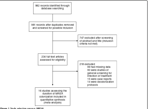
The MRSA search criteria identified 981 non-duplicate publications. Full-text review was completed for 234
publications, and each was reviewed in detail for final determination of inclusion and for data extraction (Figure 1). This procedure resulted in a total of 16 studies with 1,804 non-duplicated subjects included in the review, and 13 cohort studies included in the quality assessment (Figure 1) [10].
MRSA study characteristics
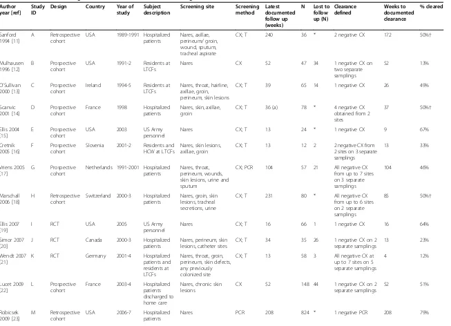
The 16 studies were diverse in design, geographic site, enrollment period, anatomic screening site, and patient location (Table 1) [11-26]. The median duration of total follow-up was 38 weeks.
For studies that included hospitalized patients, data were more frequently not provided about the subjects’residence prior to admission (i.e., home versus facility). Fourteen of 16 studies sampled subjects at least three months after documented colonization. For loss to follow-up, seven studies had less than 30%, four had more than 30%, and five provided no information.
MRSA clearance rates
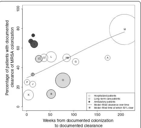
Reported clearance rates ranged from 12% [21] to 79% [23]. The time to observed clearance ranged from one [26] to 208 weeks [23]. A plot of the percentage of sub-jects documented to have cleared MRSA colonization over time demonstrates a trend toward the majority of subjects clearing colonization over the follow-up periods reported (Figure 2). Using logistic regression, we found that 50% of patients cleared colonization at 88 weeks after initial documentation of colonization. At one, two, three and four years after initial determination of MRSA colonization, the model-estimated proportions of sub-jects with documented clearance of colonization were 41, 54, 66, and 77%, respectively. Comparing the patient populations represented, long-term care, hospitalized, and ambulatory, at 26 weeks after initial documentation of colonization, the model-fitted percentages of those clearing were significantly different at 22, 36, and 68%, respectively (P < 0.0001).
The majority of studies did not provide enough infor-mation to determine if the cohorts of colonized or cleared subjects were similar. However, three studies [14,22,25] did provide these data, and in general the demographic characteristics of the subjects in the colonized and cleared groups from each cohort were similar, with some notable exceptions. Scanvic reported residence at another healthcare institution and break in the skin to be significantly associated with persistent carriage (32% vs 11% and 67.7% vs. 28%, respectively). Lucet found as-sistance with activities of daily living (ADLs) to be sig-nificantly different between the groups (57.5% vs. 49.3%, respectively). Manzur reported the presence of decubi-tus ulcers to be a risk factor for persistent colonization (27.5% vs. 13.7%, respectively).
Shenoyet al. BMC Infectious Diseases2014,14:177 Page 3 of 13
The jackknife sensitivity analysis revealed that, for all but two studies ([25], Study“O”and [23], Study“M”), the estimated median time to documented clearance would change only minimally if each study was excluded. The ex-clusion of Manzur [25] resulted in a model-fitted median time to clearance of 68 weeks. Separately, the exclusion of Robicsek [23] resulted in a model-fitted median clearance time >172 weeks after documented colonization.
The impact of antibiotic exposure on persistence of colonization was either not reported or found to have no significant association with duration of colonization for the majority of studies. Only one study reported that anti-biotic exposure in colonized patients was significantly associated with persistence of colonization [18].
MRSA study quality
The 13 cohort studies were assessed using the modified NOS. Of these, only Lucet [22] met all quality criteria based on the modified NOS. The remainder of the studies were missing quality criteria in either comparability, ap-propriate time to follow-up or loss of follow-up; overall, these studies were of moderate quality, with the mean
score of 4.8/6 (range 3–6). As the NOS results lacked vari-ation and were applicable only to cohort studies, they were not used as weights in the pooled analyses.
VRE
VRE study identification
The search criteria identified 278 non-duplicate screened publications. Full-text review was completed for 108 pub-lications, all of which were reviewed in detail for final determination of inclusion and data extraction. This procedure resulted in 13 studies included in the review, and 12 cohort studies included in the quality assessment (Figure 3) [10].
VRE study characteristics
The 13 included studies, including a total of 1,936 non-duplicated patients varied in design, geographic site, en-rollment period, and anatomic screening site; the vast majority included hospitalized patients (Table 2) [27-39]. The median duration of total follow-up was 25 weeks. Seven of the 13 studies reported on E. faecium alone,
982 records identified through database searching
981 records after duplicates removed and screened for possible inclusion
747 excluded after screening of abstract and title (inclusion criteria not met)
234 full-text articles assessed for eligibility
218 excluded
99 had missing data 90 were studies of general screening for infection or treatment 15 were case reports 14 were decolonization protocols
16 studies assessing the duration of MRSA colonization included in
[image:4.595.62.540.89.441.2]quantitative synthesis (meta-analysis)
Figure 1Study selection process (MRSA).
Shenoyet al. BMC Infectious Diseases2014,14:177 Page 4 of 13
Table 1 Studies documenting duration of MRSA colonization meeting inclusion criteria
Author year [ref]
Study ID
Design Country Year of study
Subject description
Screening site Screening method
Latest documented follow up (weeks)
N Lost to follow up (N) Clearance defined Weeks to documented clearance % cleared Sanford 1994 [11]
A Retrospective
cohort
USA 1989-1991 Hospitalized
patients
Nares, axillae, perineum/ groin, wound, sputum, tracheal aspirate
CX; T 240 36 * 2 negative CX 172 50%†
Mulhausen 1996 [12]
B Prospective
cohort
USA 1991-2 Residents at
LTCFs
Nares CX 52 47 34 1 negative CX on
two separate samplings
52 13%
O’Sullivan 2000 [13]
C Prospective
cohort
Ireland 1994-5 Residents at
LTCFs
Nares, throat, hairline, axillae, groin, perineum, skin lesions
CX; T 39 65 14 1 negative CX 26 49%
Scanvic 2001 [14]
D Prospective
cohort
France 1998 Hospitalized
patients
Nares, skin, axillae, groin
CX; T 36 (a) 78 * 4 negative CX
obtained from 2 sites
37 50%†
Ellis 2004 [15]
E Prospective
cohort
USA 2003 US Army
personnel
Nares CX; T 13 24 * 1 negative CX 9 67%
Cretnik 2005 [16]
F Prospective
cohort
Slovenia 2001-2 Residents and
HCW at LTCFs
Nares, skin lesions, axillae, groin
CX; T 13 12 2 2 negative CX from
2 sites on 3 separate samplings
13 33%
Vriens 2005 [17]
G Prospective
cohort
Netherlands 1991-2001 Hospitalized patients
Nares, throat, perineum, wounds, skin lesions, urine and sputum
CX; PCR 104 57 21 All negative CX
from up to 7 sites on 3 separate samplings
104 46%
Marschall 2006 [18]
H Retrospective
cohort
Switzerland 2000-3 Hospitalized patients
Nares, groin, skin lesions, tracheal secretions, urine
CX; T 231 80 * All negative CX
from up to 6 sites on 2 separate samplings
85 50%†
Ellis 2007 [19]
I RCT USA 2005 US Army
personnel
Nares CX; T 16 66 1 1 negative CX 16 64%
Simor 2007 [20]
J RCT Canada 2000-3 Hospitalized
patients
Nares, perineum, skin lesions, catheter sites
CX; T 34 35 26 1 negative CX on 2
separate samplings
13 23%
Wendt 2007 [21]
K RCT Germany 2001-4 Hospitalized
patients and residents at LTCFs
Nares, throat, groin, perineum, skin defects, any previously colonized site
CX; T 13 58 3 All negative CX at
up to 7 sites on 5 separate samplings
4 12%
Lucet 2009 [22]
L Prospective
cohort
France 2003-4 Hospitalized
patients discharged to home care
Nares, chronic skin lesions
CX 52 148 44 1 negative CX on 2
separate samplings
52 51%
Robicsek 2009 [23]
M Retrospective
cohort
USA 2006-7 Hospitalized
patients
Nares PCR 208 824 * 1 negative PCR 208 79%
Table 1 Studies documenting duration of MRSA colonization meeting inclusion criteria(Continued)
Lautenbach 2010 [24]
N Prospective
cohort
USA 2008 Ambulatory
patients and household contacts
Nares, axilla, throat; groin and perineum
CX; PCR; T 14 11 0 All negative cultures
from up to 5 sites on 6 separate collections
14 73%
Manzur 2010 [25]
O Prospective
cohort
Spain 2005-7 Residents at
LTCFs
Nares, decubitus ulcers CX; T 77 231 104 1 negative CX on 2
separate collections
77 27%
Van Velzen 2011 [26]
P Retrospective
cohort
Scotland 2010 Hospitalized
patients
Nares, perineum, axillae, throat, wounds and devices
CX; PCR; T 4 32 0 All negative cultures
from up to 6 sites on 2 separate occasions
1 25%
(a) Follow up at least 13 weeks since hospitalization; median clearance at 37 weeks reported; mean time since hospital discharge 36 weeks. *Not documented;†Kaplan Meier estimates which do not provide information on those lost to follow up.
HCW: health care worker; LTCF: long term care facility; MRSA: methicillin resistantStaphylococcus aureus; N: number; CX: culture; T: typing of strain performed; PCR: polymerase chain reaction; RCT: randomized controlled trial.
Shenoy
et
al.
BMC
Infectious
Diseases
2014,
14
:177
Page
6
o
f
1
3
http://ww
w.biomedce
ntral.com/1
while six of 13 made no distinction between E. faecalis andE. faecium.
VRE clearance rates
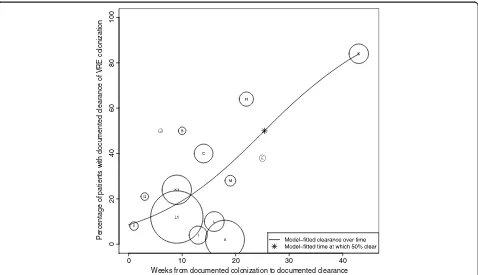
Reported time to clearance ranged from one [32] to 43 weeks [37], with documented clearance rates of zero [30] to 84% [37]. A plot of the percentage of subjects doc-umented to have cleared VRE colonization over time demonstrates a trend toward the majority of subjects clearing colonization during the period of observation (Figure 4). Using logistic regression, we found that 50% of subjects cleared colonization at 25 weeks after initial documentation of colonization. At 10, 20, 30 and 40 weeks after initial determination of VRE colonization, the model estimated that 19, 38, 61, and 80% of subjects had docu-mented clearance of colonization, respectively. Since the vast majority of subjects were hospitalized patients, we were unable to assess the effect of patient population type (inpatient versus LTCF resident) on time to documented clearance.
Only two of the studies included, Park [38] and Yoon [39] provided sufficient demographic data to formally evaluate variables associated with either persistence or clearance of colonization. Park [38] found three variables, age (odds ratio [OR]: 0.99; P = 0.05), duration of glycopep-tide use prior to VRE positivity (OR: 2.16; P = 0.003), and
length of hospital stay (OR 1.01; P = 0.001) associated with subjects having three consecutive negative rectal cultures. Mean duration of glycopetide use was reported with respect to hemodialysis; patients on chronic HD were observed to be exposed to 12.7 days while patients on non-chronic HD were observed to be exposed to 4.5 days (P = 0.001). Yoon [39] compared subjects who cleared colonization early (< three weeks) to those who cleared later (≥five weeks) and found that they differed on several characteristics. The early group was more likely to be younger (P = 0.01) and to have a shorter length of stay in an ICU (P = 0.03). This group was also less likely to have had prior exposure to medical devices, including central venous catheters and endotracheal intubation (P = 0.04 and P = 0.04, respectively), and less likely to have received selected antibiotics after colonization, including carbapen-ems (P = 0.01), vancomycin (P < 0.001), or fluoroquinolones (P = 0.04). In multivariable logistic regression analysis, they found that vancomycin use after VREF colonization was significantly associated with prolonged carriage (OR 4.1; P = 0.02).
Unlike for the MRSA analysis, the VRE jackknife sensi-tivity analysis revealed that the estimated median time to documented clearance did not appear substantially differ-ent with the exclusion of any one study.
Antibiotic exposure was reported to be significantly and positively associated with longer duration of carriage in several of the studies [28,34,39], however others found no such association [33,36,38]. The remaining studies did not report on the relationship.
VRE study quality
The 12 cohort studies were assessed using the modified NOS. No study fulfilled all quality criteria for the modified NOS; the majority were of moderate quality with the mean score of 4.6/6 (range 3–5). As the NOS results lacked variation and were applicable only to cohort studies, they were not used as weights in the pooled analyses.
Comparison of MRSA and VRE pooled clearance rates
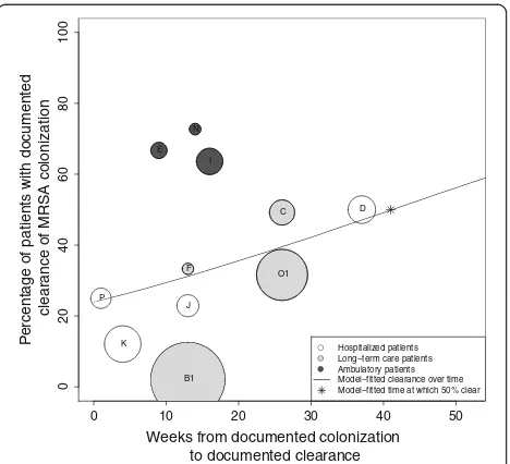
Clearance rates for MRSA and VRE were compared by restricting the time to follow-up for the MRSA studies to the maximum observed time point for VRE studies (43 weeks). Figure 5 shows that with this restriction, which results in the exclusion of five studies (A, G, H, L, M) and the inclusion of earlier-reported time intervals from two studies (Manzur, “O1” and Mulhausen, “B1”), the model-fitted median time to documented clearance would occur at 41 weeks.
Discussion
We reviewed the published literature on the natural his-tory of MRSA and VRE colonization. There is substantial
0 50 100 150 200
0
2
04
06
08
0
1
0
0
Weeks from documented colonization to documented clearance
P
e
rcentage of patients with documented clear
ance of MRSA colonization
P E
K N
I
F
J
C D
B
L H
O
M
A G
[image:7.595.58.291.88.300.2]Hospitalized patients Long−term care patients Ambulatory patients Model−fitted clearance over time Model−fitted time at which 50% clear
Figure 2Analysis of median time to documented clearance of MRSA colonization.The X-axis represents time (in weeks) from documented colonization and the Y-axis represents the proportion of patients with documented clearance of colonization. This proportion included the initial subject population minus those lost to follow-up. Each circle represents a single study (A-P). The size of the circle is inversely proportional to its standard error. The line represents the model-fitted clearance over time, with 50% of subjects clearing MRSA at 88 weeks from time of documented colonization (*).
Shenoyet al. BMC Infectious Diseases2014,14:177 Page 7 of 13
heterogeneity among the included studies. This hetero-geneity both identifies a clear need for further investi-gation and tempers our interpretation of the pooled estimates of time to clearance of colonization. Based on the studies meeting our inclusion criteria, our system-atic review demonstrates that persistent colonization decreases over time, with clearance of colonization in half of patients at 88 weeks for MRSA and 26 weeks for VRE.
While the weight of the existing data, and clinical experience, suggest that clearance of MRSA and VRE colonization increases over time, precision around the time to clearance is not possible due to the major limita-tions of the studies in this domain. These limitalimita-tions not only hinder our interpretation of the pooled findings, but also highlight the need for additional studies. Lack of a consistent definition of clearance, uncertainty re-garding the time of initial colonization, variation in fre-quency of sampling for persistent colonization, and variation in duration of follow-up and loss to follow-up all impose substantial constraints on our interpretation of the median time to clearance.
Because subjects are not screened prospectively and continuously, the initial date of colonization is assumed to be the time MRSA or VRE were discovered and docu-mented (based on either a clinical infection or a surveil-lance screening). There is almost certainly variation in the lag time between actual colonization and the identification of a subject as being colonized. Under these inevitable circumstances, calculations of duration of colonization may underestimate the true duration of colonization and the pattern of clearance. On the other hand, if the time interval between initial documentation of colonization and re-screening is prolonged, duration of colonization may also be overestimated. A striking observation from the combined reviews is the relatively short time to clearance for VRE, as compared to MRSA. The longer median time to clearance estimated for MRSA is in part a reflection of the length of follow-up, as evidenced by the reduction from 88 weeks to 41 weeks observed when follow-up was restricted to the same duration as the VRE studies.
We did not impose a universal definition of clearance as a prerequisite for inclusion in our review, and across
284 records identified through database searching
278 records after duplicates removed and screened for possible inclusion
170 excluded after screening of abstract and title (inclusion criteria not met)
108 full-text articles assessed for eligibility
95 excluded
39 had missing data 50 were studies of general screening for infection or treatment 4 were decolonization protocols
1 were case reports 1 unrelated to VRE colonization 13 studies assessing the
duration of VRE colonization included in
[image:8.595.57.543.88.440.2]quantitative synthesis (meta-analysis)
Figure 3Study selection process (VRE).
Shenoyet al. BMC Infectious Diseases2014,14:177 Page 8 of 13
Table 2 Studies documenting duration of VRE colonization meeting inclusion criteria
Author year [ref]
VRE Study ID
Design Country Year of study Subject description Screening site Screening method Latest documented follow up (weeks)
N Lost to follow up (N)
Clearance defined Weeks to documented clearance
% cleared
Montecalvo 1995 [27]
VREF A Prospective
cohort
USA 1993-4 Hospitalized
patients
Perianal CX; T 18 86 50 1 negative CX on admission
and weekly negative culture during admission
18 2%
Brennen 1998 [28]
VREF B Prospective
cohort
USA 1993-4 Residents in
LTCFs
Rectal CX 25 36 * 2 negative CX on 2 separate
samplings
10 50%†
Goetz 1998 [29]
VREF C Prospective
cohort
USA 1994-6 Hospitalized
patients
Rectal or stool
CX * 210 61 1 negative CX on 2 separate
samplings
14 40%†
Bhorade 1999 [30]
ND D Prospective
cohort
USA 1996-8 Hospitalized
patients
Rectal or stool
CX 2 10 6 1 negative CX on 5 separate
samplings
N/A 0%
Weinstein 1999 [31]
VREF E Prospective
cohort
Canada 1995 Hospitalized
patients
Rectal CX 25 24 0 1 negative CX on at least
3 separate samplings
25 38%
D’Agata 2001 [32]
ND F Prospective
cohort
USA 1998 Hospitalized
patients
Rectal CX 3 13 6 1 negative culture on
at least 2 separate samplings
1 8%
Wong 2001 [33]
ND G RCT USA * Hospitalized
patients and residents of LTCFs
Rectal CX 3 24 4 1 negative CX on 3 separate
samplings
3 21%
Byers 2002 [34]
ND H Retrospective
cohort
USA 1994-6 Hospitalized
patients
Rectal CX; T 86 116 0 1 negative CX on 3 separate
samplings
22 64%
Hachem and Raad 2002 [35]
VREF I Prospective
cohort
USA 1997 Hospitalized
patients
Stool CX 13 28 0 1 negative CX on at least 2
separate samplings
13 4%
Mascini 2003 [36]
VREF J Prospective
cohort
Netherlands 2000 Hospitalized patients
Rectal CX; PCR;T 26 11 (a) * 3 negative CX on at least 3
separate samplings
6 50%†
Huang 2007 [37]
ND K1 Retrospective
cohort
USA 2002-4 Hospitalized
patients
Rectal CX; PCR (b) 52 394 (c) * 1 negative CX 9 24%
Huang 2007 [37]
ND K Retrospective
cohort
USA 2002-4 Hospitalized
patients
Rectal CX; PCR (b) 52 126 (d) * 1 negative CX 43 84%
Park 2011 [38]
ND L1 Retrospective
cohort South Korea 2003-10 Hospitalized patients on chronic HD
Rectal CX 39 89 20 1 negative CX on 3 separate
samplings
16 10%
Park 2011 [38]
ND L Retrospective
cohort
South Korea
2003-10 Hospitalized patients on non-chronic HD
Rectal CX 35 723 339 1 negative CX on 3 separate
samplings
9 12%
Yoon 2011 [39]
VREF M Retrospective
cohort
South Korea
2006-9 Hospitalized patients
Rectal CX 19 58 * 1 negative CX on 3 separate
samplings
19 28%
(a) Only patients with non-epidemic strain were included in analysis. (b) One of four participating sites used both culture and PCR; the others used culture only. (c) Subset of patients admitted≤60 days from last known positive culture. (d) Subset of patients admitted > 300 days from last known positive culture.
*Not documented;†Kaplan Meier estimates which do not provide information on those lost to follow up.
HCW: health care worker; HD: Hemodialysis; LTCF: long term care facility; VRE: vancomycin resistant enterococcus; VREF:E. faecium; ND: no distinction made betweenE. faecalisandE faecium; N: number; CX: culture; T; strain typing performed; PCR: polymerase chain reaction; RCT: randomized controlled trial.
studies, the definition of clearance varied in part due to the absence of guidelines that define clearance of colonization [40]. The diversity of results that were considered evidence of clearance across the studies is a reflection of the lack of consensus on this point, and limits our interpretation of the pooled estimates. The frequency of re-sampling and duration of follow up varied as well. These factors would be expected to affect reported time to clearance and thus add reasons to be cautious in the interpret-ation of the systematic review. Several of the MRSA studies [11,14,18,22,23,25] showed an initial brisk de-cline in colonization followed by a stabilization of the pool of colonized (in those studies following patients for extended periods), supporting the general consensus that there are likely sub-groups among colonized pa-tients including those who are transiently, intermittently or persistently colonized. While some of the studies did make such distinctions, again, the definition of each carrier-state varied.
Beyond the concepts of transient, intermittent or per-sistent colonization, isolates identified in the screening studies may represent an initial colonizing strain or a second (or third) isolate. Some studies performed add-itional analysis to identify strain types. In the absence of strain-typing, it is not possible to conclude that a patient
who remains persistently colonized is in fact colonized with the endogenous strain, or intermittently colonized with different strains. From the perspective of infection control implementation, such distinctions may not be meaningful in terms of the practical implementation of CP measures, and those cases in which cohorting is per-mitted [40,41]. Given currently available assays, docu-mented clearance of colonization may in fact represent a level of colonization below the limits of detection (with the same strain or different strain).
A conceptual limitation of our review relates to colonization dynamics and specifically the clearance of the colonizing strain or re-colonization with a new strain, which may be particularly relevant in the setting of select-ive antibiotic pressure. In the VRE analysis, one clinical variable, prior antibiotic use, was associated with a trend toward early clearance of colonization, supporting the ob-servation that concurrent antibiotic therapy affects the sensitivity of surveillance cultures for VRE [42,43]. The issue of test sensitivity is particularly relevant in the set-ting of selective antibiotic pressure, as has been demon-strated in the case of VRE [44-47]. A detailed analysis of the impact of antibiotics on colonization dynamics was not directly within the scope of the study, however, is an important area for further investigation, especially with
0 10 20 30 40
0
2
0
4
0
6
0
8
0
100
Weeks from documented colonization to documented clearance
[image:10.595.60.538.88.363.2]Percentage of patients with documented clearance of VRE colonization
Figure 4Analysis of median time to documented clearance of VRE colonization.The X-axis represents time (in weeks) from documented colonization and the Y-axis represents the proportion of patients with documented clearance of colonization. This proportion included the initial subject population minus those lost to follow-up. Each circle represents a single study (A-M). The size of the circle is inversely proportional to its standard error. The line represents the model-fitted clearance over time, with 50% of subjects clearing VRE at 26 weeks from time of documented colonization (*).
Shenoyet al. BMC Infectious Diseases2014,14:177 Page 10 of 13
respect to VRE. In the VRE studies included in this review, the lack of universal definitions of clearance and carriage-type again limit our interpretation. Finally, the years in-cluded span close to 20 years for both MRSA and VRE, during which changes in epidemiology, screening prac-tices, and technology have taken place, raises challenges for comparison across studies and interpretation of the pooled clearance estimates.
Additional limitations are second-order compared to the fundamental deficiencies described above. Most stud-ies provided insufficient details regarding the demographic characteristics of subjects who cleared versus those who remained persistently colonized. However, one demo-graphic variable for MRSA-colonized patients, ambula-tory patient status versus inpatient or LTCF patient status, was associated with a trend toward early clear-ance of colonization in three studies. It is possible that the persistence of colonization in hospitalized and LTCF patients may be the result of re-colonization. Based on our quality assessment of cohort studies, the majority were of moderate quality.
Our analysis was also limited by the use of aggregate data rather than patient-level data, which would have permitted a survival analysis approach. Most studies screened using microbiological methods that relied on standard culture techniques. Although molecular assays
are more costly on a per-test basis, their greater sensitiv-ity may allow reduction in repetitive testing and more ef-fective implementation [48]. Additional research employing molecular methods in studies of the natural history of colonization is needed. Even with the more widespread use of molecular assays however, it is still possible that colonized patients may fail to be identified, if the level of colonization is below the limit of the sampling methods. In terms of practical implications of false negative screens, patients with low levels of colonization may pose a lower transmission risk. Finally, we are not able to address the risk of publication bias in the inclusion of studies.
Conclusions
Our study is the first systematic review to address the topic of time to clearance of MRSA and VRE colonization. Our review highlights a substantial degree of heterogen-eity across the studies, beyond those common in such analyses. The fundamental differences across studies in-cluding definition of clearance of colonization, frequency of sampling, assays implemented and duration of follow up, highlight the gaps in the available data and caution the interpretation of estimates of clearance derived from pool-ing the studies included. Despite the strengths and weak-nesses of the existing literature and the methodological challenges of interpreting pooled results across a hetero-geneous group of studies, the data suggest a decline in colonization over time. While there is uncertainty about the complete duration of colonization, the types of data in the included studies are those generally available in clin-ical settings and thus can set a time frame for clearance in most patients. The analysis presented brings to the fore the inconsistencies with infection control policies that as-sume colonization is life-long.
The duration of colonization has important implica-tions for patient care, infection control policy, and re-source utilization. Once a patient is known to have a clinical infection or to be colonized with MRSA or VRE, he or she is usually placed on contact precautions (CP) based on guidelines from the Centers for Disease Con-trol and Prevention (CDC) [41]. Many institutions docu-ment such patients as MRSA or VRE carriers, so that on readmission they are again placed on CP. CDC has not provided guidance on when or under what testing cir-cumstances CP may be discontinued, resulting in nation-wide variation in policies and procedures regarding the duration of CP for MRSA and VRE [49]. If patients la-beled as carriers have in fact cleared colonization, they are likely exposed to the various adverse consequences of CP with additional costs [5,6,50-56].
Further research is needed to address the lag time from initial colonization to documented colonization and to ad-dress the issue of sampling bias (both frequency and dur-ation of follow-up). Prospective studies of the natural
0 10 20 30 40 50
0
2
0
4
0
6
0
8
0
100
Weeks from documented colonization to documented clearance
P
e
rcentage of patients with documented clear
ance of MRSA colonization
P E
K N
I
F
J
C D
B1
O1
[image:11.595.58.292.88.301.2]Hospitalized patients Long−term care patients Ambulatory patients Model−fitted clearance over time Model−fitted time at which 50% clear
Figure 5Analysis of median time to documented clearance of MRSA colonization, restricted follow-up period.The X-axis represents time (in weeks) from documented colonization and the Y-axis represents the proportion of patients with documented clearance of colonization. This proportion included the initial subject population minus those lost to follow-up. Each circle represents a single study as in Figure 2, excluding studies A, G and H). The size of the circle is inversely proportional to its standard error. The line represents the model-fitted clearance over time, with 50% of subjects clearing MRSA at 41 weeks from time of documented colonization (*).
Shenoyet al. BMC Infectious Diseases2014,14:177 Page 11 of 13
history of colonization, based on consensus definitions of colonization and clearance, are needed. Such studies will be critical for informing screening policies for identifying those patients no longer colonized with MRSA or VRE and to support guidelines on duration of contact precautions.
Abbreviations
MRSA:Methicillin-resistantStaphylococcus aureus; VRE: Vancomycin-resistant Enterococcus; LTCF: Long-term care facility; CP: Contact precautions.
Competing interests
The authors declare that they have no competing interests.
Authors’contributions
ESS and DCH conceived of the study. ESS, MLP, FN, RPW and DCH participated in the design of the study. ESS, MLP and FN performed the analysis. ESS, MLP, FN, RPW and DCH analyzed the data. ESS and MLP wrote the first draft of the manuscript and ESS, MLP, FN, RPW and DCH contributed to the writing of the manuscript and approved the final manuscript.
Acknowledgements
The authors thank Carole Foxman, MA, MS, AHIP and Daniel McClosky, MLIS from Treadwell Library at the Massachusetts General Hospital for their assistance. The authors also thank Lynn Simpson, MPH, for her assistance with electronic data capture using REDCAP.
Funding sources
Support for this work provided by: The Institute for Health Technology Studies; NIH T32 A107061; 2010 MGH Clinical Innovation Award; Departmental Funds; CFAR (NIH P30 AI060354) and the Harvard Catalyst | The Harvard Clinical and Translational Science Center (National Center for Research Resources and the National Center for Advancing Translational Sciences, National Institutes of Health Awards 8UL1TR000170-05 and 1 UL1 RR02578). The funders had no role in study design, data collection and analysis, decision to publish, or preparation of the manuscript.
Author details
1Division of Infectious Diseases, Infection Control Unit and Medical Practice
Evaluation Center, Massachusetts General Hospital and Harvard Medical School, Boston, MA, USA.2Division of Infectious Diseases, Massachusetts General Hospital and Harvard Medical School, Boston, MA, USA.3The Institute for Clinical Research and Health Policy Studies, Tufts Medical Center, Tufts Clinical and Translational Science Institute and Tufts University, Boston, MA, USA.4Divisions of Infectious Diseases, Massachusetts General and Brigham and Women’s Hospital; Medical Practice Evaluation Center, Massachusetts General Hospital and Harvard Medical School, Boston, MA, USA.5Division of Infectious Diseases, Infection Control Unit, Massachusetts General Hospital and Harvard Medical School, Boston, MA, USA.
Received: 8 October 2013 Accepted: 19 March 2014 Published: 31 March 2014
References
1. Jarvis WR, Jarvis AA, Chinn RY:National prevalence of methicillin-resistant
Staphylococcus aureusin inpatients at United States health care facilities,
2010.Am J Infect Control2012,40(3):194–200.
2. Zilberberg MD, Shorr AF, Kollef MH:Growth and geographic variation in hospitalizations with resistant infections, United States, 2000–2005.
Emerg Infect Dis2008,14(11):1756–1758.
3. Ramsey AM, Zilberberg MD:Secular trends of hospitalization with vancomycin-resistant enterococcus infection in the United States, 2000–2006.Infect Control Hosp Epidemiol2009,30(2):184–186.
4. Klein E, Smith DL, Laxminarayan R:Hospitalizations and deaths caused by methicillin-resistantStaphylococcus aureus, United States, 1999–2005.
Emerg Infect Dis2007,13(12):1840–1846.
5. Shenoy ES, Walensky RP, Lee H, Orcutt B, Hooper DC:Resource burden associated with contact precautions for methicillin-resistant
Staphylococcus aureusand vancomycin-resistant Enterococcus: the
patient access Managers’perspective.Infect Control Hosp Epidemiol2012,
33(8):849–852.
6. Stelfox HT, Bates DW, Redelmeier DA:Safety of patients isolated for infection control.JAMA2003,290(14):1899–1905.
7. Conterno LO, Shymanski J, Ramotar K, Toye B, van Walraven C, Coyle D, Roth VR:Real-time polymerase chain reaction detection of
methicillin-resistantStaphylococcus aureus: impact on nosocomial
transmission and costs.Infect Control Hosp Epidemiol2007,
28(10):1134–1141.
8. Greenwood M:The natural duration of cancer.Rep Public Health Med Subj 1926,33:1–26.
9. Wells GA, Shea B, O’Connell D, Peterson J, Welch V, Losos M, Tugwell P:The Newcastle-Ottawa Scale (NOS) for assessing the quality of nonrandomized studies in meta-analyses.Ottowa Health Research Institute; L’Institut De Recherche en Sante D’Ottowa Web site; 2014. http://www.ohri.ca/programs/ clinical_epidemiology/oxford.htm.
10. Moher D, Liberati A, Tetzlaff J, Altman DG, PRISMA Group:Preferred reporting items for systematic reviews and meta-analyses: the PRISMA
statement.BMJ2009,339:b2535.
11. Sanford MD, Widmer AF, Bale MJ, Jones RN, Wenzel RP:Efficient detection and long-term persistence of the carriage of methicillin-resistant
Staphylococcus aureus.Clin Infect Dis1994,19(6):1123–1128.
12. Mulhausen PL, Harrell LJ, Weinberger M, Kochersberger GG, Feussner JR:
Contrasting methicillin-resistantStaphylococcus aureuscolonization in
Veterans Affairs and community nursing homes.Am J Med1996,
100(1):24–31.
13. O’Sullivan NR, Keane CT:The prevalence of methicillin-resistant
Staphylococcus aureusamong the residents of six nursing homes for the
elderly.J Hosp Infect2000,45(4):322–329.
14. Scanvic A, Denic L, Gaillon S, Giry P, Andremont A, Lucet JC:Duration of colonization by methicillin-resistantStaphylococcus aureusafter hospital discharge and risk factors for prolonged carriage.Clin Infect Dis2001,
32(10):1393–1398.
15. Ellis MW, Hospenthal DR, Dooley DP, Gray PJ, Murray CK:Natural history of
community-acquired methicillin-resistantStaphylococcus aureus
colonization and infection in soldiers.Clin Infect Dis2004,39(7):971–979. 16. Cretnik TZ, Vovko P, Retelj M, Jutersek B, Harlander T, Kolman J, Gubina M:
Prevalence and nosocomial spread of methicillin-resistant
Staphylococcus aureusin a long-term-care facility in Slovenia.Infect
Control Hosp Epidemiol2005,26(2):184–190.
17. Vriens MR, Blok HE, Gigengack-Baars AC, Mascini EM, van der Werken C, Verhoef J, Troelstra A:Methicillin-resistantStaphylococcus aureuscarriage among patients after hospital discharge.Infect Control Hosp Epidemiol 2005,26(7):629–633.
18. Marschall J, Muhlemann K:Duration of methicillin-resistantStaphylococcus
aureuscarriage, according to risk factors for acquisition.Infect Control
Hosp Epidemiol2006,27(11):1206–1212.
19. Ellis MW, Griffith ME, Dooley DP, McLean JC, Jorgensen JH, Patterson JE, Davis KA, Hawley JS, Regules JA, Rivard RG, Gray PJ, Ceremuga JM, Dejoseph MA, Hospenthal DR:Targeted intranasal mupirocin to prevent colonization and infection by community-associated methicillin-resistant
Staphylococcus aureusstrains in soldiers: a cluster randomized controlled
trial.Antimicrob Agents Chemother2007,51(10):3591–3598.
20. Simor AE, Phillips E, McGeer A, Konvalinka A, Loeb M, Devlin HR, Kiss A:
Randomized controlled trial of chlorhexidine gluconate for washing, intranasal mupirocin, and rifampin and doxycycline versus no treatment for the eradication of methicillin-resistantStaphylococcus aureus
colonization.Clin Infect Dis2007,44(2):178–185.
21. Wendt C, Schinke S, Wurttemberger M, Oberdorfer K, Bock-Hensley O, von Baum H:Value of whole-body washing with chlorhexidine for the eradication of methicillin-resistantStaphylococcus aureus: a randomized, placebo-controlled, double-blind clinical trial.Infect Control Hosp Epidemiol 2007,28(9):1036–1043.
22. Lucet JC, Paoletti X, Demontpion C, Degrave M, Vanjak D, Vincent C, Andremont A, Jarlier V, Mentre F, Nicolas-Chanoine MH, Staphylococcus aureus Resistant a la Meticilline en Hospitalisation A Domicile (SARM HAD) Study Group:Carriage of methicillin-resistantStaphylococcus aureusin home care settings: prevalence, duration, and transmission to household members.Arch Intern Med2009,169(15):1372–1378.
23. Robicsek A, Beaumont JL, Peterson LR:Duration of colonization with methicillin-resistantStaphylococcus aureus.Clin Infect Dis2009,48(7):910–913. 24. Lautenbach E, Tolomeo P, Nachamkin I, Hu B, Zaoutis TE:The impact of
household transmission on duration of outpatient colonization with
Shenoyet al. BMC Infectious Diseases2014,14:177 Page 12 of 13
methicillin-resistantStaphylococcus aureus.Epidemiol Infect2010,
138(5):683–685.
25. Manzur A, Dominguez MA, Ruiz De Gopegui E, Mariscal D, Gavalda L, Segura F, Perez JL, Pujol M, Spanish Network for Research in Infectious Diseases:Natural history of methicillin-resistantStaphylococcus aureus
colonization among residents in community long term care facilities in Spain.J Hosp Infect2010,76(3):215–219.
26. van Velzen EV, Reilly JS, Kavanagh K, Leanord A, Edwards GF, Girvan EK, Gould IM, Mackenzie FM, Masterton R:A retrospective cohort study into acquisition of MRSA and associated risk factors after implementation of universal screening in Scottish hospitals.Infect Control Hosp Epidemiol 2011,32(9):889.
27. Montecalvo MA, de Lencastre H, Carraher M, Gedris C, Chung M, VanHorn K, Wormser GP:Natural history of colonization with vancomycin-resistant Enterococcus faecium.Infect Control Hosp Epidemiol1995,16(12):680–685. 28. Brennen C, Wagener MM, Muder RR:Vancomycin-resistant Enterococcus
faecium in a long-term care facility.J Am Geriatr Soc1998,46(2):157–160. 29. Goetz AM, Rihs JD, Wagener MM, Muder RR:Infection and colonization
with vancomycin-resistant Enterococcus faecium in an acute care Veterans Affairs Medical Center: a 2-year survey.Am J Infect Control1998,
26(6):558–562.
30. Bhorade SM, Christenson J, Pohlman AS, Arnow PM, Hall JB:The incidence of and clinical variables associated with vancomycin-resistant enterococcal
colonization in mechanically ventilated patients.Chest1999,
115(4):1085–1091.
31. Weinstein MR, Dedier H, Brunton J, Campbell I, Conly JM:Lack of efficacy of oral bacitracin plus doxycycline for the eradication of stool
colonization with vancomycin-resistant Enterococcus faecium.Clin Infect
Dis1999,29(2):361–366.
32. D’Agata EM, Green WK, Schulman G, Li H, Tang YW, Schaffner W:
Vancomycin-Resistant Enterococci among chronic hemodialysis patients: a prospective study of acquisition.Clin Infect Dis2001,32(1):23–29. 33. Wong MT, Kauffman CA, Standiford HC, Linden P, Fort G, Fuchs HJ, Porter SB,
Wenzel RP, Ramoplanin VRE2 Clinival Stury Group:Effective suppression of vancomycin-resistant Enterococcus species in asymptomatic gastrointestinal carriers by a novel glycolipodepsipeptide, ramoplanin.Clin Infect Dis2001,33(9):1476–1482.
34. Byers KE, Anglim AM, Anneski CJ, Farr BM:Duration of colonization with
vancomycin-resistant Enterococcus.Infect Control Hosp Epidemiol2002,
23(4):207–211.
35. Hachem R, Raad I:Failure of oral antimicrobial agents in eradicating gastrointestinal colonization with vancomycin-resistant enterococci.
Infect Control Hosp Epidemiol2002,23(1):43–44.
36. Mascini EM, Jalink KP, Kamp-Hopmans TE, Blok HE, Verhoef J, Bonten MJ, Troelstra A:Acquisition and duration of vancomycin-resistant enterococcal carriage in relation to strain type.J Clin Microbiol2003,41(12):5377–5383. 37. Huang SS, Rifas-Shiman SL, Pottinger JM, Herwaldt LA, Zembower TR,
Noskin GA, Cosgrove SE, Perl TM, Curtis AB, Tokars JL, Diekema DJ, Jernigan JA, Hinrichsen VL, Yokoe DS, Platt R, Centers for Disease Control and Prevention Epicenters Program:Improving the assessment of
vancomycin-resistant enterococci by routine screening.J Infect Dis
2007,195(3):339–346.
38. Park I, Park RW, Lim SK, Lee W, Shin JS, Yu S, Shin GT, Kim H:Rectal culture screening for vancomycin-resistant Enterococcus in chronic haemodialysis patients: false-negative rates and duration of colonization.J Hosp Infect 2011,79(2):147–150.
39. Yoon YK, Lee SE, Lee J, Kim HJ, Kim JY, Park DW, Sohn JW, Kim MJ:Risk factors for prolonged carriage of vancomycin-resistant Enterococcus faecium among patients in intensive care units: a case–control study.
J Antimicrob Chemother2011,66(8):1831–1838.
40. Siegel JD, Rhinehart E, Jackson M, Chiarello L, Health Care Infection Control Practices Advisory Committee:2007 Guideline for isolation precautions:
preventing transmission of infectious agents in health care settings.Am
J Infect Control2007,35(10 Suppl 2):S65–S164.
41. Siegel JD, Rhinehart E, Jackson M, Chiarello L, Healthcare Infection Control Practices Advisory Committee:Management of multidrug-resistant organisms in health care settings, 2006.Am J Infect Control2007,
35(10 Suppl 2):S165–S193.
42. Donskey CJ, Chowdhry TK, Hecker MT, Hoyen CK, Hanrahan JA, Hujer AM, Hutton-Thomas RA, Whalen CC, Bonomo RA, Rice LB:Effect of antibiotic
therapy on the density of vancomycin-resistant enterococci in the stool of colonized patients.N Engl J Med2000,343(26):1925–1932.
43. Bhalla A, Pultz NJ, Ray AJ, Hoyen CK, Eckstein EC, Donskey CJ:Antianaerobic antibiotic therapy promotes overgrowth of antibiotic-resistant, gram-negative bacilli and vancomycin-resistant enterococci in the stool of colonized patients.Infect Control Hosp Epidemiol2003,24(9):644–649. 44. Drees M, Snydman DR, Schmid CH, Barefoot L, Hansjosten K, Vue PM,
Cronin M, Nasraway SA, Golan Y:Antibiotic exposure and room contamination among patients colonized with vancomycin-resistant enterococci.Infect Control Hosp Epidemiol2008,29(8):709–715. 45. Donskey CJ, Hoyen CK, Das SM, Helfand MS, Hecker MT:Recurrence of
vancomycin-resistant Enterococcus stool colonization during antibiotic therapy.Infect Control Hosp Epidemiol2002,23(8):436–440.
46. Karki S, Houston L, Land G, Bass P, Kehoe R, Borrell S, Watson K, Spelman D, Kennon J, Harrington G, Cheng AC:Prevalence and risk factors for VRE colonisation in a tertiary hospital in Melbourne, Australia: a cross sectional study.Antimicrob Resist Infect Control2012,1(1):31. 47. Iosifidis E, Evdoridou I, Agakidou E, Chochliourou E, Protonotariou E,
Karakoula K, Stathis I, Sofianou D, Drossou-Agakidou V, Pournaras S, Roilides E:
Vancomycin-resistant Enterococcus outbreak in a neonatal intensive care unit: epidemiology, molecular analysis and risk factors.Am J Infect Control 2013,41(10):857–861.
48. Shenoy ES, Kim J, Rosenberg ES, Cotter JA, Lee H, Walensky RP, Hooper DC:
Discontinuation of contact precautions for methicillin-resistant Staphylococcus
aureus: a randomized controlled trial comparing passive and active
screening with culture and polymerase chain reaction.Clin Infect Dis2013,
57(2):176–184.
49. Shenoy ES, Hsu H, Noubary F, Hooper DC, Walensky RP:National survey of infection preventionists: policies for discontinuation of contact
precautions for methicillin-resistantStaphylococcus aureusand
vancomycin-resistant Enterococcus.Infect Control Hosp Epidemiol2012,
33(12):1272–1275.
50. Doebbeling BN, Wenzel RP:The direct costs of universal precautions in a teaching hospital.JAMA1990,264(16):2083–2087.
51. Kim T, Oh PI, Simor AE:The economic impact of methicillin-resistant
Staphylococcus aureusin Canadian hospitals.Infect Control Hosp Epidemiol
2001,22(2):99–104.
52. Conterno LO, Shymanski J, Ramotar K, Toye B, Zvonar R, Roth V:Impact and cost of infection control measures to reduce nosocomial transmission of extended-spectrum beta-lactamase-producing organisms in a non-outbreak setting.J Hosp Infect2007,65(4):354–360.
53. Morgan DJ, Diekema DJ, Sepkowitz K, Perencevich EN:Adverse outcomes
associated with contact precautions: a review of the literature.Am J
Infect Control2009,37(2):85–93.
54. Day HR, Morgan DJ, Himelhoch S, Young A, Perencevich EN:Association between depression and contact precautions in veterans at hospital admission.Am J Infect Control2011,39(2):163–165.
55. McLemore A, Bearman G, Edmond MB:Effect of contact precautions on wait time from emergency room disposition to inpatient admission.
Infect Control Hosp Epidemiol2011,32(3):298–299.
56. Harris AD, Furuno JP, Roghmann MC, Johnson JK, Conway LJ, Venezia RA, Standiford HC, Schweizer ML, Hebden JN, Moore AC, Perencevich EN:
Targeted surveillance of methicillin-resistantStaphylococcus aureusand
its potential use to guide empiric antibiotic therapy.Antimicrob Agents Chemother2010,54(8):3143–3148.
doi:10.1186/1471-2334-14-177
Cite this article as:Shenoyet al.:Natural history of colonization with methicillin-resistantStaphylococcus aureus(MRSA) and vancomycin-resistantEnterococcus(VRE): a systematic review.BMC Infectious Diseases201414:177.
Shenoyet al. BMC Infectious Diseases2014,14:177 Page 13 of 13
