Iowa State University Patents
Iowa State University Research Foundation, Inc.
5-13-1997
Method for measuring muscle mass
Steven L. Nissen
Iowa State University
John A. Rathmacher
Iowa State University
Paul J. Flakoll
Vanderbilt University
Follow this and additional works at:
http://lib.dr.iastate.edu/patents
Part of the
Agriculture Commons
, and the
Animal Sciences Commons
This Patent is brought to you for free and open access by the Iowa State University Research Foundation, Inc. at Iowa State University Digital Repository. It has been accepted for inclusion in Iowa State University Patents by an authorized administrator of Iowa State University Digital Repository. For more information, please [email protected].
Recommended Citation
Nissen, Steven L.; Rathmacher, John A.; and Flakoll, Paul J., "Method for measuring muscle mass" (1997).Iowa State University Patents. 43.
Method for measuring muscle mass
Abstract
A method for determining muscle mass in a human subject useful for monitoring athletic conditioning,
weight loss programs, nutritional deficiencies, and disease states which cause muscle wasting is provided
comprising administration of a bolus dose of a metabolic marker for 3-methylhistidine, the use of a
three-compartment model to describe data from blood samples collected periodically thereafter, and calculation of
muscle mass as a function of specific values generated by the model.
Keywords
Animal Science
Disciplines
Agriculture | Animal Sciences
United States Patent
[19]Nissen et al.
[54] METHOD FOR MEASURING MUSCLE MASS
[75] Inventors: Steven L. Nissen, Ames; John A. Rathmacher, Nevada, both of Iowa; Paul J. Flakoll, Old Hickory, Tenn.
[73] Assignees: Iowa State University Research Foundation, Ames, Iowa; Vanderbilt University, Nashville, Tenn.
[21] Appl. No.: 428,052
[22] Filed: Apr. 25, 1995
[51] fut. CI. 6 ... A61B 5/103 [52]
u.s. c1 ...
128n74 [58] Field of Search ... 1281774, 782[56]
4,144,763 4,184,371 4,616,658 4,699,887 5,105,825 5,209,919
References Cited
U.S. PATENT DOCUMENTS
3/1979 Vogelman ... 73/433 111980 Brachet ... 73/433 10/1986 Shell et al ... 128/691 10/1987 Abbott et al ... 436no 4/1992 Dempster ... 128n74 5/1993 Turteltaub et al. ... 424/1.1
OTHER PUBUCATIONS
Abumrad, N.N. et al., "Use of a Heated Superficial Hand Vein as an Alternative Site for the Measurements of Amino Acid Concentrations and for the Study of Glucose and Alanine Kinetics in Man," Metabolism (1981) 30:936-940. Bates, P.C. et al., "Myofibrillar protein turnover: synthesis of protein-bound 3-methylhistidine, actin, myosin heavy chain and aldolase in rat skeletal muscle in the fed and starved states," Biochem. J. (1983) 214:593-605.
*
D
3
.3M H
URINE
111111
1111111111111111111111111111111111111111111111111111111111111
US005628328A
[11]
Patent Number:
[45]
Date of Patent:
5,628,328
May 13, 1997
Berman, M. and Weiss, M.F., SAAM Manual, US Depart-ment of HEW Publication No. (NIH) 78-180. US GPO, Washington, D.C. (1978).
Blackburn, G.L. et al., ''Nutritional and metabolic assess-ment of hospitalized patients," J. Parenter. Enteral. Nutr. (1977) 1:11-22.
Brenner, U. et al. (1987), "Der Einfluss des Dnnndarms auf den 3-Methylhistidin-staffwechsel des Menschen (The effect of the small intestine on 3-methylhistidine metabo-lism in the human)", Infusionther. Klin. Ernahr. 14:248-251. Brenner, U. et al., ''The contribution of small gut to the 3-methylhistidine metabolism in the adult rat," Metabolism (1987) 36:416-418.
Carraro, F. (1990), "Effect of exercise and recovery on muscle protein synthesis in human subjects," Am. J. Physiol. 259E470-476.
(List continued on next page.)
Primary Examiner-Max Hindenburg
Attorney, Agent, or Firm-Greenlee, Winner and Sullivan,
P.C.
[57] ABSTRACT
A method for determining muscle mass in a human subject useful for monitoring athletic conditioning, weight loss programs, nutritional deficiencies, and disease states which cause muscle wasting is provided comprising administration of a bolus dose of a metabolic marker for 3-methylhistidine, the use of a three-compartment model to describe data from blood samples collected periodically thereafter, and calcu-lation of muscle mass as a function of specific values generated by the model.
20 Claims, 5 Drawing Sheets
DE NOVO
5,628,328
Page 2
OTHER PUBLICATIONS
Cobelli, et al., "Models to interpret kinetic data in stable isotope tracer studies," Am. J. Physiol. (Endrocrinol. Metab.) (1987) 253:E551-E564.
Cobelli, C. and Toffolo, G., "Compartmental versus non-compartmental modeling for two accessible pools," Am. J. Physiol. (Endocrinol. Metab.) 247: R488-R496, 1984. Goldman, R.F. and Buskirk, E.R., "Body volume measure-ment by underwater weighing: description of a method", In:
Techniques for Measuring Body Composition, J. Brozek (Ed.), Washington, D.C.: Nat'lAcademy of Sciences (1961), 78-79.
Goodman, M.N., "Differential effects of acute changes in cell Ca2+ concentration on myofibrillar and non-myofibril-lar protein breakdown in the rat extensor digitorum longus muscle in vitro: Assessment by production of tyrosine and N-tau-methylhistidine," Biochem. J. (1987) 241:121-127. Goodman, M.N. and Gomez, M.D.P., "Decreased myofibril-Jar proteolysis after refeeding requires dietary protein or amino acids," Am. Physiological Soc. (1987), pp. E52-E58. Harris, C. I. and G. Milne, 'The urinary excretion of Nt-methyl histidine in sheep: an invalid index of muscle protein breakdown," Br.J.Nutr. (1980) 44: 129-140. Harris, C.l and Milne, G., "The urinary excretion of N-tau--methyl histidine by cattle: validation as an index of muscle protein breakdown," Br. J. Nutr. (1981) 45:411-422. Harris, C. I. and G. Milne, 'The inadequacy of urinary (N-tau)-methyl histidine excretion in the pig as a measure of muscle protein breakdown," Br.J.Nutr. (1981) 45:423-429.
Harris, C.I. et al., "3-Methylhistidine as a measure of skeletal-muscle protein catabolism in the adult New Zealand white rabbit," Biochem. Soc. Trans. (1977) 5:706-708.
Harris, C.l and Milne, G., 'The identification of the N-methyl histidine-containing dipeptide, balenine, in muscle extracts from various mammals and the chicken," Comp. Biochem. PhysioL (1987) 86B(2):273-279. Haverberg, L.N. et al., "Nt-Methylhistidine content of mixed proteins in various rat tissues," Biochem. Biophys. Acta (1975) 405:67-71.
Horswill, C.A. et al., "Total-body electrical conductivity (TOBEC): relationship to estimates of muscle mass, fat-free weight, and lean body mass," Am. J. Clin. Nutr. (1989) 49:593-598.
Johnson, P. et al., "3-Methylhistidine in actin and other muscle proteins," Biochem. J. (1967) 105:361-370. Jones, R.H. et al., "Statistical identification of compartmen-tal models with application to plasma protein kinetics," Comp. Biomed. Res. (1984) 17:277-288.
Link, G.A. (1991), "A comprehensive approach to
describ-ing protein turnover in lambs," Ph.D. thesis, Department of Animal Science, Iowa State University.
Lohman, T.G. et al., "Bone mineral measurements and their relation to body density in children, youth and adults," Hum. Biol. (1984) 56:667-679.
Long, C.L. et al., "Metabolism of 3-methylhistidine in man," Metabolism (1975) 24:929-935.
Lowell, B.B. et al., "Regulation of myofibrillar protein degradation in rat skeletal muscle during brief and pro-longed starvation," Metabolism (1986) 35:1121-1127. Lowell, B.B. et al., "Evidence that lysosomes are not involved in the degradation of myofibrillar proteins in rat skeletal muscle," Biochem. J. (1986) 234:237-240.
Lukaski, H. C. et al., "Relationship between endogenous 3-methylhistidine excretion and body composition," Am. J.
Physiol. (Endocrinol. Metab.)(1981) 240(3):E302-E307. Lukaski, H.C. and Mendez, J., "Relationship between fat--free weight and urinary 3-methylhistidine excretion in man," Metabolism (1980) 29:758-761.
Mendez, J. et al., "Fat-free mass as a function of maximal oxygen consumption an 24-hour urinary creatinine, and 3-methylhistidine excretion," Am. J. Clin. Nutr. (1984) 39:710-714.
Millward, D.J. et al., "Quantitative importance of non-skel-etal-muscle sources of N-tau-methyl-histidine in urine," Biochem. J. (1980) 190:225-228.
Millward, D. J. andP. C. Bates, "3-Methylhistidine turnover in the whole body, and the contribution of skeletal muscle and intestine to urinary 3-methylhistidine excretion in the adult rat," Biochem. J. (1983) 214:607-615.
Nishizawa, M. et al., "Fractional catabolic rates of myosin and actin estimated by urinary excretion of N-methyl his-tidine: the effect of dietary protein level on catabolic rates under conditions of restricted food intake," Br. J. Nutr. (1977) 37:345-353.
Pencharz, P.B. et al., 'The effect of an energy-restricted diet on the protein metabolism of obese adolescents: nitrogen--balance and whole-body nitrogen turnover," Clin. Sci. (1980) 59:13-18.
Rathmacher, J.A., "Comparative evaluation of muscle pro-teolysis by a compartmental model of 3-methylhistidine," Ames:Ph.D. Thesis, Iowa State University, 1994.
Rathmacher, J .A. et al., "A compartmental model of 3-meth-ylhistidine metabolism in humans," Am. Physiological Soc. (1995), pp. E193-E198.
Rathmacher, J.A. et al., "Estimation of 3-methylhistidine production in swine by compartmental analysis," Ann. Meeting Animal Sci. & International Soc. of Applied Ethol-ogy (Aug. 1992) abstract.
Rathmacher, J.A. et al., "Relationship between de novo 3-methylhistidine metabolism and body composition in pigs," J. Anim. Sci (1995).
Rathmacher, J.A. et al., "A compartmental model to measure 3-methylhistidine production in dogs following surgery," J. Nutr. (1994).
Rathmacher, J. et al., 'The use of compartmental models of 3-methylhistidine flux to evaluate skeletal muscle protein turnover in implanted steers," J. Anim. Sci. (1993) 71:135 (Abstract).
Rathmacher, J.A. et al., '"fechnical Note: The use of a compartmental model to estimate the de novo production rate of Nt-methylhistidine in cattle," J. Anim. Sci. (1992) 70:2104-2108.
Rathmacher, J.A. et al., "Measurement of 3-methylhistidine production in lambs by using compartmental-kinetic
analy-sis," Br. J. Nutr. (1992) 69:743-755.
Rathmacher, J.A. et al., "Estimation of 3-methylhistidine production in swine by compartmental analysis," J. Anim. Sci. (1992) Abstract, 70:194.
Rathmacher, J.A., et al., "Gas chromatographic-mass spec-trometric analysis of stable isotopes of 3-methylhistidine in biological fluids: application to plasma kinetics in vivo," Biol. Mass Spectrom. (1992) 21:560-566.
5,628,328
Page3
Sjolin, J. et al., "Exchange of 3-methylhistidine in the splanchnic region in human infection," Am. J. Clin. Nutr. (1989) 50:1407-1414.
Sjolin, J. et al., "Total and net muscle protein breakdown in
infection determined by amino acid effluxes," Am. J.
Physiol. (Endocrinol. Metab.) (1990) 258:E856-E863. Wolfe, R., "Radioactive and stable isotopes tracers in bio-medicine: Principles and practice of kinetic analysis," New York: Wiley-Liss (1992) pp. 145-165.
Young, V. R. and H. N. Munro, "Nt-Methylhistidine (3-methylhistidine) and muscle protein turnover: an over-view," Fed. Proc. (1978) 37:2291-2300.
U.S. Patent
0
>
0
z
UJ~
c
:I:
:E
(f)
I C')
c
•
May 13, 1997
,..
..
N
..J
Sheet 1 of 5
5,628,328
,..
•
CJ
-
LL.
"!.
,..
....1
,..
w
-0
....1
z
-
a:
U.S.
Patent
"0 CD
~ CD CD "0
en
o
.0
::E
0
<J
May 13, 1997
T""
c:i
Sheet 2 of 5
T"" 0
c:i
OI.L VH
33~\ftl.lO.L
ti3~Vti.LT""
0
0
c:i
5,628,328
-
0 0 0C\1
~
N
,....
0
z
•
-o
::E_
;LL
1-U.S. Patent
May 13, 1997
Sheet 3 of 5
5~---~
'
"t?
4
....
'C)
E
.
0
E
~
.
0
3
E
~
2
1
0.3
0.25
0.2
0.15
0.1
0.05
A
B
c
D
MEAN
SUBJECTS
FIG. 3A
A
B
c
D
MEAN
SUBJECTS
FIG. 38
5,628,328
0
MODEL
E)
URINE
0
MODEL
U.S. Patent
May 13, 1997
Sheet 4 of 5
5,628,328
URINE
3MH
URINE
3MH
FIG. 4A
DE NOVO
PRODUCTION OF
3MH
l
DE NOVO
/
PRODUCTION OF 3MH
6.
FIG.
48
DE NOVO
PRODUCTION OF 3MH
/
.,.
· U.S.
Patent
45
(!)
a.
a:
z
w
0
a_
i=
40
w
()_J
w
()
en
en en
:::>
0
~
~
35
u.
0
0
en
w
~
a:
<(
:::>
a:
en
(!)
w
<(30
g
::!E
~
25
>
rD
70
0
w
z
~a
60
wen
~~50
~&1
It
q:
40
oi=
w..J
~~
30
:::>
25
May 13, 1997
Sheet 5 of 5
30
35
40
KILOGRAMS OF MUSCLE PREDICTED
FROM THE REGRESSION MODEL
FIG. 5
R
2=
0.979
5,628,328
45
g
C3
20~----~~----~---r---~---~20
30
40
50
60
70
OBSERVED FFM DETERMINED BY UNDERWATER WEIGHING
5,628,328
1
METHOD FOR MEASURING MUSCLE MASS
This invention was made, at least in part, with funding from the National Institutes of Health (Grants DK-26657, DK-20593, RR-00095 and DK-43290) and the United States Government may have certain rights therein.
FIELD OF THE INVENTION
This invention concerns the field of medicine and athletics and involves the administration of a metabolic marker for 3-methylhistidine to a subject, measurement of plasma con-centration of marker and 3-methylhistidine over time, and use of these measurements to calculate muscle mass by means of a 3-compartment mathematical model.
BACKGROUND OF THE INVENTION
Techniques to measure body composition are numerous and include measurements based on: total body water, total body potassium, urinary creatinine excretion, underwater weighing (Goldman, R. F. and Buskirk, E. R., "Body volume measurement by underwater weighing: description of a
method", In: Techniques for Measuring Body Composition,
J. Brozek (ed.), Washington, D.C.: Nat'l Academy of
Sci-ences (1961), 78-79; Lohman, T. G. et al., "Bone and mineral measurements and their relation to body density in
2
white fibers in skeletal muscle contain the unique amino acid 3-methylhistidine (Johnson, P. et al., "3-Methylhistidine in actin and other muscle proteins," Biochem. J. (1967) 105:361-370). Following degradation of muscle proteins,
5 free 3-methylhistidine is released. Yet, 3-methylhistidine is
not reutilized for protein synthesis because it does not have a specific tRNA (Young, V. R. et al, "Metabolism of admin-istered 3-methylhistidine: Lack of muscle transfer ribo-nucleic acid charging and quantitative excretion as
10 3-methylhistidine and its N-acetyl derivative," J. Bioi. Chern. (1972) 217:3592-3600). 3-Methylhistidine is quan-titatively excreted in the urine of man, rat, cattle and rabbit (Long, C. L. et al., "Metabolism of 3-methylhistidine in
man," Metabolism (1975) 24:929-935; Young, V. R. et al.,
15 "Metabolism of administered 3-methylhistidine: Lack of
muscle transfer ribonucleic acid charging and quantitative excretion as 3-methylhistidine and its N-acetyl derivative,"
J. Biol. Chern. (1972) 217:3592-3600; Harris, C. 1 and
Milne, G., "The urinary excretion of N-tau-methyl histidine
20 by cattle: validation as an index of muscle protein
breakdown," Br. J. Nutr. (1981) 45:411-422; Harris, C. I. et al., "3-Methylhistidine as a measure of skeletal-muscle protein catabolism in the adult New Zealand white rabbit," Biochem. Soc. Trans. (1977) 5:706-708). Therefore, it is
25 thought to be a marker of skeletal muscle protein
break-down.
children, youth and adults," Hum. Biol. (1984) 56:667-679), Urine 3-methylhistidine estimation of muscle proteolysis
and neutron activation analysis conductivity and bioelectri- depends on quantitative collection and accurate
measure-cal impedance (Wolfe, R., "Radioactive and stable isotopes ment of urinary 3-methylhistidine. It is assumed that no
tracers in biomedicine: Principles and practice of kinetic 30 metabolism of 3-methylhistidine occurs in vivo, which is
analysis," New York: Wiley-Liss (1992) p. 145-165). Of the true in most species (Harris,
c.
I. and Milne, G., 'Thecurrent methods available to measure body composition, identification of the N-methyl histidine-containing
estimates are made of fat and fat free mass (FFM). None of dipeptide, balenine, in muscle extracts from various
roam-the current body composition methods estimate muscle mass mals and the chicken," Comp. Biochem. PhysioL (1987)
directly. All the methods relate back to underwater weighing 35 86B(2):273-279). However, in the rat 3-methylhistidine is
which is in turn related to previous limited dissection of transported to liver and is acetylated. The
N-acetyl-3-muscle in human cadavers. Also, such tests only estimate methylhistidine is the major form excreted in the rat (Young,
lean tissue, which includes bone, liver and skin.
v.
R. et al., "Metabolism of administered 3-methylhistidine:There is great interest in the athletic population in accu- Lack of muscle transfer ribonucleic acid charging and
quan-rately knowing muscle mass. There is also a need for an 40 titative excretion as 3-methylhistidine and its N-acetyl
accurate method for measurement of muscle mass in order derivative," J. Biol. Chern. (1972) 217:3592-3600), whereas
to monitor medical conditions such as nutritional deficien- in the adult human, N-acetyl-3-methyl-histidine accounts for
cies and wasting diseases. less than 5% of the daily 3-methylhistidine excreted (Long,
There have been many models proposed to measure in C. L. et al., "Metabolism of 3-methylhistidine in man,"
vivo amino acid/protein kinetics. Associated with each 45 Metabolism (1975) 24:929-935). In sheep (Harris, C. I. and
model are both theoretical and practical problems that must G. Milne, 'The urinary excretion of Nt-methyl histidine in
be addressed. Noncompartmental models are widely used sheep: an invalid index of muscle protein breakdown,"
but are limited greatly by the necessary assumption that Br.J.Nutr. (1980) 44: 129-140) and pigs (Harris, C. I. and G.
production does not occur in the sampling compartment into Milne, 'The inadequacy of urinary (N-tau)-methyl histidine
which the tracer is administered (Cobelli, C. and G. Toffolo. 50 excretion in the pig as a measure of muscle protein
Compartmental versus noncompartmental modeling for two breakdown," Br .J.Nu tr. ( 19 81) 45:423-42 9)
accessible pools. Am. J. Physiol. (Endocrinol. Metab.) 247: 3-methylhistidine is not quantitatively excreted in urine but
R488-R496, 1984). In contrast, compartmental modeling is retained in muscle as a dipeptide balenine (Harris, C. I.
requires certain physiological assumptions, generally and G. Milne, 'The identification of theN-methyl
histidine-assigning compartments to model various components of a 55 containing dipeptide, balenine, in muscle extracts from
metabolic system. The model is constructed from an under- various mammals and the chicken," Comp. Biochem.
standing of the metabolic system under study and is devel- Physiol. (1987) 86B(2):273-279). Hence, urinary
oped based on relationships between mathematical functions 3-methylhistidine excretion cannot be used to estimate
describing the isotopic decay curve and the metabolic sys- muscle protein breakdown in these species.
tern (Wolfe, R., "Radioactive and stable isotopes tracers in 60 A compartmental model for swine or sheep must include
biomedicine: Principles and practice of kinetic analysis," a compartment for 3-methylhistidine metabolism other than
New York:Wiley-Liss (1992) 145-165). excretion into a urine compartment Swine excrete less than
3-Methylhistidine has been used as a noninvasive marker 2% of 3-methylhistidine from muscle metabolism into the
of muscle proteolysis in vivo (Young, V. R. and H. N. urine with the majority being retained in muscle as the
Munro, "Nt-Methylhistidine (3-methylhistidine) and muscle 65 dipeptide balenine. Therefore, swine not only have a large
protein turnover: an overview," Fed. Proc. (1978) pool of free 3-methylhistidine in muscle but also a large
bale-5,628,328
3
4
nine. Likewise, sheep excrete approximately 15% of 14:248-251. Nishizawa et al. (Nishizawa, M. et al.,
"Frac-3-methylhistidine in the urine with the remainder being tional catabolic rates of myosin and actin estimated by
retained in muscle as the dipeptide balenine. Hence a urinary excretion ofN-methyl histidine: the effect of dietary
compartmental model describing the metabolism of protein level on catabolic rates under conditions of restricted
3-methylhistidine in these two species must incorporate 5 food intake," Br. J. Nutr. (1977) 37:345-353) concluded that
these metabolic differences as compared to humans, cattle, the skin and intestine contributed up to 10% of the total body
rats and rabbits. Although there is a substantial body of pool of 3-methylhistidine. Comparative turnover studies of
literature on the metabolism of 3-methylhistidine, few 3-methylhistidine-containing proteins in intestine, skin and
reports have actually measured daily variability of endog- skeletal muscle suggest that the skin and the intestine
enous 3-methylhistidine excretion. Lukaski et al. (Lukaski, 10 contributed 17% of the 3-methylhistidine excreted per day in
H. C. et al., "Relationship between endogenous the urine (Millward, D. J. and P. C. Bates,
3-methylhistidine excretion and body composition." Am. J. "3-Methylhistidine turnover in the whole body, and the
Physiol. (Endocrinol. Metab.)(1981) 240(3):E302-E307) contribution of skeletal muscle and intestine to urinary
reported an intra-individual coefficient of variation of 4.5% 3-methylhistidine excretion in the adult rat," Biochem. J.
(range 2.2 to 7.0%). Similar intra-individual variation (5%) 15 (1983) 214:607-615). These calculated values are based on
was reported by Sjolin et al. (Sjolin, J. et al., "Urinary the fitting of exponential regression lines to the specific
excretion of 1-methylhistidine: A qualitative indicator of activity of 3-methylhistidine from the various tissues after
exogenous 3-methylhistidine and intake of meats from vari- giving a dose [methyl-3H]methionine. The accuracy of this
ous sources," Metabolism (1987) 36:1175-1184), whereas estimate is uncertain because the labeling technique used
interindividual variation (20%) is usually of a much higher 20 may be confounded by the reutilization of labeled
methion-magnitude even with large and homogeneous subject popu- ine (Young, V. R. and H. N. Munro, "Nt-Methylhistidine
lations (Sjolin, J. et al., "Urinary excretion of (3-methylhistidine) and muscle protein turnover: an
1-methylhistidine: A qualitative indicator of exogenous overview," Fed. Proc. (1978) 37:2291-2300). Millward et
3-methylhistidine and intake of meats from various al. (Millward, D. J. et al., "Quantitative importance of
sources," Metabolism (1987) 36:1175-1184). 25 non-skeletal-muscle sources of N-tau-methyl-histidine in
A relationship between fat free mass and urinary urine," Biochem. J. (1980) 190:225-228) have also
con-3-methylhistidine has been demonstrated in humans eluded that skeletal muscle, skin, and gastrointestinal tract
(Lukaski, H. C. and Mendez, J., "Relationship between contribute only 25, 7 and 10%, respectively, of the
fat-free weight and urinary 3-methylhistidine excretion in 3-methylhistidine excreted in the adult rat, with the
remain-man." Metabolism (1980) 29:758-761; Lukaski, H. C. et al., 30 der being excreted by some unknown organ. These values
"Relationship between endogenous 3-methylhistidine excre- were based on a measurement of a decay curve of labeled
tion and body composition," Am. J. Physiol. (Endocrinol. 3-methylhistidine, after an injection of labeled methionine.
Metab.) (1981) 240(3):E302-E307; Mendez, J. et al., ''Fat- In contrast, following a duodenoileostomy in rats which
free mass as a function of maximal oxygen consumption and left only 8% to 10% of small gut intact, it was concluded that
24-hour urinary creatinine, and 3-methylhistidine 35 the small intestine did not make a significant contribution to
excretion," Am. J. Clin. Nutr. (1984) 39:710-714). Urinary a 24-hour urinary excretion of 3-methylhistidine (Brenner,
creatinine has been suggested as an index of muscle mass U. et al., "The contribution of small gut to the
and strong correlations have been demonstrated between 3-methylhistidine metabolism in the adult rat," Metabolism
urinary creatinine and 3-methylhistidine excretion (r-=0.87, (1987) 36:416-418). A similar study with short-bowel
P<0.001) and between urinary creatinine and muscle mass 40 humans indicated that skeletal muscle was the major source
(Lukaski, H.C et al., "Relationship between endogenous of urinary 3-methylhistidine. In human patients with varying
3-methylhistidine excretion and body composition," Am. J. degrees of infection (Sjolin, J. et al., "Exchange of
Physiol. (Endocrinol. Metab.) (1981) 240(3):E302-E307). 3-methylhistidine in the splanchnic region in human
Previous studies have attempted to validate the quantitative infection," Am. J. Clin. Nutr. (1989) 50:1407-1414) it was
urinary excretion of 3-methylhistidine. 14C-labeled 45 concluded that urinary 3-methylhistidine was a valid marker
3-methylhistidine was injected, but its isotopic dilution was of myofibrillar protein breakdown because it was correlated
not described (Long, C. L. et al., "Metabolism of with the release of 3-methylhistidine from the leg.
3-methylhistidine in man," Metabolism (1975) Furthermore, it was later shown with additional patients
24:929-935). (Sjolin, J. et al., '"Total and net muscle protein breakdown in
There is a controversy as to whether urinary 50 infection determined by amino acid effluxes," Am. J.
3-methylhistidine is primarily a product of skeletal muscle Physiol. (Endocrinol. Metab.) (1990) 258:E856-E863) that
protein turnover or whether other tissues might contribute a there was a significant linear relationship between the
significant amount to the daily production. Haverberg et al. effluxes of tyrosine and phenylalanine and the efflux and
(Haverberg, L. N. et al., "Nt-Methylhistidine content of urinary excretion of 3-methylhistidine.
mixed proteins in various rat tissues," Biochem. Biophys. 55 A method to describe 3-methylhistidine metabolism in
Acta (1975) 405:67-71) showed that the mixed proteins in cattle has been described by using a compartmental model
all of the organs sampled contained detectable levels of (Rathmacher, J. A. et al., '"Technical Note: The use of a
bound 3-methylhistidine. However, when examining each compartmental model to estimate the de novo production
organ as a whole, it was found that skeletal muscle contained rate of Nt-methylhistidine in cattle," J. Anim. Sci. (1992)
the majority (98%) of the total amount. This study did not 60 70:2104-2108). The model showed similar results from
include an assessment of the amount of 3-methylhistidine in plasma and urine. The model estimated 3-methylhistidine
skin and intestine. Intestinal tissue is also a source of production. Fractional muscle breakdown was calculated
3-methylhistidine production in humans as measured in from the production rate by assuming that muscle mass
urine. Brenner, U. et al. (1987), "Der Ein:fluss des accounted for 33% of total mass of the steers. (Rathmacher,
Dunndarrns auf den 3-Methylhistidin-sta:ffwechsel des Men- 65 J. A. et al., 'Technical Note: The use of a compartmental
schen (The effect of the small intestine on 3-methylhistidine model to estimate the de novo production rate of
5,628,328
5
6
As an alternative to measuring urinary 3-methylhistidine, the difference between plasma 3-methylhistidine measure-ments of venous and arterial flows in humans has been used to determine 3-methylhistidine afflux in patients with myo-70:2104-2108). Cattle quantitatively excrete
3-methylhistidine into the urine, as do humans (Harris, C. 1
and Milne, G., ''The urinary excretion of N-tau-methyl histidine by cattle: validation as an index of muscle protein
breakdown," Br. J. Nutr. (1981) 45:411-422).
3-methylhistidine production can be described by the model. s tonic dystrophy; this difference was found not to correlate with the presence of myotonic dystrophy, suggesting that myofibrillar protein degradation is not increased in myotonic dystrophy.
A three-compartment mathematical model for analysis of plasma measurements of isotopic and natural 3-methylhistidine in lambs has been tested, resulting in the
suggestion that pool (compartment) 3 may be a valid indi- 10
cator of muscle mass in sheep. (Link, G. A. (1991), "A comprehensive approach to describing protein turnover in
lambs," Ph.D. thesis, Department of Animal Science, Iowa
State University.) However, sheep metabolize 3-methylhistidine differently than humans, i.e., not all of it is excreted in the urine but, as discussed above, part is 15
All publications referred to herein are incorporated in
their entirety by reference.
There is thus a need in the art for the measurement of muscle mass in humans which has greater accuracy and is subject to less interference by extraneous factors than prior art tests.
converted to balenine and stored in the muscles. BRIEF DESCRIPTION OF THE DRAWINGS
A similar kinetic approach can be utilized in sheep and FIG. 1. Schematic of a 3-compartment model used to
swine (Rathmacher, J. A. et al., "Measurement of al th ki ti fdi tri'b ti' t b li dd
3 -met y iStl me pro uctwn m am s h lh . 'd' d · . 1 b b y usmg . an yze e ne cs o od cti f 3 th 1hi s tidin (3 u on, me a o sm, an th 1hi tidin ) M e novo
compartmental-kinetic analysis," Br. J. Nutr. (1992) 20 pr u on
°
-me Y s e -me Y s ..e:
.1'69:743-755; Rathmacher, J.A. et al., "Estimation of M2, and M3 represent the mas~ of 3-methylhistidine m
3-methylhistidine production in swine by compartmental compartments 1, 2 and 3, respectively. L2,1, L1,2, Lo,1• L3,2•
analysis," J. Anim. Sci. (1992) Abstract, 70:194) which, and L2,3 . ~e. frac~o~al transfer rate coefficient~ of
unlike humans, retain 3-methylhistidine in muscle as the 3-methylhistidine Within the system. The tracer, 3-[
H3-dipeptide balenine (Harris, C. 1 and Milne, G., ''The urinary 25 methyl]-methylhistidine (D3-3-methylhistidine), was
excretion of Nt-methyl histidine in sheep: an invalid index injected into compartment 1. Sampling was performed from
of muscle protein breakdown," Br. J. Nutr. (1980) compartment 1. De novo production of 3-methylhistidine
44:129-140; Harris, C. 1 and Milne, G., ''The inadequacy of was into compartment 3.
urinary (N-tau)-methyl histidine excretion in the pig as a FIG. 2. Disappearance of tracer,
3-[2H3-methyl]-measure of muscle protein breakdown," Br. J. Nutr. (1981) 30 methylhistidine, as a ratio of 3-[ 2H
3-methyl]-45:423-429). methylhistidine:3-methylhistidine in plasma as described by
Muscle is degraded in response to many metabolic situ- the 3-compartment model of 3-methylhistidine. Symbols
ations including: starvation, infection, surgery, diabetes, (.._)represent observed data, and the line(-) represents best
nutrition level, hormonal and stress conditions. Scientific fit generated by the model.
~dvances in these cli~cal areas are ~ted due to ~tations 35 FIG. 3A: Daily 3-methylhistidine production expressed as m me~odology availa~le to quantitate myofibrillar pr'; J.IID.Ol· kg-1.d-1 for each individual subject and a mean off our
teolys1s versus proteolysis of ~uscle as a whole. The ~o!em individuals as calculated from urinary excretion (hatched
rese~e of skele~ muscl~ iS composed of two distinct bars) and by a 3-compartment model of 3-methylhistidine
fractions; m~ofibrillar protem, the structural component, ~d production (solid bars). There was no mean difference
non-my~fibrillar, non-structural component. The my~fibril- 40 between urinary and model 3-methylhistidine production,
lar protem makes up 60% of the s~eletal m~scle protem and P>0.30. FIG. 3B: Daily 3-methylhistidine:creatinine ratio,
turn?ver slow~ than non.-myofibrillar protem ~Bates, P. C: et J.IID.Ol·mg-1 for each individual subject and a mean of the
al., 'Myofibrillar protem turnover: synthesis of protem- four individuals as calculated from urinary excretion
bound 3~methylhistidine, acti~, myosin heavy chain an~ (hatched bars) and by a 3-compartment model of
al~olase m rat skeletal muscle m the fed a~d starve~ states, 45 3-methylhistidine production (solid bars). There was no
B10chem. J. (1983).214:593-605). There _IS also eVIdence to mean difference between urinary and model
show that myofibrillar and non-myofibrillar are not under 3-methylhistidine:creatinine ratio, P>0.30.
the s~e metabolic control n~~ degra~ed by the s~e FIG. 4A-C. A comparison of 3-compartment model
struc-mech.amsm (Lo~ell, .B. B. et al., Regulation °~ myo~brillar tures of 3-methylhistidine metabolism among cattle
protem degradation m rat skeletal muscle dunng bnef and 50 • '
prolonged starvation," Metabolism (1986) 35:1121_1127; humans, sheep and swme. FIG. 4A shows the model useful
Lo . we ' . · 11 B B · et al "E 'd ., . V1 ence th t a . 1 ysosome~ ar~ no t for cattle and humans. FIG. 4B shows the sheep model and FIG. 4C shows the swine model. mvolved m the degradation of myofibrillar protems m rat
skeletal muscle," Biochem. J. (1986) 234:237-240; Goodman, M. N., "Differential effects of acute changes in cell Ca2+ concentration on myofibrillar and non-myofibrillar protein breakdown in the rat extensor digitorum longus muscle in vitro: Assessment by production of
tyrosine and N-tau-methylhistidine," Biochem. J. (1987)
FIG. 5. A plot of muscle mass as predicted by the method
55 of this invention compared to observed muscle mass in
swine.
241:121-127). 60
An insignificant increase in 3-methylhistidine excretion in
humans during exercise and a significant increase during recovery from exercise has been reported, suggesting that exercise does not result in significant depletion of muscle mass. Carraro, F. (1990), "Effect of exercise and recovery on 65 muscle protein synthesis in human subjects," Am. J. Physiol. 259E470-476.
FIG. 6. A plot of fat-free mass determined by the method of this invention as compared to fat free mass as determined by underwater weighing.
SUMMARY OF THE INVENTION
5,628,328
7
"tracer" herein) for 3-methylhistidine and the use of a three-compartment kinetic model to describe data collected from blood samples collected periodically thereafter.
8
erably by gas chromatography mass spectroscopy utilizing 1,1-e80
2)1-methylhistidine as an internal standard (meat
diet) or 1-methylhistidine as an internal standard (meat-free diet).
A three-compartment mathematical model is then gener-ated from these measurements, preferably using a Simulation, Analysis and Modeling (SAAM) computer pro-gram as more fully described hereinafter. The three-compartment mathematical model of this invention is An important advantage of the compartmental model is
that it provides additional information about the metabolism 5
and distribution of 3-methylhistidine which urinary 3-methylhistidine measurements cannot provide. This method does not necessitate quantitative urine collection; reduces error due to the frequency of plasma sampling vs. the infrequency of urine collection; and in providing detailed information about compartment size and transfer rates, allows more accurate and efficient determinations of muscle mass than previously-known methods.
10 depicted in FIG. 1. Applicants have discovered that the
three-compartment model most accurately and simply describes the system and provides data for accurate mea-surement of muscle mass. A two-compartment model was found to be inadequate, while little or no gain in accuracy The method of this invention for measuring muscle mass
in a human subject comprises:
(a) obtaining the total body weight of the subject; (b) administering to said subject a known amount of a
metabolic marker for 3-methylhistidine;
(c) periodically removing blood or urine samples from said subject and recording the time to the nearest second;
15 was obtained using a four-compartment model. While the
compartments are not literally described as corresponding to particular locations in the body, applicants have determined
that compartment 1 (M1) may be roughly thought of as
corresponding to the plasma and extracellular space and
(d) measuring the amount of said marker and of 3-methylhistidine in each such sample;
20 compartments 2 (M2) and 3 (M3) may be roughly thought of
as corresponding to intracellular space (within the muscles).
(e) generating a three-compartment mathematical model from said measurements comprising values for the functional transfer rates in and out of said compartments, values for the mass of 3-methylhistidine
Muscle mass may be calculated using values generated from using the three-compartment model. Muscle mass may be calculated using total body weight and the mass of
25 3-methylhistidine in compartment 2 by the equation:
in each compartment and values for the mass transfer rates of 3-methylhistidine in and out of said
compart-ments; 30
Muscle (kg)=-31.96+D.0000027(M2)+0.80 (wt)
where
M2 is the mass of 3-methylhistidine in compartment 2.
and wt is body weight (kg).
(f) calculating muscle mass as a function of the numerical
value of at least one of said values and the total body weight of the subject.
As used herein, M1 , M2 AND M3 are mass of
3-methylhistidine in nmol; Rii is the mass transfer rate in nmollmin; and Lii is the fraction/min transferred.
Alternatively, muscle mass may be calculated using the fractional transfer rate of 3-methylhistidine from
compart-ment 1 into urine and the mass of 3-methylhistidine in
35 compartment 2 using the equation:
Metabolic markers for 3-methylhistidine may be any markers known to the art including radioactive isotopes of
3-methylhistidine such as 14C-labelled 3-methylhistidine,
and stable, non-radioactive isotopes of 3-methylhistidine 40
such as 3-[ 2
H3-methyl]-methylhistidine (03
-3-methylhistidine) and 3-[13C]methylhistidine. 03
-3-methylhistidine is a preferred marker of this invention. Administration may be oral or intravenous, and is
pref-erably intravenous. Any site may be used for administration 45
of the marker, as is known to the art.
The amount of marker administered should be between about 0.2 nmollkg and about 0.8 nmollkg body weight, preferably between about 0.2 and about 0.6, and most
preferably between about 0.2 and about 0.3. When admin- 50
istered orally, the does is usually at the higher end of the
range. 03-3-methylhistidine administered in such small
quantities is non-toxic and is quantitatively excreted in urine.
Blood or urine samples are periodically taken from the 55
patient, as is known to the art, preferably in an amount of between about 2.5 and about 10 mL per sample. Samples are preferably taken over a period sufficient to achieve steady state decay. Generally, samples should be taken over a
period of at least about 48 hours, preferably between about 60
48 and about 72 hours, and more preferably about 60 hours. At least about 12 blood or urine samples should be taken over this period, preferably evenly spaced over the period. More preferably between about 10 and about 18 samples are
taken, and most preferably about 14 samples are taken. 65
The amount of marker and 3-methylhistidine in each sample is measured by any means known to the art,
pref-Muscle (kg)=-9.90+3039.5(L,,1)+0.000155(M2)
where
L0 1 is the fractional transfer rate (min-1) for
3-methylhistidine from compartment 1 (M1) into urine
(0), and M2 is the mass of 3-methylhistidine in
com-partment 2 (nmol).
Additional accuracy is possible when body weight is also used to calculate muscle mass. The following equation is used:
Muscle mass (kg)=-32.39 -0.00035(M2)-783.194(L0 ,1)+0.927
(wt)
where wt is body weight (kg).
Additional accuracy is possible when the mass transfer
rate of 3-methylhistidine from compartment 2 into
compart-ment 1 (R1,2 ) is used. The following equation is used:
where
Muscle mass
(kg)=-40.16--0.000031(M2)-1000.69(L,,1)-0.0021(R1,2)+1.15 (wt)
R1, 2 is the mass transfer rate from compartment 2 to
compartment 1 (nmol·min-1
).
Alternatively, muscle mass is calculated as a function of the following values: L1,2 , L0 , 1, M1 , ~.3 and wt using the equation:
where
Muscle mass (kg)=-22.02-255.13(L1,2)-2498.4(L0 ,
5,628,328
9
L1,2 is the fractional transfer rate of 3-methylhistidine
from compartment 2 to compartment 1 (min-1
),
L0 , 1 is the fractional transfer rate of 3-methylhistidine
from compartment 1 into urine (0) (min-1
),
M1 is the mass of 3-methylhistidine in compartment 1
(nmol), and
R2.3 is the mass transfer rate of 3-methylhistidine from
compartment 3 to compartment 2 (nmol·min-1
).
Prior to testing, the subject is preferably placed on a meat-free diet for at least about 72 hours. Alternatively, the subject may be asked to fast for at least about 12 hours prior to the test. Preferably, meat is excluded from the diet for at least about three days and the subject is asked to fast overnight prior to the test.
DETAILED DESCRIPTION OF THE PREFERRED EMBODIMENTS
A preferred method of measuring muscle mass provided herein involves administration of a bolus dose of D3
-3-methylhistidine of about 0.20 J.l1lllkg administered intrave-nously into the foreann vein of the dominant ann. Blood samples are taken at regular intervals over a twelve hour period, more frequently during the first 90 minutes, e.g. at 0, 2, 5, 10, 15, 30, 45, and 90 minutes, followed by further samples at 150, 210, 270, 330, 720, 1440, 2160, 2880 and 3600 minutes.
The concentrations of 3-methylhistidine and D3
-3-methylhistidine are measured by gas chromatography mass spectroscopy in each sample, and the ratio of isotopic to natural 3-methylhistidine in each sample entered into a SAAM modeling program on personal computer to generate
10
catheter was inserted into the contralateral foreann vein for
the infusion of L-3-[methyl-2H
3]-histidine (MSD Isotopes,
Montreal, Canada). Finally, another catheter was threaded retrograde into the brachial vein of the dominant ann and
5 used for sampling of the foreann tissue.
Each study was four days in duration. Starting at
t=O
(8.00hours), subjects were administered a bolus infusion of
L-3-[methyl2H
3]-histidine (D3-3-methylhistidine) at 0.13
J.IffiOI/kg into the foreann vein of the dominant arm. The
10 infusate was passed through a Millex-GS sterilizing filter (0.22 Jllll; Millipore Products Divisions, Bedford, Mass.) before the infusion. Blood samples were obtained from the superficial hand vein and from the deep foreann vein at 0, 1, 2, 5, 10, 15, 30, 45, 90, 150, 210, 270, 330, and 720 min. postinjection. During this time, the subjects were required to
15 stay at the Clinical Research Center, although they were
allowed to ambulate freely within their rooms. Free access
to food and drink was allowed after the 330-min sample.
After 720 min, the catheters were removed and the patients were allowed to go home. For the following three days, the
20 subjects returned to the CRC each morning before eating for
further blood sampling by percutaneous venous puncture at 1,440, 2,880, and 4,320 min postinjection. Urine was col-lected over 24 hour periods the day before and for three days after tracer infusion.
25 Blood was collected in heparinized syringes, transferred
to tubes containing N~EDTA (15 mg/tube), and
centri-fuged. The plasma obtained was immediately placed on ice. An aliquot of the plasma was processed for determination of 3-methylhistidine concentration as well as the
L-3-[methyl-30 2H3]-histidine:3-methylhistidine isotope enrichment. The
remainder of the plasma was frozen at -70° C. The urine obtained was processed for the determination of creatinine both as a measure of the completeness of the collection a three-compartment model. The three-compartment model
provides values for the mass of 3-methylhistidine in each
compartment and for the mass transfer rates of 35
3-methylhistidine in and out of each compartment.
(Pencharz, P. B. et al., 'The effect of an energy-restricted diet on the protein metabolism of obese adolescents: nitro-gen-balance and whole-body nitrogen turnover," Clin. Sci. (1980) 59:13-18), and for the estimation of lean body mass (Blackburn, G. L. et al., "Nutritional and metabolic assess-ment of hospitalized patients," J. Parenter. Enteral. Nutr. The values are then used to calculate muscle mass for
each subject.
EXAMPLE 1 40 (1977) 1:11-22). Urinary 3-methylhistidine concentration
and production were also measured (Rathmacher, J. A. et al.,
Determination of Fat Free Mass as a Function of M2 • "Gas chromatographic-mass spectrometric analysis of stable
Normal volunteers with no evidence or history of diabetes isotopes of 3-methylhistidine in biological fluids:
awlica-mellitus or of cardiac, liver, renal or pulmonary diseases tion to plasma kinetics in vivo," Bioi. Mass Spectrom.
were studied. The subjects consisted of 3 males and 1 45 (1992) 21:560-566).
female, ranging in age from 31 to 37 years, in body weight Body composition was determined for each subject from
from 47 to 92 kg, and in body mass index (BMI) from 19.2 the measurements of body density estimated by underwater
to 25.5. The purpose and possible risks of the study were weighing (Goldman, R. F. and Buskirk, E. R., "Body volume
explained to all subjects, and their voluntary written consent measurement by underwater weighing: description of a
was obtained. The study protocol was approved by the 50 method," In: Techniques for measuring body composition, J.
Committee for the Protection of Human Subjects of the Brozek (ed) Washington D.C., National Academy of
Sci-Institutional Review Board at Vanderbilt University School ences (1961) 78-79). Body weights in air and underwater
of Medicine. All studies were performed at the Clinical were measured to the nearest 25 g by using Detecto (Webb
Research Center (CRC). Subjects were placed on balanced City, Mo.) and Chatilion Spring (New Gardens, N.Y.) scales,
weight-maintenance diets for at least one week before the 55 respectively. Residual lung volume was determined
(simul-start of the study. Meat was excluded from the subjects' diets taneously with underwater weighing) with a closed-circuit,
for three days before the study and during the study. nitrogen-dilution method (Pencharz, P.B. et al., 'The effect
All studies were performed after a 10-12 hour overnight of an energy-restricted diet on the protein metabolism of
fast. On the morning of the experiment ( 6.30 to 7.00 hours), obese adolescents: nitrogen-balance and whole-body
nitro-an 18-gauge Angiocatheter™ (Benton Dickinson, Snitro-andy, 60 gen turnover," Clin. Sci. (1980) 59:13-18). Nitrogen
con-Utah) was placed in a superficial hand vein of the nondomi- centration during rebreathing was measured with a
Med-nant ann for blood sampling. The hand was heated to 55° C. Science 505-D Nitralizer (St. Louis, Mo.). The percentage of
to ensure complete arterialization (Abumrad, N.N. et al., body fat was estimated from body density by using the
"Use of a heated superficial hand vein as an alternative site revised equation of Lohman et al. (Lohman, T. G. et al.,
for the measurement of amino acid concentrations and for 65 "Bone and mineral measurements and their relation to body
the study of glucose and alanine kinetics in man," Metabo- density in children, youth and adults," Hum. Biol. (1984)
5,628,328
11
The enrichment of D3-3-methylhistidine was quantitated
in blood over time. The baseline enrichment was subtracted
from experimental samples. D3-3-methylhistidine decay
from plasma was evaluated using a compartmental model developed through the use of the Simulation, Analysis and
Modeling program (SAAM/CONSAM-31.0~) (Berman, M.
and Weiss, M. F., SAAM Manual, US Department of HEW
Publication No. (NTII) 78-180. US GPO, Washington, D.C. (1978), incorporated herein by reference; Boston, R. C. et al., "Conversational SAAM-an inter-reactive program for kinetic analysis of biological systems," Comp. Prog. Biomed (1981) 13: 111, incorporated herein by reference) on
a personal computer. The model, illustrated in FIG. 1, was
configured by entering the isotope ratio in plasma of the
D3-3-methylhistidine:natural 3-methylhistidine into
com-partment 1 over time as described for models using stable
isotope kinetic data (Cobelli, et al., "Models to interpret kinetic data in stable isotope tracer studies," Am. J. Physiol. (Endocrinol. Metab.) (1987) 253:E551-E564). The ratio was standardized by dividing the isotope ratio by the dose of tracer in the bolus injection. The model was solved using SAAM and allowed to converge on the observed tracer data, and produced transfer coefficients that minimized the weighted total sum of squares between observed and calcu-lated data points. Model code 10, which uses a set of linear differential equations having constant coefficients, was used to explain the tracer data A minimum of three compartments were needed to accurately describe the kinetics and metabo-lism of 3-methylhistidine. The model consisted of three compartments or pools of 3-methylhistidine; the only exit from model was out of pool 1. A steady-state solution was also obtained and initialized by setting the mass of com-partment 1 equal to the mean concentration of natural 3-methylhistidine in the plasma multiplied by the space of distribution in compartment 1. Compartmental masses and flux of 3-methylhistidine between compartments were also acquired.
In this model, compartment 1 is assumed to represent plasma and extracellular fluid, while compartments 2 and 3 are probably tissue pools of intracellular 3-methylhistidine. The predicted irreversible loss into urine from compartment
1 was derived from the de novo production of
3-methylhis-tidine as it appears into an intracellular tissue compartment
3.
Steady state masses and transport rates were calculated, and the de novo production rate was calculated and used to calculate a fractional degradation rate for the myofibrillar proteins. Steady state calculations were initialized by mul-tiplying plasma 3-methylhistidine concentration (nmol/mL) by the initial space of compartment 1. The initial space of distribution is calculated experimentally from the kinetic data as a proportionality constant. The remainder of com-partment masses and fluxes of 3-methylhistidine were resolved form the three differential equations that describe the model. No meat was fed to the subjects, so the only source of 3-methylhistidine entering the model was from de novo production which is depicted in the model as an arrow into compartment 3. Models were evaluated by performing an F test on the sum of squares for each model (Jones, R.H.
12
based calculations were compared by at-test (Steele, R.G.D. and Torrie, J. H., Principles and Procedures of Statistics: A
Biofnetrical Approach, New York: McGraw-Hill Book Co.,
1980). Pearson correlation coefficients were used to evaluate
5 the relationship between model parameters and body
com-position using the SAS statistical software (SAS Inst., Inc., Cary, N.C.).
Table 1 presents measurements of body composition,
including estimates of fat (kg), body mass index, kg/m2
10 (BMI), % fat, and fat free mass (FFM, kg) for each indi-vidual subject. Lean body mass was correlated to
compart-ments of the model (Table 3). The compartment 2 mass (M2 •
nmoles) was positively correlated with FFM (P=0.9), and
the model estimate of 3-methylhistidine production was
15 correlated with FFM but not significantly (r=0.74, P=0.25),
whereas urinary creatinine was not correlated with FFM
(r-=0.36, P=0.64). BMI and fat were positively correlated
with M3 (P=0.16 and P=0.08).
Plasma levels of 3-methylhistidine did not change over 20 the four days of the study, so steady state was assumed. A
representative decay of D3
_3-methylhistidine:3-methylhisti-dine is presented in FIG. 2. The decay curve is characterized by a rapid decrease in the D3 _3-methylhistidine:3-methyl-histidine ratio during the first 5 hours followed by a slow 25 linear (sernilog plot) decay over the three days of blood
sampling.
The kinetic model parameters (Lii) are presented in Table
2. Variations of the parameters were evaluated by the coefficient of variation (%CV=100xSD+mean) of each
indi-30 vidual parameter. Ranges for %CV were from 16 to 43% for
L2 •1 , 24 to 46% for L1,2 , 9 to 136% for
Lo.
1 , 12 to 49% forL3 2 , and 15 to 115% for L2 3 • These parameters were within
th~ acceptable range, except for L0 •1 and L2,3 of subject A which were above 100%. A %CV greater than 100% is not
35 acceptable for a model parameter estimation (Wolfe, Robert
R., Radioactive and Stable Isotopes Tracer in Biomedicine:
Principles and Practice of Kinetic Analysis, New York:
Wiley-Uss (1992) 145-165). In previous studies using
cattle, the %CV's for the Lii were less than 50%. The larger
40 %CV for parameters L0 •1 and L2•3 , particularly for subject A,
may be explained by too few points between 300 and 1440 min; additional data points will be needed to lower the variation. The 3-methylhistidine model compartment mass (Mi) and mass transfer rates (Rii) are presented in Table 2.
45 Compartment 2 is 1.5 times larger than compartment 1, and
compartment 3 is 13 times larger than compartment 1.
The calculated de novo production rate of 3-methylhisti-dine and the nrinary 3-methylhisti3-methylhisti-dine excretion is presented in FIG. 3 expressed as J.lillOl-kg-1·d-I, and they were also
50 expressed as a 3-methylhistidine:creatinine ratio
(J.Iillol·rng-1). In both cases the model estimate of 3-methylhistidine production was not different (P>0.30) from urinary 3-me-thylhistidine production. Urinary 3-me3-me-thylhistidine produc-tion was lower than the model estimate of 3-methylhistidine
55 production in each subject, but these variables were highly
correlated (r-=0.97, P=0.033).
et al., "Statistical identification of compartmental models
with application to plasma protein kinetics," Comp. Biomed. 60
Res. (1984) 17:277-288). Individual model parameters were evaluated by the coefficient of variation. A coefficient of variation of less than 50% was determined adequate for each parameter.
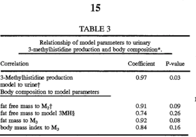
Table 3 presents the relationship of model parameters to urinary 3-methylhistidine and body composition and shows M2 to be highly correlated to fat free mass.
An objective of the present study was to determine whether a de novo production rate of 3-methylhistidine as estimated by the isotope model was similar to urinary 3-methylhistidine production. The slope of curve (FIG. 2) was similar to that of other species evaluated with this model Values are presented for each individual subject as well as
mean ±SE. Means for 3-methylhistidine production and for 3-methylhistidine to creatinine ratio for model- and
urine-65 (Rathmacher, J.A. et al. (1992), 'Technical Note: the use of
a compartmental model to estimate the de novo production
5,628,328
13
14
A. et al. ( 1992), ''Technical Note: the use of a compartmental
model to estimate the de novo production rate of N-meth-ylhistidine in cattle," J. Anim. Sci. 70:2104-2108) where
model and urinary 3-methylhistidine production results were
70:2104-2108; Rathmacher, J. A. et al. (1993),
"Measure-ment of 3-methylhistidine production in lambs by using
compartmental-kinetic analysis," Brit J. Nutrition
69:743-755; Rathmacher, J. A. et al., "Estimation of
3-me-thylhistidine production in swine by compartmental
analy-sis," J. Anim. Sci. (1992) 70:194 (Abstract)). The in vivo kinetics of 3-methylhistidine in human subjects can be described by means of a simple serial model of 3-compart-ments and sampling of only plasma. The kinetic parameters, L;j, compartment mass M; and mass transfer rates Rij are presented as a reference for future modeling of 3-methyl-histidine metabolism.
5
similar but tended to be higher than urine values. This could
be due to minor losses in urine or small amounts of 3-me-thylhistidine metabolism/conjugation in humans.
10 TABLE 1
Subject characteristics
15 Subjects
Parameter A B c D
20
Age, years 35 37 31 31
Sex 0 !i! 0 0
Weight, kg 92 47 78 69
25 Body Mass Index, kg!m
2* 25.5 19.2 21.6 24.4
Fat,%* 32.3 28.7 13.5 22.4
Fat, kg* 29.1 13.7 10.0 18.13
Fat-free mass, kg* 61.1 34.0 64.3 50.8
30 The Lij parameters for human subjects were similar to
those reported for cattle except for L2.1 which was lower for
humans (0.18 in cattle versus 0.07 min-1 in humans). Table
5 provides a summary of three studies: Rathmacher, J. A. et
al., "Evaluation of muscle protein turnover in steers differing in mature size (1993), unpublished; Rathmacher, J. et al., 'The use of compartmental models of3-methylhistidine flux to evaluate skeletal muscle protein turnover in implanted steers," J. Anim. Sci. (1993) 71:135 (Abstract); and
Rath-macher, J. A. et al., ''Technical Note: The use of a
compart-mental model to estimate the de novo production rate of
Nt-methylhistidine in cattle," J. Anim. Sci. (1992)
70:2104-2108. Based on the size of compartment 1, M1 has
a mass similar to extracellular water space and M2 and M3
are most consistent with intracellular pools of 3-methylhis-tidine (fable 2). Muscle biopsy data from lambs (Rathma-cher, J. A. et al. (1993), "Measurement of 3-methylhistidine production in lambs by using compartmental-kinetic analy-sis," Brit. J. Nutrition 69:743-755) indicate that compart-ments 2 and 3 appear to be muscle specific for 3-methyl-histidine in muscle.
The present study confirmed that model estimates of
3-methylhistidine production correspond to urinary 3-meth- 35
ylhistidine production. Means were not different when expressed on body weight basis or on urinary creatinine basis, but they were 16% higher than urinary estimates.
Similar correlations were measured in cattle (Rathmacher, J.
*Body composition was determined for each subject from the measurements of body density estimated by underwater weighing (Goldman, R. F. and
Buskirk, E. R., "Body volume measurement by underwater weighing:
description of a method," In: Techniques for Measuring Body Composition
(Brozek, J. ed.) Washington, D.C.: Nat'lAcad. of Sciences (1961) p. 78-79).
The percentage of body fat was estimated from body density by using the revised equation of Lohman et a!. (Lohman, T. G. et a!., "Bone and mineral measurements and their relation to body density in children, youth and adults," Hum. Bioi. (1984) 56:667--679).
TABLE2
Kinetic parameters of 3-methylhistidine metabolism and 3-methylhistidine model masses and mass transfer rates.*
Subjects
Parameter A B
c
D Mean SEtL,,., min-'; 0.0509 0.1573 0.0196 0.1372 0.0759 0.0686
L,,2, min-I 0.0224 0.1505 0.0111 0.1887 0.0614 0.0944
J..,,l> min-I 0.0048 0.0051 0.0024 0.0035 0.0041 0.0017
I., . -1 0.0051 0.0141 0.0074 0.0148 0.0089 0.0074
.2•mm
!..,,3, min-I 0.0004 0.0017 0.0029 0.0016 0.0017 0.0008
M., nmol . kg-1f 383 390 858 780 603 109
M,, nmol · kg-1 952 421 1692 581 912 245
M3 , nmol · kg-1 15856 3584 5070 7240 7938 2377
u3, nmol . kg-!. min-1** 1.83 2.00 2.04 2.72 2.15 0.17
R,,., nmol · kg-1
• min-'tt 19.49 61.36 16.83 10694 51.15 18.37
R1,2, nmol · kg-1· min-1 21.31 63.35 18.87 109.66 53.30 18.52
Ro,., nmol · kg-1
• min-1 1.83 2.00 2.04 2.72 2.15 0.17
R3,2, nmol · kg-1• min-1 4.86 595 12.50 8.62 798 1.47
R,,3 nmol · kg-'. min-1 6.69 595 14.55 11.34 19.63 1.75
*The kinetic parameters for a 3-rompartment model of 3-methylhistidine metabolism are presented for each subject along with the mean and tstandard error. ;L,;, fractional transfer
from compartment i to compartment j. §M;, mass of compartment i; **U3 , de novo
production into compartment 3; ttR;;, mass transfer rate from compartment j to compartment
5,628,328
15
TABLE3
Relationship of model parameters to urinacy 3-methylhistidine production and body composition*.
Correlation Coefficient P-value
3-Methylhistidine production model to urinet
Body composition to model parameters
fat free mass to M2 t
fat free mass to model 3MH§ fat mass to M3
body mass index to M3
0.97 0.91 0.74 0.92 0.84 0.03 0.09 0.26 0.08 0.16
*Model is a 3-compartment model of 3-methylhistidine production. t3-Me-thylhistidine production for model and urine have the units J.U110l · d-1
• :j:Fat
free mass has the units of kg, and M2 is mass of compartment 2 in nmoles.
§Model 3-methylhistidine is the estimate of production expressed as J.U110l ·
o1
. **Fat mass has the units of kg, and M3 is the mass of compartment 3 in nmoles.
EXAMPLE2
Comparison of Human and Animal 3-methylhistidine Mod-els.
The differences between 3-methylhistidine metabolism in humans as found in Example 1 and other animals is shown in Table 5. For cattle, the same three-compartment model was used as described herein. With respect to sheep, the model differs from that shown in FIG. 1 in that de novo production of 3-methylhistidine is shown in compartment 2 and additionally, exit from compartment 3 occurs as well as from compartment 1 into urine. With respect to swine, again, de novo production of 3-methylhistidine is shown in com-partment 2, and exit to urine is from compartment 3.
TABLE4
Summacy of 3-methylhistidine metabolism as described by a 3-compartment model.
Included are mass of compartments and mass transfer rates.
Parameter Human Cattle* Pigst Sheep§ Dogs'{
n 4 36 20 36 5
M1 , nmol · kg-1**
603 806 1110 4693 3227
M2 , nmol · kg-• 912 2106 2857 11634 7973
M3 , nmol · kg-• 7938 7803 6151 16494 9261
R,1, nmol · kg-1
• min-' 51 103 247 928 319 R12, nmol ·
kg-1
• min-' 53 107 247 930 329
R32, nmol · kg-'· min-1 7.9 5.4 37 84 56
R,, nmol · kg-1• min-• 9.6 9.6 32 80 56
R,1 , nmol· kg-1• min-1 2.2 4.2 NA 1.4 9
Ro,, nmol · kg-1• min-' NA NA 5.0 5.7 NA
3MH Production, 3.1 6.2 7.2 10.1 12
jJlilOl · kg-1.d-1
*Data obtained from cattle experiments (reported in Rathmacber, J. A. et a!. (1992), "Technical Note: the use of a compartmental model to estimate the de novo production rate of N-methylhistidine in cattle," J. Anim. sci. 70:2104-2108; Rathmacber, J. et a!., "'The use of compartmental models of
3-methylhistidine flux to evaluate skeletal muscle protein turnover in
implanted steers," J. Anim. Sci. (1993) 71:135 (Abstract); Rathmacher, J. A.
eta!., "Evaluation of muscle protein turnover in steers differing in mature size (1993), unpublished).
tData obtained from finishing swine experiment (Rathmacher, J. A. et a!., "Estimation of 3-methylhistidine production in pigs by compartmental analy-sis," J. Anim. Sci (1995) In Press).
§Data summarized from Rathmacher, J. A., "Comparative evaluation of muscle proteolysis by a compartmental model of 3-methylhistidine," Ames: Ph. D. Thesis, Iowa State University, 1994.
'!Data obtained from dog experiment (Rathmacher, J. A. eta!., "A
compart-mental model to measure 3-methylhistidine production in dogs following
surgery," J. Nutr. (1994) submitted).
5
16
TABLE 4-continued
Summacy of 3-methylhistidine metabolism as described by a 3-compartment model. Included are mass of compartments and mass transfer rates.
Parameter Human Cattle* Pigst Sheep§ Dogs'{
**M; = compartment mass i; R = mass transfer rate between compartment j
and i; 3-methylhistidine production was obtained from the model (See FIG.
10 4A, 4B, and 4C for model illustration).
Urinary 3-methylhistidine may be used in cattle and humans as an index of muscle protein breakdown but is invalid for use in pigs and lambs. 3-methylhistidine is produced in these species but is not quantitatively excreted
15 in the urine. Previously, in validating urinary 3-methylhis-tidine as an index of muscle proteolysis, researchers have injected 14C-3-methylhistidine intravenously and recovered the tracer in urine but have never described its decay in plasma. In constructing the 3-compartment model, we kept
20 in mind known physiology of 3-methylhistidine. It has been established that there are pools of 3-methylhistidine in plasma, in other extracellular fluid pools, within muscle and in other tissues. The primary fate of 3-methylhistidine