UDC 577.2:576.32/.36:611.018.04:602.9
Generation of optimized preparations of bone
morphoGenetic proteins for bone reGeneration
Kh. V. MalySheVa1,2, I. M. SpaSyUK3, O. K. paVleNKO3, R. S. StOIKa1, O. G. KORchyNSKyI1,4
1Institute of cell Biology, National academy of Sciences of Ukraine, lviv; 2Insitute of animal Biology, National academy of agrarian Sciences of Ukraine, lviv;
3Ivan Franko National University of lviv, Ukraine; 4centre for Innovative Research in Medical and Natural Sciences,
Medical Faculty of Rzeszow University, poland; e-mail: [email protected]
Correction of inherited skeletal abnormalities, traumas affecting wide bone areas and non-healing fractures require efficient bone formation and regeneration. Bone morphogenetic proteins (BMPs) are signa-ling molecules that play a crucial role in bone and cartilage formation and regeneration. Osteoinductive properties of existing hydroxyapatite-based osteoplastic materials are frequently insufficient for efficient bone regeneration, thus raising a requirement for novel matrices involving BMPs for highly efficient local induction of bone formation at the area of the bone defect. The aim of this study was conducting in vitro op
-timization of osteoinductive properties of recombinant BMPs preparations to be used in bone regenerative practice. Recombinant BMPs were produced in human embryonic kidney 293 cells upon their transfection or co-transfection with plasmids expressing BMP2 and BMP7 at different ratios. A quality of BMP preps was validated based on their ability to induce in vitro osteoblast differentiation of C2C12 cells. Alkaline phos
-phatase that is widely used as a marker of osteoblast differentiation was measured spectrophotometrically. We found that the most effective inducer of osteoblast differentiation was recombinant BMP preparation pro
-duced upon cotransfection of 85% of BMP2 and 15% of BMP7 plasmids, that is most likely due to generation of conditions most favorable for formation of BMP2/7 heterodimers. Frozen BMP2/7 preparations stored for 3 h in experimental setup and for several weeks in routine work do not lose their osteoinductive properties compared with freshly prepared BMP2/7 preparations and can be successfully used for generation of highly efficient bone regenerative matrices.
K e y w o r d s: bone morphogenetic proteins, bone regeneration, osteogenesis, mouse mesenchymal stem cells, alkaline phosphatase.
G
enetic diseases, non-jointing fractures and other traumas (including military) oftenlead to large bone defects. Therefore, effi -cient high-quality bone grafting is critically impor-tant in the healing of bone defects that occur as their result. In addition, 10% of hip replacement opera-tions (primarily – osteoporosis patients) have failed due osteolysis at the site of prosthesis integration. Apart of that, it is often necessary to increase the bone density and thickness in the patient’s jaw to be
sufficient for an effective dental implant integration
and maxillofacial surgery. On the other hand, the bone morphogenetic proteins (BMPs) are inducers of bone formation during embryogenesis, and they are essential for maintenance of the skeletal homeostasis
[1]. Thus, usage of osteoplastic materials incuding BMPs for local induction of bone formation in the area of the bone defect is highly promising.
The history of BMPs study began in the mid-dle of 1960s with the observation that demineralized bone matrix is able to induce ectopic bone forma-tion in subcutaneous and intramuscular pockets in rodents [2, 3]. However, it was not possible until the
late 1980s, when the first BMPs were charac terized
and cloned, that individual BMPs could be studied biochemically [4]. Many studies have since demon-strated the ability of BMPs to induce endochondral/
intramembranous ossification and chondrogenesis, by inducing mesenchymal stem cells differentia -tion towards the osteoblastic lineage, being critical
for maintenance of the skele tal integrity and in bone fracture healing. BMPs are synthesized by osteopro-genitor cells, osteoblasts, chondrocytes and platelets but their production is not restricted to bone, since they also play an essential role in development and cell functions in other organs [4, 5].
BMPs belong to a group of multifunctional signaling molecules that belong to the transforming
growth factor-β (TGF-β) superfamily of proteins.
BMPs synthesized as precursor proteins with an N-terminal signal peptide, a pro-domain for folding and secretion, and a C-terminal mature peptide. Pre-cursors are formed in the cytoplasm as dimeric pro-protein complexes, which are cleaved by pro-pro-protein convertases to generate N- and C-terminal frag-ments. The C-terminal mature fragment is capable of binding to its receptor, with the non-covalently associated pro-domain playing an important regula-tory role [4, 6].
BMPs can perform signaling functions through both canonical and non-canonical pathways. In the canonical signaling pathway, they initiate the sig-nal transduction cascade by binding to cell surface receptors and forming a heterotetrameric complex comprised of two dimers of type I and type II serine/ threonine kinase receptors [7]. Both receptor types have a short extracellular domain, a single trans-membrane domain, and an intracellular domain with serine/threonine kinase activity. There are seven
type I receptors (ALK1-7) for the TGF-β family of
ligands, four of which bind BMPs: type 1A BMP receptor (BMPR-1A or ALK3), type 1B BMP tor (BMPR-1B or ALK6), type 1A activin recep-tor (ActR-1A or ALK2) and ALK1 receprecep-tor, which binds BMP9. There are four type II receptors for
the TGF-β family, three of which can interact with
BMPs: type 2 BMP receptor (BMPR-2), type 2A activin receptor (ActR-2A), and type 2B activin re-ceptor (ActR-2B). While BMPR-1A, BMPR-1B, and
BMPR-2 are specific to BMPs, ActR-1A, ActR-2A,
and ActR-2B can function as receptors for activins,
which are also members of the TGF-β
superfami-ly. The mechanism of the heterotetrameric signaling complex formation can vary. The existence of pre-formed oligomeric complexes adds an additional layer of intricacy; indeed, binding to preformed ceptor complexes versus BMP-induced receptor
re-cruitment can activate different pathways [4, 8, 9].
Upon formation of a heterotetrameric complex, the constitutively active type II receptor trans-phos-phorylates the type I receptor at a glycine–serine
rich motif known as the GS domain. This activates the type I receptor and allows phosphorylation of the immediately downstream substrate proteins known as the receptor-regulated Smads (R-Smads) at a C-terminal SSXS motif. The R-Smads involved in BMP signaling are Smad1, Smad5, and Smad8 (Smad1/5/8). R-Smads then associate with the co-mediator Smad (co-Smad) Smad4, and this complex translocates to the nucleus where it functions as a transcription factor with coactivators and corepres-sors to regulate gene expression. Inhibitory Smads (I-Smads), Smad6 and Smad7 (Smad6/7), are in-volved in feedback inhibition of the signaling path-way [4, 10, 11].
Interestingly, BMPs heterodimers contai-ning subunits of various groups (e.g. BMP2/7 or
BMP4/6) more effectively activate BMP signaling
pathways and, as a result, exhibit higher osteoinduc-tive proper ties [12].
BMPs are crucial growth factors regulating nu-merous processes of skeletal formation,
hematopoie-sis, neurogenehematopoie-sis, as well as defining the differen -tiation map of cells during embryonic development [13].
More than fifty years after the discovery by
Marshal R. Urist at least 20 human BMPs were
iden-tified and they possess varying degrees of the induc -tive activity. Two of these, BMP2 and BMP7, have become the subject of extensive research aimed at developing therapeutic strategies for the restoration and treatment of skeletal conditions [14, 15]. Thus, a main goal of this study was generation of optimized recombinant BMPs preparations that can be used in bone regenerative practice.
materials and methods
Reagents and ligands. Plasmids expressing BMP2 and BMP7 full-length cDNA were purchased from Open Biosystems/GE Healthcare (Lafayette, CO, USA). pShuttle-CMV vector was kindly pro-vided by Dr. Bert Vogelstein. Human recombinant BMP2 and BMP7 were purchased from PeproTech (Rocky Hill, NJ, USA). Heterodimeric human re-combinant BMP2/7 was purchased from R&D Sys-tems (Minneapolis, MN, USA).
cloning strategy. Full-length human BMP2 cDNA was digested with EcoRI and NotI enzymes in an order to re-clone it from non-expressing MGC (Mammalian Gene Collection) vector pINCY into
-structs were digested with KpnI and NotI enzymes in order to reclone full-length cDNAs into pShuttle-CMV expression plasmids that confer kana mycin resistance gene.
cell culture. Our studies were p erformed using immortalized mouse mesenchymal precursor cells of
C2C12 line, human embryonic kidney 293 (НЕК293)
cells and human mesenchymal stem cells (hMSC) were isolated from bone marrow aspirate of the gical material obtained upon hip replacement sur-gery under informed patient’s consent. A pluripoten-cy of hMSC was evaluated based on their capacity to
efficiently differentiate into osteoblast, adipocyte and
chondrocyte lineages upon proper stimulation and conditions. C2C12 and НЕК293 cells were cultured
in Dulbecco’s modified Eagle’s medium (DMEM, Biowest, France) containing 10% fetal bovine se
-rum (FBS, Biowest, France). hMSC were cultured
in low-glucose (1 mg/ml) DMEM supplemented with
10 ng/ml of basic fibroblast growth factor. All cells
were grown in a 5% CO2-containing atmosphere at 37 °C and 100% humidity. Culture medium was re-freshed every 2-3 days. Before subcultivation cells
were washed with phosphate-buffe red saline (PBS)
that contained 137 mM NaCl, 2.7 mM KCl, 4.3 mM Na2HPO4 and 1.4 mM KH2PO4 with pH 7.4.
Transfection of НЕК293 cells. HEK293 were
seeded at 60-70% of confluency in 6 or 9 cm Petri
dishes “Anumbra”. Next day, cells were transfected with plasmid constructs expressing BMP2 and BMP7 and control plasmid – pcDNA3 (correspondi-ngly 5 or 20 µg/ml of total DNA per dish). Trans-fection was carried out using transTrans-fection reagent
polyethyleniminе (PEI, Polysciences Inc., War -rington, PA, USA) following the manufacturer’s pro-tocol. After transfection (4-7 h), culture medium was replaced with a fresh complete medium. After 48 h of transfection, we collected conditioned medium, which contained BMP2 and BMP7 homo- or hetero-dimers. Depending on the type of experiment, the conditioned medium was stored at 4 °C in refrigera-tor (Samsung, Seoul, Korea) or at –70 °C in ultra low temperature freezer (NuAire, Plymouth, MN, USA) in sample holders. Due to the absence of alive cells in these preparations no cryoprotectors or special cryoconservation programs were employed.
Luciferase reporter assays. hMSC cells were
split at a density of 1․104 cells per cm2 in 12-well plates. The following day, cells were transiently
transfected with a BMP-specific luciferase reporter
adenovirus we generated before at a multiplicity of
infection, MOI of 190) in a combination with β-Gal
construct (MOI = 10) to be used as an internal
con-trol for the efficacy of transduction. Six h later a
culture medium was refreshed. After 36 h of
trans-fection, cells were starved for 12 h in a 0.1% FCS-DME, re-fed with the fresh 0.1% FCS-DME and
treated with indicated ligands for the next 12-15 h.
β-Galactosidase activity was quantified in 100 mM
Na2HPO4/NaH2PO4 , 1 mM MgCl2, 100 mM 2-mer-captoethanol, and 0.67 mg/ml O-nitrophenylgalacto-pyranoside spectrophotometrically. Both luciferase
reporter and β-galactosidase activity assays were
conducted using luciferase reporter assay reagent (Promega, Madison, WI, USA) on a Victor 3 ma-chine (Perkin Elmer , Waltham, MA, USA) [16]. Each transduction was carried out in triplicate and repeated at least twice.
Induction of osteoblast differentiation. Cells of C2C12 line can be induced to differentiate into os
-teoblasts by different BMPs, including BMP2 and BMP7. Cells were split at a density of 1.5∙104 cells per cm2 in 12- or 24-well plates, respectively. Next day, cells were treated with fresh differentiation-sup -porting medium supplemented with 50 mg/ml ascor-bic acid complete culture medium in a 1 : 1 ratio with conditioned medium, which contained BMP2 and BMP7 in appropriate proportions. In a current study, we addressed the evaluation of early
osteo-blast differentiation, therefore other components, like β-glycerophospate that is required as a source
of calcium for matrix mineralization, were not nece-ssary. On the third day after induction, C2C12 cells were repetitively treated with conditioned medium. During osteogenesis assay, C2C12 cells were cul-tured for 4 days.
Alkaline phosphatase assay. The
alka-line phosphatase activity produced by C2C12 was analyzed spectrophotometrically using a
π-nitrophenylphosphate (π-NPP), as a substrate [17]. Four days after induction of osteogenesis, the cells
were washed twice with 0.4 ml of 1X PBS per well. Afterwards, cells were lysed in 0.2 ml of alkaline
phosphatase (ALP) lysis buffer (10 mM glycine,
100 µM MgCl2, 10 µM ZnCl2, 0.1% Triton X-100) per well and agitated gently for 5 min. Then, 10 µl aliquot of cell lysate was placed into a 96-well plate and ALP activity was revealed with 90 µl/well of
ALP assay buffer (100 mM glycine, 1 mM MgCl2, 100 µM ZnCl2) supplemented with 6 mM π-NPP
(Pierce-Thermo Fisher Scientific, Grand Island, NY,
at room temperature until color developed. The ab-sorption was measured at 405 nm in a 96-well plate reader (BioTek, Winooski, VT, USA). The level of expression of the enzyme is directly proportional to the intensity of mesenchymal stem cells osteogenic
differentiation [17].
All experiments were repeated twice using three parallel wells in each variant. Results of spec-trophotometric measurements of ALP activity are expressed as mean ± standard deviation. Data were analyzed using GraphPad Prism 6 program.
Statisti-cal differences between experimental variants were
assessed by Student’s t-test. Appropriate p values
were shown in graphs to demonstrate the signifi
-cance of the results. Only differences with p-values
lower than 0.05 were regarded as significant.
results and discussion
The heterodimers consisting of subunits that
belong to different groups of BMPs (e.g. BMP2/7 or
BMP4/6) activate BMP signaling pathway more ef-fectively and, as a result, exhibit higher osteoinduc-tive properties due to recruiting subunits belonging
to different BMP type I receptor subclasses [12]. We
carried out a comparative study of the osteoinductive properties of the recombinant BMP2, BMP6, and
BMP2/7 heterodimers. As osteoinductive properties
of different BMPs directly depend on the intensity
of BMP signaling pathway activation, we used in our work luciferase reporter assays that allow quan-tifying the level of activation BMP-Smad signaling pathway in a wide linear range.
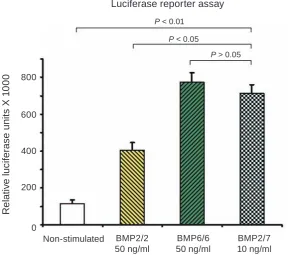
As shown in Fig. 1, the activation of the BMP
signaling pathway in hMSC using 50 ng/ml of BMP6/6 was observed. Twice higher activation of the BMP-sensitive BRE-Luc reporter system than activation in cells treated with BMP2/2 at the same
concentration was found. For such activation of BMP
signaling pathway, we used BMP7/7 at three-fold higher concentration (data not shown here). Probab-ly, this recombinant protein was partially inactiva-ted because of its interaction with secreinactiva-ted protein product of its target gene Noggin. The mechanism
of such effect was established recently [18]. At the
same time, BMP2/7 heterodimers at a concentration
of 10 ng/ml show comparable effect with BMP6/6 at five-fold higher concentration.
A search for recombinant BMPs prepara
-tions with the best osteoinductive properties. The main goal of our work was to choose the optimal combinations of recombinant BMPs preparations
demon strating the best osteoinductive effect. Thus,
Fig. 1. The BMP2/7 heterodimers at a concentration of 10 ng/ml are more potent activators of BMP signaling
pathway than BMP2/2 and equally potent with BMP6/6 homodimers at a concentration of 50 ng/ml each. Lu
-ciferase reporter assay was carried out with human mesenchymal stem cells Non-stimulated BMP2/2
50 ng/ml
BMP6/6 50 ng/ml
BMP2/7 10 ng/ml 800
600
400
200
0
R
e
la
ti
v
e lu
c
if
e
ras
e u
n
it
s X 1
0
0
0
P < 0.01
P < 0.05
we carried out a study of the combination of BMP2
and BMP7 action in different proportions. For this,
we used a commonly accepted model of cell line HEK293, which is widely used for various research
purposes due to its high efficiency, in particular, for
the production of recombinant proteins (in our case BMPs).
HEK293 cells were transfected with plasmid constructs expressing BMP2 and BMP7 and control plasmid – pcDNA3, that does not contain the coding
sequence of the genes which products can affect the
results of the experiment. The transfection (or co-transfection) with indicated plasmids was carried out in the following proportions (Table 1).
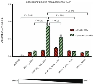
To validate the efficacy of combined action of
BMP2 and BMP7 in a comparison with a separate use of each ligand we re-cloned both cDNAs into a similar vector pShuttle that confers a kanamycin
re-sistance. After 48 h of transfection of НЕK293 cells,
we collected conditioned medium (CM) containing secreted BMP2 and BMP7 homo- or hetero dimers in appropriate proportions. We mixed this CM in a 1 : 1 ratio with fresh culture medium. On the same day, the induction of C2C12 was carried out. Two days after induction re-addition of CM was performed as one-time stimulation resulted in low induction
of C2C12 osteoblast differentiation. Analysis of the
experiment results was performed by spectropho-tometric measurement of ALP activity, which is
widely used as a marker of osteoblast differentiation (Fig. 2). As one can see from Fig. 2, both BMP2/2
and BMP7/7 expressed from this vector had a rela-tively low osteoinductive activity: respecrela-tively 2.5 and 2.4 times higher than a control vehicle. Mixture of these constructs in equimolar concentrations was
1.8 times more effective. While taking into account a
low osteoinductive activity of these preparation, we generated the optimized version of BMP2 expression
plasmid upon its re-cloning into pDEF vector that
confers a high expression level for recombinant pro-teins [16] and used in a parallel with a BMP7
sub-cloned into a pSport6.1 vector. Figure 3 shows the
results of the comparison between recombinant
plas-mids based on different vectors used in the study.
As it is clear from such comparison made side-by-side into one experiment, optimized versions of
vec-tors (hBMP2-pDEF in a combination with
hBMP7-pSport6.1) in a case of optimal ratio between BMP2
and BMP7 was almost three times more efficient that original variant (pShuttle-CMV vector). Most effi -cient combination appeared to be 75% BMP2/25% BMP7.
Ta b l e 1. The proportions of plasmids expressing BMP2 and BMP7 (crude evaluation)
BMP2, % 100 75 50 25 0
BMP7, % 0 25 50 75 100
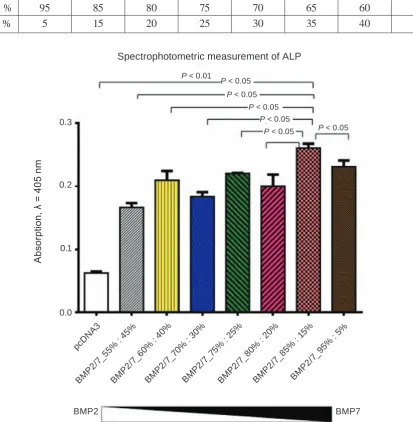
The next experiment was carried out for more accurate determination of recombinant BMP2 and BMP7 concentrations in the preparation,
which showed the best osteoinductive effect in the
preceding study.
This experiment was carried out similarly to the previous one. The HEK293 cells transfection (or co-transfection) with plasmid constructs expressing BMP2 and BMP7 was performed in the following proportions (Table 2).
The CM that already contained recombinant BMP2/7 in appropriate proportions was collected 48 h after transfection. On the same day, the
induc-tion of C2C12 osteoblast differentiainduc-tion was carried
out similarly to previous experiment Analysis of the experimental results was performed by a
spectropho-tometric measurement of the ALP activity (Fig. 3).
It was shown that BMPs preparation produced upon co-transfection of 85% of BMP2 and 15% of
BMP7 is the most effective inducer of C2C12 osteo
-blast differentiation.
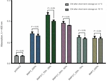
The study of recombinant BMP preparations osteoinductive properties after their storage at low temperature. It is well known that freezing-thawing can lead to the destruction of proteins, particularly their complexes. Therefore in our next experiment we studied the possibility of maintaining the osteo-inductive properties of recombinant BMPs during their short-term low-temperature storage.
This experiment was carried out similarly to the previous studies. We collected the CM (48 h after transfection), which included recombinant BMP2/7 in ratios that were described in Table 1. One part of CM aliquots we stored at 4 °C for 3 h, and the other part – at –70 °C during the same period of time.
On the same day, the fresh culture medium was mixed in a 1 : 1 ratio with CM and the induction of
С2С12 cells was carried out in two versions: A – CM after term storage at 4 ºС, B – CM after short-term storage at –70 ºС.
Two days after osteoblast differentiation induc -tion, CM re-addition was performed in an appropria-te way. The results of spectrophotometric
Fig. 2. The BMPs preparation generated upon transfection of 75% of BMP2 and 25% of BMP7 plasmids into producer HEK293 cells was the most effective inducer of osteoblast differentiation in C2C12 mesenchymal cells (crude evaluation). Producer cells of HEK293 line were transfected with plasmids expressing BMP2 and BMP7 at indicated ratios. Two days after transfection, the CM with secreted BMPs was collected, and mixed with fresh culture medium. The osteogenic properties of BMP preparations were tested using mesenchymal C2C12 cells. The intensity of osteogenesis was evaluated spectrophotometricaly by measuring the ALP activity
pcD NA
3
BM P2_1
00%
1.5
1.0
0.5
0.0
A
bs
or
pt
io
n, λ =
4
05 nm P < 0.05
Spectrophotometric measurement of ALP
P < 0.001
BM P2/
7_75 % : 2
5%
BM P2/
7_50 % : 5
0%
BM P2/
7_25% : 7
5%
BM P7_
100%
BMP2 BMP7
pShuttle-CMV
Optimized plasmids P < 0.001 P < 0.001
P < 0.001
In next experiment, we used a similar CM stored at –20 °C for 3 weeks, and also did not reveal a loss of the osteogenic properties (data not shown).
It was shown that the recombinant BMP prepa-rations kept at low temperature (–70 °C) for a short period of time practically did not lose their
osteoin-duction effectiveness compared to fresh recombinant BMP preparations after short-term storage at 4 °С.
Currently in our experiments we successfully use such preparations stored for weeks at –70 °C or even at –20 °C (data not shown).
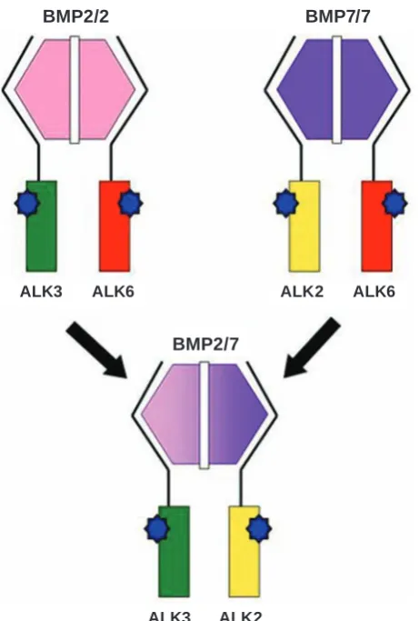
BMP2 and BMP7 trigger their signaling path-way via recruiting a minimally heterotetrameric complex, which consists of BMP receptor types I and II with minimally two subunits of each type into a complex [19]. Our experiments, as well as in vitro
and in vivo studies of other scientists, demonstrated the best osteoinductive properties of BMP2/7 hetero-dimers compared with the corresponding homodi-mers [12]. BMP2 mainly binds to BMPRIA/ALK3 and BMPRIB/ALK6 receptors, while BMP7 selec-tively binds to ALK2 receptor in a combination with BMPRIB/ALK6 receptors, the best osteoinductive properties of BMP2/7 heterodimer can be caused by combining of ALK3 and ALK2 into a one complex
(Fig. 5).
Preparations of the bone morphogenetic protein were generated upon overexpression of optimized
versions of expression vectors. Freezing and storage of these preparations for 3 h do not affect
Ta b l e 2. The proportions of plasmids expressing BMP2 and BMP7 (fine evaluation)
BMP2, % 95 85 80 75 70 65 60 55
BMP7, % 5 15 20 25 30 35 40 45
Fig. 3. The BMPs preparation generated upon transfection of 85% of BMP2 and 15% of BMP7 plasmids into producer HEK293 cells was the most effective inducer of osteoblast differentiation of C2C12 cells. Producer cells of HEK293 line were transfected with plasmids expressing BMP2 and BMP7 in given ratios. Two days after transfection, the CM with secreted BMP2/7 was collected, and mixed with fresh culture medium. The osteogenic properties of BMPs preparations were tested on C2C12 cell line. The intensity of osteogenesis were evaluated spectrophotometricaly by measuring the ALP activity
pcD NA
3
BM P2/7
_55% : 45
%
0.3
0.2
0.1
0.0
A
bs
or
pt
io
n, λ =
4
05 nm
Spectrophotometric measurement of ALP
P < 0.05
BM P2/
7_60 % : 4
0%
BM P2/
7_70 % : 3
0%
BM P2/
7_75 % : 2
5%
BMP2 BMP7
P < 0.05 P < 0.05 P < 0.01
P < 0.05
P < 0.05 P < 0.05
BM P2/
7_85% : 1
5%
BM P2/
7_95 % : 5%
BM P2/
7_80 % : 2
0%
preparations can be successfully used for generation
of highly efficient bone regenerative matrices or in
other biological applications.
acknowlegments
This research was supported in a part by the Molecular & Cellular Biotechnologies Grant of the
Fig. 4. The freezing-thawing of the BMP2/7 preparations does not reduce their osteoinductive properties. Producer cells of HEK293 line were transfected with plasmids expressing BMP2 and BMP7 in given ratios. Two days after transfection, the CM with secreted BMP2/7 was collected, and divided into two parts (one part
was stored at 4 °C for 3 h and another one at –70 °C during the same period of time). Then CM were mixed with in a 1 : 1 ratio with fresh culture medium. Its osteogenic properties were tested on C2C12 cell line. The intensity of osteoblast differentiation were evaluated spectrophotometricaly by measuring the ALP activity
pcD NA
3
0.3
0.2
0.1
0.0
A
bs
or
pt
io
n, λ =
4
05 nm
Spectrophotometric measurement of ALP
BM P2/
7_75 % : 2
5%
BM P2_1
00%
P > 0.05
BM P2/
7_25% : 7
5%
BM P7_
100%
BM P2/
7_50 % : 5
0%
CM after short-term storage at +4 °C
P > 0.05
P > 0.05 P > 0.05 P > 0.05
P > 0.05 CM after short-term storage at -70 °C
Financial support
The study was supported in a part by the Mo-lecular & Cellular Biotechnologies Grant of the
Fig. 5. Formation and activation of heteromeric BMP type I receptor complex triggered by the BMP2/7
hete-rodimers
BMP2/2 BMP7/7
BMP2/7
ALK3 ALK6 ALK2 ALK6
ALK3 ALK2
Створення оптимізованих препаратів морфогенних протеїнів кіСтки для регенерації кіСткової тканини
Х. В. Малишева1,2, И. М. Спасюк3,
О. К. Павленко3, Р. С. Стойка1,
О. Г. Корчинський1,4
1Інститут біології клітини НАН України, Львів;
2Інститут біології тварин НААН України, Львів;
3Львівський національний університет
імені Івана Франка, Україна;
4Центр інноваційних досліджень в галузі
медицини та природничих наук, медичний факультет, Жешувський університет, Польща;
e-mail: [email protected]
Корекція спадкових аномалій скелета, травми обширних ділянок кістки, переломи, що тривалий час не загоюються, вимагають ефективного відновлення кісткової ткани
-ни. Морфогенні протеїни кістки (МБК) – це сигнальні молекули, які відіграють важливу роль у формуванні та регенерації кісткової і хрящової тканин. Остеоіндуктивні властивості вже існуючих кістковопластичних матеріалів на основі гідроксіапатиту часто бувають не
-придатними для ефективної регенерації кісткової тканини, і тому зростають вимо
-ги до нових матриксів, що містять МБК для високоефективної локальної регенерації кістки в дефектній ділянці. Метою цього дослідження була оптимізація in vitro остеоіндуктивних вла
-стивостей препаратів рекомбінантних МБК, які будуть використані на практиці для регенерації кісткової тканини. Рекомбінантні МБК були продуковані ембріональними клітинами ни
-рок людини (НЕК293) після їх трансфекції або сумісної трансфекції плазмідами, що експресу
диференціацію клітин лінії С2С12. Активність лужної фосфатази, що широко використовується як маркер остеобластної диференціації, вимірювали за допомогою спектрофотометра. Виявлено, що найефективнішим індуктором остеобластної диференціації був препа
-рат рекомбінантного МБК, що утворюється за сумісної трансфекції плазмідами МБК2 і МБК7 у співвідношенні 85 до 15% відповідно. Цей результат, імовірно, пояснюється створен
-ням найсприятливіших умов для формування МБК2/7 гетеродимерів. Під час зберігання замо
-рожених препаратів МБК2/7 протягом 3 годин в ході експериментів або декількох тижнів в ході щоденної роботи вони практично не призводили до втрати їхніх остеоіндуктивних властивостей в порівнянні зі свіжовиділеними препаратами МБК2/7 і тому можуть успішно використову
-ватися у створенні високоефективних регенера
-тивних кісткових матриксів.
К л ю ч о в і с л о в а: морфогенні протеїни кістки, регенерація кісткової тканини, остеоге
-нез, мезенхімні стовбурові клітини миші, лужна фосфатаза.
Создание
оптимизированных
препаратов морфогенных протеинов коСти для
регенерации коСтной ткани
Х. В. Малышева1,2, И. М. Спасюк3,
А. К. Павленко3, Р. С. Стойка1,
А. Г. Корчинский1,4
1Институт биологии клетки НАН Украины, Львов;
2Институт биологии животных
НААН Украины, Львов;
3Львовский национальный университет
имени Ивана Франко, Украина;
4Центр инновационных исследований в области
медицины и естественных наук, медицинский факультет, Жешувский университет, Польша;
e-mail: [email protected]
Коррекция наследственных аномалий скелета, длительно незаживающие переломы, травмы, затрагивающие широкие области ко
-сти, требуют эффективного восстановления костной ткани. Морфогенные протеины кости (МБК) являются сигнальными молекулами, ко
-торые играют решающую роль в формирова
-нии и регенерации костной и хрящевой тканей. Остеоиндуктивные свойства существующих костнопластических материалов на основе ги
-дроксиапатита часто непригодны для эффектив
-ной регенерации кост-ной ткани, следовательно повышаются требования к новым матриксам, содержащим МБК для высокоэффективной ло
-кальной регенерации кости в дефектной обла
-сти. Целью данного исследования было опти
-мизирование in vitro остеоиндуктивных свойств препаратов рекомбинантных МБК, которые бу
-дут использованы на практике для регенерации костной ткани. Рекомбинантные МБК были про
-дуцированы эмбриональными клетками почек человека (НЕК293) после их трансфекции либо совместной трансфекции плазмидами, экспрес
-сирующими МБК2 и МБК7 в различных соот
-ношениях. Качество препаратов МБК было под
-тверждено их способностью индуцировать in vitro остеобластную дифференциацию клеток линии С2С12. Активность щелочной фосфатазы, которая широко используется в качестве марке
-ра остеобластной дифференциации, измеряли с помощью спектрофотометра. Установлено, что наиболее эффективным индуктором остео
-бластной дифференциации был препарат реком
-бинантного МБК, образующийся при совмест
-ной трансфекции плазмидами МБК2 и МБК7 в соотношении 85 к 15% соответственно. Данный результат, предположительно, объясняется соз
-данием наиболее благоприятных условий для формирования МБК2/7 гетеродимеров. При хранении замороженных препаратов МБК2/7 в течении 3 часов в ходе экспериментов, или в течение нескольких недель при ежедневной ра
-боте, они практически не теряли своих остеоин
-дуктивных свойств по сравнению со свежевыде
-ленными препаратами МБК2/7 и поэтому могут быть успешно использованы для производства высокоэффективных регенеративных костных матриксов.
К л ю ч е в ы е с л о в а: морфогенные про
-теины кости, регенерация костной ткани, остео-генез, мезенхимные стволовые клетки мыши, щелочная фосфатаза.
references
1. Chuva de Sousa Lopes SM, Feijen A, Korving J,
Doevendans P, Mummery CL. Connective tissue growth factor expression and Smad signaling during mouse heart development and myocardial infarction. Dev Dyn. 2004; 231(3): 542-550. 2. Urist MR. Bone: formation by autoinduction.
Science. 1965; 150(3698): 893-899.
3. Groeneveld EH, Burger EH. Bone morphogenetic proteins in human bone regeneration. eur J endocrinol. 2000; 142(1): 9-21.
4. Wang RN, Green J, Wang Z, Deng Y, Qiao M, Peabody M, Zhang Q, Ye J, Yan Z, Denduluri S, Idowu O, Li M, Shen C, Hu A, Haydon RC, Kang R, Mok J, Lee MJ, Luu HL, Shi LL. Bone Morphogenetic Protein (BMP) signaling in development and human diseases. Genes Dis. 2014; 1(1): 87-105.
5. Carreira AC, Alves GG, Zambuzzi WF,
Sogayar MC, Granjeiro JM. Bone Morphogenetic Proteins: structure, biological function and therapeutic applications. Arch Biochem Biophys. 2014; 561: 64-73.
6. Harrison CA, Al-Musawi SL, Walton KL. Prodomains regulate the synthesis, extracellular
localisation and activity of TGF-β superfamily ligands. Growth Factors. 2011; 29(5): 174-186. 7. Heldin CH, Miyazono K, ten Dijke P.
TGF-beta signalling from cell membrane to nucleus through SMAD proteins. Nature. 1997; 390(6659): 465-471.
8. de Caestecker M. The transforming growth factor-beta superfamily of receptors. cytokine Growth Factor Rev. 2004; 15(1): 1-11.
9. Nohe A, Hassel S, Ehrlich M, Neubauer F,
Sebald W, Henis YI, Knaus P. The mode of bone morphogenetic protein (BMP) receptor
oligomerization determines different BMP-2
signaling pathways. J Biol chem. 2002; 277(7): 5330-5338.
10. Horbelt D, Denkis A, Knaus P. A portrait of
Transforming Growth Factor β superfamily
signalling: Background matters. Int J Biochem cell Biol. 2012; 44(3): 469-474.
11. Heldin CH, Moustakas A. Role of Smads in
TGFβ signaling. cell tissue Res. 2012; 347(1): 21-36.
12. Aoki H, Fujii M, Imamura T, Yagi K, Takehara K, Kato M, Miyazono K. Synergistic effects of different bone morphogenetic protein type I
receptors on alkaline phosphatase induction. J cell Sci. 2001; 114(Pt 8): 1483-1489.
13. Bragdon B, Moseychuk O, Saldanha S, King D, Julian J, Nohe A. Bone morphogenetic proteins: a critical review. cell Signal. 2011; 23(4): 609-620.
14. Gautschi OP, Frey SP, Zellweger R. Bone morphogenetic proteins in clinical applications. aNZ J Surg. 2007; 77(8): 626-631.
15. Bishop GB, Einhorn TA. Current and future clinical applications of bone morphogenetic proteins in orthopaedic trauma surgery. Int
Orthop. 2007; 31(6): 721-727.
16. Korchynskyi O, ten Dijke P. Identification and
functional characterization of distinct critically
important bone morphogenetic protein-specific
response elements in the Id1 promoter. J Biol chem. 2002; 277(7): 4883-4891.
17. van der Horst G, van Bezooijen RL, Deckers MM, Hoogendam J, Visser A,
Löwik CW, Karperien M. Differentiation of
murine preosteoblastic KS483 cells depends on autocrine bone morphogenetic protein signa-ling during all phases of osteoblast formation. Bone. 2002; 31(6): 661-669.
18. Krause C, Korchynskyi O, de Rooij K, Weidauer SE, de Gorter DJ, van Bezooijen RL, Hatsell S, Economides AN, Mueller TD, Löwik CW, ten Dijke P. Distinct modes of inhibition by sclerostin on bone morphogenetic protein and Wnt signaling pathways. J Biol chem. 2010; 285(53): 41614-41626.
19. Wrana JL, Attisano L, Wieser R, Ventura F,
Massagué J. Mechanism of activation of the
TGF-beta receptor. Nature. 1994; 370(6488): 341-347.