*Corresponding author:Haseena Ummer ISSN: 0976-3031
Case Report
PRESERVATION OF MANDIBULAR MOLARS WITH HEMISECTION
A SERIES OF INTERDISCIPLINARY CASE REPORTS
Haseena Ummer
1, Rizleena Majeed
2, Suneetha MP
3, Ashraf K
4and Shabana Basheer
51
Department of Prosthodontics and Crown & Bridge Kannur Dental College,
Anjarakandy, Kannur, Kerala, India
2
Departments of Periodontics Kannur Dental College, Anjarakandy, Kannur, Kerala, India
3,4,5
Department of Conservative dentistry and Endodontics Kannur dental college
Anjarakandy Kannur Kerala India
DOI: http://dx.doi.org/10.24327/ijrsr.2019.1002.3156
ARTICLE INFO ABSTRACT
Hemisection has been recognized as an effective procedure to retain tooth structure, while removing the unrestorable root and crown portion which may be affected by periodontal or endodontic disease. case reports are presented in this article, where hemisection was performed on mandibular molars for different etiologies, to preserve the tooth. The first case report details the management of a vertical root fracture while the second deals with furcal iatrogenic perforation during endodontic procedure. In both the cases, Endodontic therapy and prosthetic rehabilitation had a successful review due to the ameliorate interdisciplinary treatment approach.
INTRODUCTION
In certain clinical scenario, extractions were the only choice of treatment. We now live in an era where using advances is possible to maintain a functional dentition for a lifetime. One of the conservative ways of preserving a tooth is ‘hemisection’. Synonymous with ‘root amputation’ or ‘bisection’, hemisection is a treatment modality which allows the preservation of tooth structure, alveolar bone and cost saving over the other treatment modalities.[1]In the process of hemisection, the involved crown and root is removed to preserve the remaining tooth rather than sacrificing it.
Indications
Following indications have been suggested by Weine for tooth resection: [2]
Periodontal indications
1. Severe vertical bone loss involving only one root of multi-rooted teeth
2. Through and through furcation destruction
3. Unfavourable proximity of roots of adjacent teeth, preventing adequate hygiene maintenance in proximal areas
4. Severe root exposure due to dehiscence Endodontic and restorative indications
1. Prosthetic failure of abutments within a splint. 2. Endodontic failure:
Hemisection is useful in cases in which there is perforation through the floor of the pulp chamber, or pulp canal of one of the roots of an endodontically involved tooth which cannot be instrumented.
3. Vertical fracture of one root: 4. Severe destructive process:
This may occur as a result of furcation or subgingival caries, traumatic injury, and large root perforation during endodontic therapy.
Contraindications
1. Poorly shaped roots or fused roots
Available Online at http://www.recentscientific.com
International Journal of
Recent Scientific
Research
International Journal of Recent Scientific Research
Vol. 10, Issue, 02(D), pp. 30935-30939, February, 2019
Copyright © Haseena Ummer et al, 2019, this is an open-access article distributed under the terms of the Creative Commons Attribution License, which permits unrestricted use, distribution and reproduction in any medium, provided the original work is properly cited.
DOI: 10.24327/IJRSR CODEN: IJRSFP (USA)
Article History:
Received 06th November, 2018 Received in revised form 14th December, 2018
Accepted 23rd January, 2018 Published online 28th February, 2019
Key Words:
International Journal of Recent Scientif
2. Poor endodontic candidates or inoperable endodontic roots
3. Patient unwilling to undergo surgical and treatments
Hemisected teeth can fail due to root fractures; therefore an extra- coronal restoration is of utmost importance. A Hemisection, thus calls for an interdisciplinary approach. The present case reports discussed in this article
in volvement of surgical, endodontic as well as prosthetic treatment, brought in sights a successful review.
Case Report
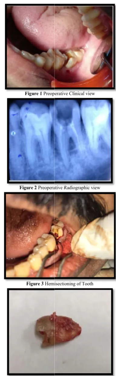
A 45 year old male patient reported with a chief complaint of pain on mastication and swelling in the lower left back region. He reported a history of root canal treatment in the concerned area five years back. On Intraoral examination, a sinus opening was detected on the buccal aspect of tooth 37, along with exudation from the same. There was a vertical root fracture extending through the floor of the tooth more towards the mesial orifice. Further examination showed a good condition of the distal root with no pocket formation or alveolar bone loss, but a periodontal pocket of probing depth 6mm was detected with respect to the mesial root [Figure 1]. Radiographic examination in relation to 37 revealed:
A coronal radiolucency involving the pulp chamber
A radiolucent line in the furcation region
incomplete obturation of the mesial root during past roo canal treatment [Figure 2].
A periapical radiolucency involving the mesial root. Thus, it was diagnosed as:
Vertical root fracture involving mesial root of 37
Chronic periapical abscess in relation to the mesial root. The treatment decided for the patient was hemisection in relation to 37 in which the amputation of ,mesial root of 37 was to be followed by obturation and prosthetic rehabilitation of the said tooth.
The first Phase of Treatment was the Periodontal
Following administration of local anaesthesia, mesial root was removed using the PRF- Choukron’s Technique. The extraction socket was then filled with PRF and bone graft. Coepack was placed following suturing in the site [Figure 3
This was Followed by an Endodontic phase
It involved re- root canal treatment for the distal root. Obturation was done using 2% Gutta percha by the lateral condensation technique. Composite was used as post endodontic restorative material.
Regular follow- up visits were scheduled for the patient for monitoring the healing of the socket. There were no post operative complications noted during these visits, and radiographic examination revealed that the site was satisfactorily healed by the end of 3 months [Figure 7]. After 3 Months of Healing, the Prosthetic Phase was Planned
It was decided to give a three unit fixed partial denture involving 36, 37 and 38 [Figure 8, 9].
International Journal of Recent Scientific Research Vol. 10, Issue, 02(D), pp. 30935-30939
Poor endodontic candidates or inoperable endodontic Patient unwilling to undergo surgical and endodontic
Hemisected teeth can fail due to root fractures; therefore an coronal restoration is of utmost importance. A Hemisection, thus calls for an interdisciplinary approach. The present case reports discussed in this article shows how the
volvement of surgical, endodontic as well as prosthetic treatment, brought in sights a successful review. [3][4]
A 45 year old male patient reported with a chief complaint of pain on mastication and swelling in the lower left back tooth region. He reported a history of root canal treatment in the concerned area five years back. On Intraoral examination, a sinus opening was detected on the buccal aspect of tooth 37, along with exudation from the same. There was a vertical root ure extending through the floor of the tooth more towards the mesial orifice. Further examination showed a good condition of the distal root with no pocket formation or alveolar bone loss, but a periodontal pocket of probing depth 6mm was pect to the mesial root [Figure 1]. Radiographic examination in relation to 37 revealed:
A coronal radiolucency involving the pulp chamber A radiolucent line in the furcation region – suggestive of incomplete obturation of the mesial root during past root A periapical radiolucency involving the mesial root. Vertical root fracture involving mesial root of 37 Chronic periapical abscess in relation to the mesial root.
ent was hemisection in relation to 37 in which the amputation of ,mesial root of 37 was to be followed by obturation and prosthetic rehabilitation of the
Periodontal phase.
Following administration of local anaesthesia, mesial root was Choukron’s Technique. The extraction socket was then filled with PRF and bone graft. Coepack was placed following suturing in the site [Figure 3- 6].
by an Endodontic phase
root canal treatment for the distal root. Obturation was done using 2% Gutta percha by the lateral condensation technique. Composite was used as post
up visits were scheduled for the patient for monitoring the healing of the socket. There were no post- operative complications noted during these visits, and radiographic examination revealed that the site was
nd of 3 months [Figure 7]. he Prosthetic Phase was Planned
It was decided to give a three unit fixed partial denture
The overall treatment resulted in the patient regaining his masticatory efficiency and was extremely satisfied with the same.
Figure 1 Preoperative Clinical view
Figure 2 Preoperative Radiographic view
Figure 3 Hemisectioning
Figure 4 Hemisectioned portion of tooth
30939, February,, 2019
The overall treatment resulted in the patient regaining his tory efficiency and was extremely satisfied with the
Preoperative Clinical view
Preoperative Radiographic view
Hemisectioning of Tooth
Figure 5 Sutures placed
Figure 6 Periodontal dressing (CoePack)
Figure 7 Post- operative radiograph after 3 months, showing healed socket, with root canal done for the distal root
Figure 8 Post- operative view with fixed prosthesis
Case Report
A 46 year old male patient reported with a chief complaint of pain in the right back tooth region since 5 days. History of root canal treatment initiation reported one month back.
examination, temporized access cavity in relation to 46 and deep cervical abrasion in relation to 44 and 43 with gingival recession was seen. [Figure 1o]. An intra oral
3
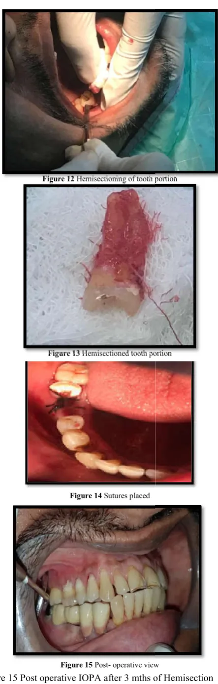
periapical radiograph revealed a case of instrument separation associated with the mesial root of 46and perforation in the floor of pulp chamber. [Figure 11]. After all investigations, treatment plan was decided. According to the plan, hemisection of the root canal treated tooth followed by prosthetic rehabilitation was the forethought. Conservation of tooth structure was better possible through this treatment plan.[Figure 10
Periodontal dressing (CoePack)
operative radiograph after 3 months, showing healed socket, with root canal done for the distal root
operative view with fixed prosthesis
Figure 9 Post – operative radiograph after 4months
Figure 10 Pre-operative tooth
Figure 11 Preoperative tooth Radiographic view
A 46 year old male patient reported with a chief complaint of the right back tooth region since 5 days. History of root canal treatment initiation reported one month back. On clinical access cavity in relation to 46 and deep cervical abrasion in relation to 44 and 43 with gingival
[Figure 1o]. An intra oral
periapical radiograph revealed a case of instrument separation associated with the mesial root of 46and perforation in the floor [Figure 11]. After all investigations, treatment rding to the plan, hemisection of the root canal treated tooth followed by prosthetic rehabilitation was the forethought. Conservation of tooth structure was better possible through this treatment plan.[Figure 10- 17].
operative radiograph after 4months
operative tooth- Clinical view
International Journal of Recent Scientif
Figure 15 Post operative IOPA after 3 mths of Hemisection prior to prosthetic rehabilitation
Figure 12 Hemisectioning of tooth portion
Figure 13 Hemisectioned tooth portion
Figure 14 Sutures placed
Figure 15 Post- operative view
International Journal of Recent Scientific Research Vol. 10, Issue, 02(D), pp. 30935-30939
mths of Hemisection
DISCUSSION
Hemisection is apredictable procedure for preservation of multi-rooted tooth as an alternative to extraction. The patient’s oral hygiene status, caries index and medical status should be considered before procedure. Root furcation must be accessible for easy separation and good bone support for remaining root is essential.[5] Endodontic evaluation and proper prosthetic designing increases the longevity of the tooth preserved. Thus, a multi-disciplinary approach enhances the prognosis of hemisectioned tooth.
Both the cases were ideal, necessitating hemisection as a treatment alternative to extraction. Also, the patients were motivated to try and save as much of the tooth as possible. In case report 1, there was a vertical root fracture involv mesial root of 37 associated with a non
sinus,necessitating extraction. Endodontic re treatment was planned for the distal root since the fracture line was involving the mesial root. So hemisection was preferred over bicuspidization. Fixed partial denture in relation to 36, 37 and 38 was done.
In case report 2, in relation to 47, the mesiobuccal canal could be negotiated to bypass the separated instrument but since the mesiolingual canal was calcified and perforation was more mesiobuccally located, hemisection was opted for the preservation of the tooth. Root canal treatment was also done for the deep cervical lesion in relation to 45, 44. Fixed partial denture in relation to 44, 45, 46, 47, 48 was done with narrow occlusal table and shallow cusps. Gingival porcelain was given in relation to 44 and 45 for esthetics.
Buhler has stated that hemisection should be considered before every molar extraction, given its advantages such as provision of a good, absolute and biological cost saving alter
good long- term success. An improperly shaped occlusal contact area may convert acceptable forces into destructive forces and predispose the tooth to trauma from occlusion and ultimate failure of hemisection
It has been concluded by Saad
mandibular molar may be a suitable treatment option when the decay is restricted to one root and the other root is healthy and remaining portion of tooth can very well act as an abutment, as in the presented cases.
Hemisectioning of tooth portion
Hemisectioned tooth portion
operative view
Figure 16 Post operative IOPA after 4mths of prosthetic rehabilitation
30939, February,, 2019
Hemisection is apredictable procedure for preservation of rooted tooth as an alternative to extraction. The patient’s oral hygiene status, caries index and medical status should be considered before procedure. Root furcation must be ccessible for easy separation and good bone support for Endodontic evaluation and proper prosthetic designing increases the longevity of the tooth disciplinary approach enhances the sectioned tooth.
Both the cases were ideal, necessitating hemisection as a treatment alternative to extraction. Also, the patients were motivated to try and save as much of the tooth as possible. In case report 1, there was a vertical root fracture involving the mesial root of 37 associated with a non-healing sinus,necessitating extraction. Endodontic re treatment was planned for the distal root since the fracture line was involving the mesial root. So hemisection was preferred over partial denture in relation to 36, 37 and
In case report 2, in relation to 47, the mesiobuccal canal could be negotiated to bypass the separated instrument but since the mesiolingual canal was calcified and perforation was more located, hemisection was opted for the preservation of the tooth. Root canal treatment was also done for the deep cervical lesion in relation to 45, 44. Fixed partial denture in relation to 44, 45, 46, 47, 48 was done with narrow cusps. Gingival porcelain was given in relation to 44 and 45 for esthetics.
Buhler has stated that hemisection should be considered before every molar extraction, given its advantages such as provision of a good, absolute and biological cost saving alternative with An improperly shaped occlusal contact area may convert acceptable forces into destructive forces and predispose the tooth to trauma from occlusion and ultimate failure of hemisection [6]
It has been concluded by Saad et al[7], that hemisection of a mandibular molar may be a suitable treatment option when the decay is restricted to one root and the other root is healthy and remaining portion of tooth can very well act as an abutment, as
Successful restoration of periodontally weakened teeth is aided by creating an occlusal scheme with canine protected occlusion, decreased vertical overlap and flattened posterior cusps.[8]
CONCLUSION
In the era of conservative dentistry, dental practitioners should be encouraged to work together in an interdisciplinary approach for the preservation and maintenance of tooth structure that can be retained. For this, timely intervention with hemisection and endodontic therapy followed by planned prosthetic rehabilitation should be adopted as one of the routine procedures.
References
1. Babaji P, Sihag T, Chaurasia VR, Senthilnathan S. Hemisection: A conservative management of periodontally involved molar tooth in a young patient. J Nat Sci Biol Med. 2015; 6(1): 253- 5.
2. Weine FS. Endodontic Therapy, 5 th ed. St. Louis: Mosby; 1996
3. Mukherjee M, Riyas AB M. Hemisection of mandibular molar: Hopeless to hoping. J Cont Med A Dent. 2017; 5(2): 79- 81.
4. Verma PK, Srivastava R, Baranwal HC, Gautam A. A ray of hope for the hopeless: Hemisection of mandibular molar with socket preservation. Dent Hypothesis. 2012; 3: 159- 63.
5. Akki S, Mahoorkar S. Tooth hemisection and restoration an alternative to extraction- A Case Report. International Journal of Dental Clinics. 2011; 3(3): 67- 8.
6. Buhler H Survival rates of hemisectioned tooth:an attempt to compare them with survival rates of alloplastic implant .Int J Periodontics Restorative Dent 1994;14:536-543.
7. Saad MN, Moreno J, Crawford C. Hemisection as an alternative treatment for decayed multirooted terminal abutment: a case report. J Can Dent Assoc. 2009;75(5):387-90. Shillinburg .Fundamentals of Fixed Prosthodontics. 4th edition
How to cite this article:
Haseena Ummer et al,. 2019,Preservation OF Mandibular Molars with Hemisectiona Series Of Interdisciplinary Case Reports. Int J Recent Sci Res. 10(02), pp.30935-30939. DOI: http://dx.doi.org/10.24327/ijrsr.2019.1002.3156