________________________________ Author for Correspondence:
Ch.Kishore,
Holy Mary Institute of Technology and Science (College of Pharmacy), Bogaram, Keesara, Ranga Reddy (DT),
Telangana State. 200253
Research Article
ISSN Print 2231 – 3648 Online 2231 – 3656
Available Online at: www.ijpir.com
Preparation and evaluation microspheres with HPMC polymer
Ch.Kishore*, N.Sriram*
Department of Pharmaceutics, Holy Mary Institute of Technology and Science (College of
Pharmacy) Bogaram, Keesara, Ranga Reddy (DT), Telangana State.
ABSTARCT
The aim of this study was to formulate and evaluate microspheres of Ranitidine. These formulations are mainly usefull in inhibition of gastric acid secretion. It shows action on histamine stimulation and gastrin stimulated acid secretion. The compatability was first checked with FTIR spectrophotometric for drug and excipients. The prepared product was subjected to various studies like drug entrapment efficiency particle size, bulk density, % yield, % buoyancy, etc. The individual peaks of each drug and the excipients were compared and the compatibility was studied and this was done for all the formulations in the preparations.
Keywords:
Floating microspheres, Ranitidine, In vitro relea-se, BioavailabilityINTRODUCTION
Floating Drug Delivery Systems (FDDS) or Hydrodynamically Balanced Systems (HBS) are among the several approaches that have been developed in order to increase the gastric residence time (GRft) of dosage forms (1–3).The efficacy of a drug in a specific application requires the maintenance of appropriate drug blood level concentration during a prolonged period of time. However the conventional administration of drugs, gives a poor control of the concentration of these substances in plasma because of variations in the concentration of the bioactive product, once a specific dose has been administered [4]. In the form of NDDS, existing drug molecule can get a new life, thereby increasing the market value and product patent
life. Controlled release, prolonged action, sustained release, extended release, depot dosage forms are terms used to identify these drug delivery systems that are designed to achieve prolonged therapeutic effect by continuously releasing medication over an extended period of time after administration of single dose [5]. Specific ranitidine uses include treatment or prevention of the following conditions:
Duodenal ulcers, Gastric ulcers (stomach ulcers), Gastroesophageal reflux disease (GERD) and Erosive esophagitis
Pathological hypersecretory conditions (in which too much stomach acid is produced), such as Zollinger-Ellison syndrome.The primary objective of zero-order release is to up-hold constant drug concentration in blood
for a prolonged period of time. Microspheres have played a vital role in the development of controlled/sustained release drug delivery systems [6-7]. Pharmaceutically acceptable techniques using hydrophobic biodegradable polymers as matrix materials include Emulsion-solvent evaporation, Phase separation (non solvent and solvent partitioning), Interfacial polymerization and Spray drying. Still, the multiple-unit dosage forms may be better suited because they are claimed to reduce the intersubject variability in absorption and lower the probability of dose dumping (6). Such a dosage form can be distributed widely throughout the gastrointestinal tract (GIft), affording the possibility of a longer lasting and more reliable release of the drug from the dosage form (7).
MATERIALS AND METHODS
Ranitidine (KAPL, Bangalore), HPMC (Merck laboratories), SLS (SD fine chemicals), Ethyl cellulose (SD fine chemicals), PVA (Merck laboratories), Chitosan, (SD fine chemicals), Tween 20 and HCl (Asha Reagents), Ethanol (SD fine-chemicals limited) and all other solvents and reagents used were of analytical grade
Formulation design of Ranitidine
Solvent diffusion method was used as a procedure for the preparation of the microspheres, Dichloromethane and ethanol was used as solvent in 1:1 ration for drug and polmer slurry solution preparation, The slurry was slowly introduced into 200 ml of water containing (0.75% w/v) polyvinyl alcohol maintained at a constant temperature of 40 °C with continuous stirring at 300 rpm using a propeller type mechanical stirrer. The solution was stirred for 2 hrs. The finely developed floating microspheres were separated by filtration washed with water & dried at room temperature in a dessicator for 24 hrs. The formulation was divided into nine batches prepared with different ratio of suitably chosen polymers as depicted in the table below.
Evaluation
Content uniformity / drug loading
The prepared microspheres were powdered and passed through sieve no (85/120). The
powder retained on the sieve 120 was taken for content uniformity studies. A weight of powder containing 100 mg of the drug was taken in a 100ml standard volumetric flask. To this of 0.1 N NaOH solutions was added and made upto the mark with 0.1 N NaOH solutions and kept overnight. The final solution was filtered using what man filter paper. From this 10 ml was pipetted out into a 100 ml standard volumetric flask and made upto the volume with 0.1 N NaOH solution and estimated spectrophotometrically for drug content.
Entrapment efficiency
To evaluate the amount of the drug inside the microspheres, an indirect method was used. Aliquots from the filtered solutions remaining after removal of the microspheres were assayed spectrophotometrically. The amount of drug entrapped was calculated from the difference between the total amount of drug added and the amount of drug found in the filtered solution. About 100 mg of microspheres were completely dissolved in 500 ml of phosphate buffer solutions (pH 7.4), and stirred for 1h. Then, 2 ml of solution was filtered and the concentration of drug was determined spectrophotometrically by UV. Efficiency of drug entrapment was calculated in terms of percentage drug entrapment (PDE) as per the following formula:
PDE = (Practical drug loading/Theoretical drug loading) ×100
Surface morphology
The surface morphology of microspheres was examined by scanning electron microscopy.
Percentage Yield
The maximum % yield was found to be 79.60% with batch F7 and minimum of 66.92% with F6 batch.
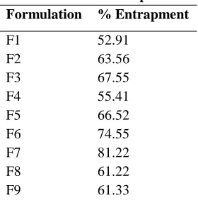
Drug Entrapment Efficiency
Table.1 % of entrapment Formulation % Entrapment
F1 F2 F3 F4 F5 F6 F7 F8 F9
52.91 63.56 67.55 55.41 66.52 74.55 81.22 61.22 61.33
In vitro dissolution study
All the release data were fitted into various kinetic models like, zero order, First order, Higuchi and Korsmeyer-peppas in order to find out the mechanism of drug release from polymeric microspheres. The correlation & diffusion coefficients were calculated as summarized in table.
Analysis of the release data as per zero order kinetic model best suited to describe the release rate of drug from the microspheres. When the release data was analyzed as per peppas equation, the release exponent ‘n’ was in the range of (0.531-0.742) with all the microspheres indicating non-fickian diffusion. Higuchi’s plots resulted in linearity (r2> 0.932) indicating non- fickian diffusion mechanism.
0 200 400 600 800
Time (min) Fig.1 % release of drug
Table 2. Correlation coefficients of different mathematical models for microspheres.
Zero order First order Higuchi
Korsem
Sl. Formulations R2 R2 R2 n r2
1 F1 0.7501 0.9628 0.9871 0.3048 0.9911
0.7641 0.9587 0.9913 0.3472 0.9934 0.7823 0.9362 0.9890 0.3681 0.9919
2 F2 0.7695 0.9719 0.9801 0.3161 0.9943
0.7825 0.9612 0.9907 0.3325 0.9907
0.7548 0.9744 0.9923 0.3475 0.9915
3 F3 0.8077 0.9816 0.9918 0.3400 0.9970
0.7941 0.9787 0.9972 0.3486 0.9956 0.7883 0.9702 0.9892 0.3952 0.9899
4 F4 0.8977 0.9847 0.9921 0.4284 0.9892
0.9004 0.9814 0.9913 0.4374 0.9905 0.8875 0.9686 0.9864 0.4473 0.9912
5 F5 0.8917 0.9739 0.9881 0.5082 0.9858
0.7926 0.9810 0.9908 0.4896 0.9935 0.8245 0.9785 0.9947 0.4976 0.9972 6 F6 0.8880 0.9611 0.9749 0.6001 0.9594 0.8754 0.9638 0.9814 0.5074 0.9845 0.8633 0.9705 0.9894 0.4921 0.9913
Table.3 % Drug release Formulation % Drug Release
F1 F2 F3 F4 F5 F6 F7 F8 F9
62.22 63.11 65.66 68.22 69.00 70.22 74.33 75.22 76.00
Scanning electron microscopy
Scanning electron microscopy (SEM) is one of the most commonly used method for characterizing drug delivery systems, owing in large part of simplicity of sample preparation and ease of operation. Scanning electron microscopy was carried out in order to characterize surface morphology of the microspheres. In this study the
morphological observations were carried out to study the surface morphology of microspheres. SEM micrographs and typical surface morphology of the microspheres are given in figures 2-4. It was observed that microspheres were spherical in nature [Fig. (2-4).], The microspheres ranged in size 150 to 408µm.
CONCLUSION
The microspheres of Ranitidine HCl were prepared with two polymers i.e. HPMC and chitosan. The particle size determination by SEM techniques revealed that the mean particle diameter was in the range of 122.60 - 189.55µm. The mean particle size were in the order of F2<F1 < F6 < F4 < F5 < F3 < F7 < F9< F8. The other physicochemical parameters determined with the microspheres were bulk density (0.23- 0.65g/ml), particle size distribution (126.80- 195.21µm), % yield (63.92%-77.60%), buoyancy % in pH 1.2 HCl buffer (53.72%- 64.45%), tapped density (0.34-0.85g/ml) and drug entrapment efficiency (51.91%-80.42%). The in vitro drug release in pH 1.2 HCl buffer ranged from 83.54%-55.10% while in simulated gastric fluid it ranged from
84.82%-56.76%. The overall determinations suggested F7 batch as the best formulation. Conclusively % yield was maximum with F7 and minimum with F6 batch. The drug entrapment efficiency was found to be of the order F1 < F7 < F4 < F8 < F2 < F9 < F5 < F3 < F6.The overall determinations suggested F7 as the best formulation. The in-vitro release of formulation F7 in pH1.2 HCl buffer and in simulated gastric fluid (SGF) were 63.12% and 58.51% respectively which showed sustained release over a period of 12 hrs. All above data satisfactorily complied with the characteristics requirements of the formulation as gastroretentive microspheres. This research work was done to attempt to prove the chances of more advantages of novel forms over conventional dosage forms.
REFERENCES
[1]. P. R. Seth and J. ftossounian, fthe hydrodynamically balanced system HBSftM: A novel drug de- livery system for oral use, Drug Dev. Ind. Pharm. 10, 1984, 313–339.
[2]. A. J. Moes, Gastroretentive dosage forms, Crit. Rev. Ther. Drug Carrier Syst. 10, 1993, 143–195. [3]. A. A. Deshpande, C. ft. Rhodes, N. H. Shah and A. W. Malick, Controlled-release drug
delivery systems for prolonged gastric residence: an overview, Drug Dev. Ind. Pharm. 22,
1996, 531–539.
[4]. Bhaskar Mazumder., Sanjib Bhattacharya., Bibhash Mohanta., Sanjay Dey., Anindya Maity. Int J
PharmTech Research, 1(3), 2009, 905-913.
[5]. Meral Yuce., Kandemir Canefe. Turk J. Pharm. Sci, 5 (3), 2008, 129-142.
[6]. Deore B.V., Mahajan H.S., Deore U.V. Int J ChemTech Research, 1(3), 2004, 634-642. [7]. Tamer Guneri., Mesut Arici., Gokhan Ertan. Fabad j. pharm. Sci, 29, 2004, 177-184.
[8]. Suranjana Roy. Manjusree Pal., Bijan K. Gupta. Pharmaceutical research, 9(9), 1992, 1132-1136. [9]. N. Rouge, J. C. Leroux, E. ft. Cole, E. Doelker and P. Buri, Prevention of the sticking
tendency of floating minitablets filled into hard gelatin capsules, Eur. J. Pharm. Biopharm.
43, 1997, 165–171.