Structural Modifications to DNA-Binding Polyamides for
Improved Biological Activity in Cell Culture
Thesis by Claire Jacobs
In Partial Fulfillment of the Requirements for the Degree of
Doctor of Philosophy
California Institute of Technology Pasadena, California
2009
Acknowledgements
I would like to thank my advisor, Professor Peter Dervan, and my fellow Dervanites for creating such a diverse and stimulating place to work. I count myself lucky to have shared a lab with Justin, Ryan, Jim, Christian, and Dave C., both for their scientific prowess and companionship, which made the late nights in lab a lot more fun and a little less frustrating. Katy graciously provided tireless proofreading services, helpful suggestions, and vital material support in the form of baked goods. Puckett knows a lot about almost everything, and I will miss dissecting current events with him. Dan provided invaluable suggestions on a manuscript and a cool head in Dervana. Anne’s sage advice during daily coffee runs is gratefully acknowledged. I would also like to thank Mareike, Nick, Fei, Sherry, Julie, Carey, Adam, John, Mike B., Dave, Ben E., Marques, Ray, Eric, Michelle, Ben L., and Dan G. It has been a pleasure working with you all, and I wish the best of luck to the newest members of the Dervan group.
I don’t have the room to thank all the people who looked out for me along the way. I do want to acknowledge Mrs. Rabin and Dr. Gordo Stonington for their early encouragement, Professor Steve Sibener for letting me into his lab as an undergraduate, and Professor Ka Yee Lee for being an inspiration. I would like to thank Kat, Russell, and Sherry for their generosity and care. I am grateful to Erika, Marten, Skumfi, and Adri for their friendship and camping companionship. PY, Tabby, Mark, Nadia, Asim; I look forward to having the time to catch up with you all.
Abstract
Table of Contents
Acknowledgements ... iii
Abstract ... v
Table of Contents ... vi
List of Figures and Tables ... vii
Chapter 1 Introduction ... 1
Chapter 2 Improved Nuclear Localization of DNA-Binding Polyamides ... 23
Chapter 3 Modulating Hypoxia-Inducible Transcription by Disrupting the HIF-1-DNA Interface ... 48
Chapter 4 Effect of Linkger, Linkage, and Tail Modifications on Biological Activity of Pyrrole/Imidazole Polyamides ... 73
Appendix A ... 109
Appendix B ... 112
List of Figures and Tables
Chapter 1
Figure 1.1 Structure of DNA ... 3
Figure 1.2 Crystal Structures of two DNA-binding Natural Products... 4
Figure 1.3 Crystal Structure of Minor-Groove Recognition by Polyamide Heterocycles... 5
Figure 1.4 Structure of Eight-Ring Hairpin Polyamide and Putative Hydrogen Bonds to DNA... 6
Figure 1.5 Polyamide Structural Motifs... 7
Figure 1.6 Schematic of Solid-Phase Polyamide Synthesis... 8
Figure 1.7 Schematic of Gene Regulation by Polyamides... 8
Figure 1.8 DNA-Binding Proteins Inhibited by Polyamides... 9
Figure 1.9 !-Enhanseosome Structure... 10
Figure 1.10 Cellular Localization of Polyamide-Fluorophore Conjugates... 11
Figure 1.11 Structure of Hairpin Polyamide Indicating C-Terminal Linker, Linkage and Tail Groups... 14
Chapter 2 Figure 2.1 Structure of Polyamide-FITC Conjugates... 26
Figure 2.2 Summary of Biological Data for Polyamide-FITC Conjugates... 30
Figure 2.3 Structures of Polyamide Cores and Tail Groups... 31
Table 2.1 Binding Affinities of Compounds... 32
Figure 2.4 DNase I Footprint Titration Data for Lead Compounds... 33
Figure 2.5 DNase I Footprint Titration Data... 34
Figure 2.7 Quantitative RT-PCR Data, Lead Compounds, HeLa Cells... 36
Figure 2.8 Confocal Microscopy Uptake Data... 37
Figure 2.9 Quantitative RT-PCR and ChIP Data, U251 Cells... 38
Table 2.2 MALDI-ToF Data... 42
Chapter 3 Figure 3.1 Project Overview... 53
Table 3.2 Binding Affinities of Compounds... 54
Figure 3.2 Quantitative DNase I Footpring Titration Data... 55
Figure 3.3 Quantitative RT-PCR Data... 57
Table 3.2 Number of Transcripts Affected by Compounds... 58
Figure 3.4 Microarray Analysis Data... 59
Table 3.3 HIF-1 Induced Genes Affected by Compounds... 61
Figure 3.5 Ven Diagram Representation of Microarray Data... 62
Figure 3.6 Chromatin Immunoprecipitation Data... 63
Figure 3.7 Analytical HPLC Data... 66
Chapter 4 Figure 4.1 Project Overview... 76
Figure 4.2 Structures of Polyamide Cores and C-Terminal Modifications... 77
Figure 4.3 Quantitative RT-PCR Data, U251 and LNCaP Cell Culture... 80
Figure 4.4 Quantitative RT-PCR Data LNCaP Cell Culture... 81
Table 4.1 Melting Temperature Data... 82
Figure 4.5 Cell Viability Data for Control Compounds... 83
Figure 4.6 Quantitative RT-PCR Data for Oxime-Linked Compounds... 87
Table 4.2 IC50 Values for Cell Viability and Gene Regulation... 88
Table 4.3 MALDI-ToF and Purity Data... 95
Figure 4.8 Structures of Polyamide Cores and C-Terminal Tail Modifications... 98
Figure 4.9 Quantitative RT-PCR Data... 99
Figure 4.10 Quantitative RT-PCR Data... 101
Figure 4.11 Quantitative RT-PCR Data... 103
Table 4.4 Melting Temperature Data... 105
Table 4.5 Melting Temperature Data... 105
Appendix A Figure Time-Course Schematics, quantitative RT-PCR... 110
Chapter 1
Introduction
The information encoding the regulation and expression of the estimated 20,000–25,000 genes in the human genome and their products is contained in the nucleotide sequence of DNA1, 2. At the simplest level, the information encoded by nucleotide sequences is expressed
Structure of DNA
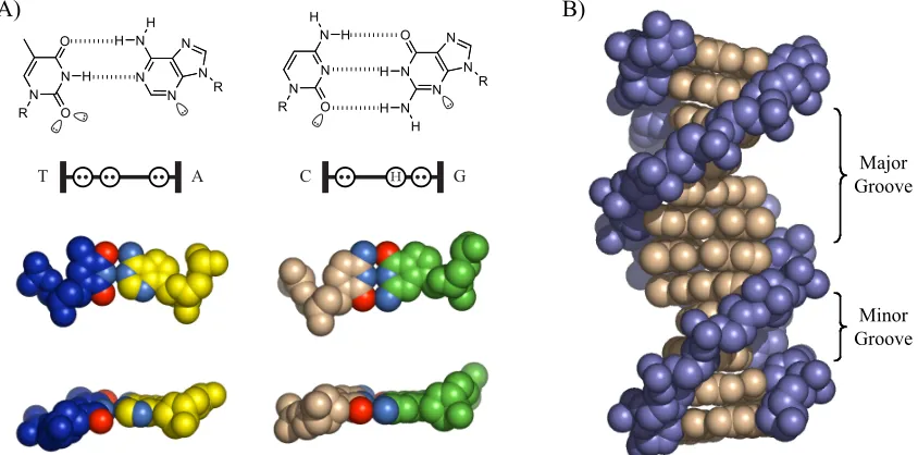
DNA is comprised of two complimentary polydeoxyribonucleotide strands paired in an antiparallel fashion through formation of hydrogen-bonding contacts between the nucleotide bases: adenine pairs with thymine, and guanine with cytosine (figure 1.1).3 Although DNA
can adopt a number of conformations, the biologically relevant is B-form DNA, in which the nucleotide pairing produces a double-helical strand with a wide and shallow major groove, and a deep, narrow minor groove between the phosphate backbones. To a rough approximation, the DNA double helix is uniform, but a series of hydrogen-bonding donors and acceptors located on the edges of nucleotide bases are presented in the major and minor grooves that function as “handholds” for DNA-binding proteins and natural products.
C H G
N N
N N O
N N
N N
O
R
R H H
H
H H
T A
N N
N N N
N N
O
O
R H
H
H
R
A)
Major Groove
Minor Groove
[image:11.612.113.535.317.526.2]B)
Figure 1.1. Schematic illustration of B-form DNA. A) Schematic of hydrogen-bond formation between a T•A basepair and a C•G basepair. Also shown is a schematic representation of the hydrogen-bonding donor and acceptor pattern presented by the basepair edges into the minor groove. Lone pairs are represented by two dots in an open circle, and hydrogen bonding donors represented by an “H” in an open circle. B) Structure of B-form DNA to illustrate the major and minor grooves of B-form DNA.
DNA-Binding Natural Products
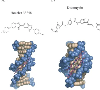
Two natural products that bind DNA with some degree of sequence specificity are Hoechst 33258 and distamycin (figure 1.2).4-7 Courtesy of their crescent shape, both these relatively
complex with DNA: the latter is illustrated in figure 1.2B). Distamycin has a preference for A,T tracts of DNA due to hydrogen-bonding contacts between the amide-bond hydrogen and minor-groove lone pairs; G•C pairs present an energetically unfavorable steric clash
between the exocyclic amine of guanine and hydrogens on the pyrrole ring. Replacement of a pyrrole ring with an N-methylimidazole ring, however, enabled G•C recognition via
the Nitrogen lone pair, which forms a hydrogen bond with the guanine exocyclic amine.8
A)
NH
N N
H N
N H N
OH Hoechst 33258
B)
N
NH O
N H
O N
N H O
N O
HN
NH2
NH2
[image:12.612.151.494.221.531.2]Distamycin
Figure 1.2. Schematic illustration of the chemical structure of Hoechst 33258 and Distamycin, and structures of DNA-Hoechst 33258 and DNA-distamycin complexes.
Polyamide Recognition of DNA
bonds.9-16 DNA binding can occur in a 1:1 or a 2:1 fashion, with polyamide strands
generally aligning N " C with the 5’ " 3’ DNA strand direction. The sickle shape of
polyamides closely matches the radius of the DNA minor groove, and allows polyamides to approach the DNA minor groove floor closely enough to engage in these hydrogen-bonding interactions.
Minor Groove
Minor Groove
Hp
Py
Py Im
A T
G C
5' 3'
5' 3'
T C T A G A
A G A T C T
+ +
[image:13.612.111.538.190.417.2]H H
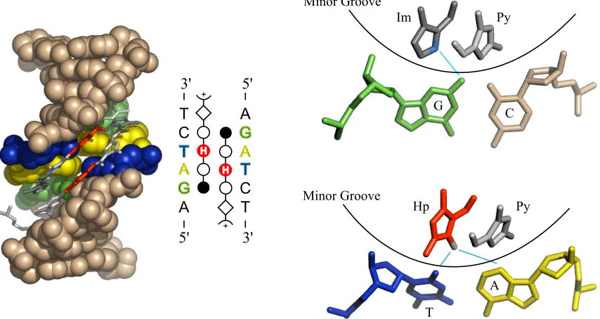
Figure 1.3. Structure of a 2:1 polyamide-DNA complex, and illustration of hydrogen-bonding interactions between the polyamide heterocycle core and the minor groove floor.
Polyamide sequence specificity is programmed through side-by-side pairings of heterocyclic amino acids in the DNA minor groove: Im/Py recognizes G•C over C•G; Py/
Py is degenerate for A•T and T•A; Hp/Py and Hz/Py distinguish T•A for A•T; and Ct/Py
prefers T•A over A•T in the cap position. These pairing guidelines were revealed through
quantitative DNase I footprinting titrations, as well as NMR and crystallographic structural data. These data have also elucidated the peculiarities of polyamide-DNA interactions that give rise to the sequence specificity. A network of hydrogen bonding interactions is created between the polyamide amide bonds and base-pair edges, while hydrogen bonding between the nitrogen lone pair of Im and the exocyclic amine of guanine forms the basis for G•C recognition (figure 1.4).17-19 Hp/Py recognizes T•A through hydrogen
shape-6
selective recognition of an asymmetric cleft between thymine-O2 and adenine-C2.17, 19 The
proposed recognition of T•A at the amino terminus by Ct/Py projection of the 3-chloro
substituent awaits structural verification.20
N N N N O N N N N O R R H H H H N N N N O N N N N O R R H H H H
G H C C H G
A T T A
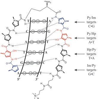
T C A T G A 5’ 3’ H H A G T A C T Py/Im targets C•G Py/Hp targets A•T Hp/Py targets T•A Im/Py targets G•C N N N N N N N O O R H H H R N N N N N N N O O R H H H R N N O N H N
O N H
[image:14.612.163.492.129.462.2]N N O H N N O H N N N O H N N O H N N O H N N H O N O H O N N H H OH HO NH3
Figure 1.4. Schematic illustration of the system of hydrogen bonding pattern between the DNA nucleotide edges and an eight-ring hairpin polyamide that gives rise to DNA recognition by polyamides.
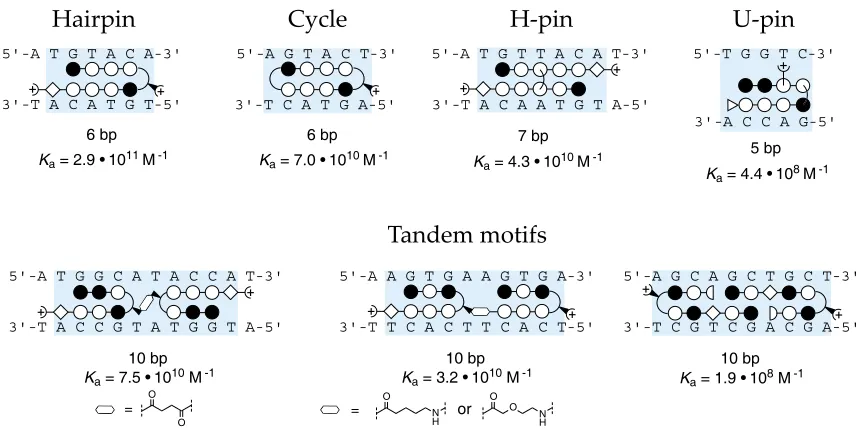
A number of polyamide architectures have been explored, and the most commonly deployed is the eight-ring hairpin polyamide with a binding site of six basepairs (figures 1.4, 1.5). Linking two antiparallel heterocycle strands through an aliphatic linker group to create a “hairpin” polyamide results in substantial gains in DNA-binding affinity (Ka(M-1)).21 This
gain in binding affinity is thought to occur as a result of reduction in the entropic cost of polyamide-DNA binding. Linking the two strands additionally enforces side-by-side heterocycle pairing rather than the slipped binding modes available to unlinked heterocycle strands.21, 22 The most commonly employed turn group has been the #-amino butyric acid,
imbues the turn residue with a strong preference for T•A and A•T pairs over G•C and C•G
pairs.21
5'-A T G T A C A-3'
3'-T A C A T G T-5'
!"#$
%&!"!#$%!&!"'(''")*' + +
5'-A G T A C T-3'
3'-T C A T G A-5'
!"#$
%&!"!'$(!&!"'('(")*' +
5'-A T G T T A C A T-3'
3'-T A C A A T G T A-5'
+"#$
%&!"!)$*!&!"'('(")*' + +
5'-T G G T C-3'
3'-A C C A G-5' ,"#$
%&!"!)$)!&!"'(-")*'"" +
.
.
5'-A T G G C A T A C C A T-3'
3'-T A C C G T A T G G T A-5'
'("#$
%&!"!'$+!&!"'('(")*'
/
+ +
5'-A G C A G C T G C T-3'
3'-T C G T C G A C G A-5'
'("#$
%&!"!,$%!&!"'(-")*' + + . . 0 1 . 0 1
5'-A A G T G A A G T G A-3'
3'-T T C A C T T C A C T-5'
'("#$
%&!"!*$#!&!"'('(")*'
23 /
+ +
!"#$%#& '()*+ !,%#& -,%#&
[image:15.612.108.538.123.338.2]."&/+01023#45
Figure 1.5. Schematic ball-and-stick representations of various polyamide structural motifs. Below each motif is given the binding site size and binding affinities (Ka (M-1)).
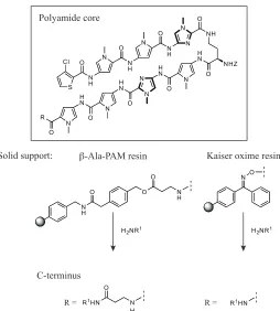
The modular nature of polyamides makes their synthesis amenable to solid-phase synthesis methods on resins including !-Ala-PAM and Kaiser oxime resin; use of !-Ala-PAM resin
results in a polyamide product with a C-terminal !-alanine moiety that is specific for A•T
basepairs (figure 1.6).24, 25 Following resin loading with the initial C-terminal, standard
Boc coupling chemistry is used to build the polyamide in the C"N direction. Aminolysis
S Cl
N H
O N N
H O N O N H N N NH O H N O NHZ N H N O N N O H N N O H N N R O Polyamide core Solid support: N O N H O O O N H
H2NR1 H2NR1
N H O
R1HN R1HN
C-terminus
!-Ala-PAM resin Kaiser oxime resin
R = R =
Figure 1.6. Schematic illustration of solid-phase synthesis of polyamides.
Regulation of Gene Expression with Polyamides:
The sequence-specificity of polyamides presents a potentially powerful tool for modulating gene expression in pursuit of disease treatment. Two possible mechansims by which gene regulation could be achieved are illustrated schematically below (figure 1.7). The first involves direct steric blockade of transcription factor binding in the promoter region of a gene, and the second mechanism is based on recruiting transcriptional machinery to the promoter through use of activator domains.
A
Response Element gene
X
TF TF
R + X
B
Response Element gene +
[image:16.612.187.441.70.352.2]TF TF
The binding affinity of most eight-ring hairpin polyamides is on the order of 109-1010 M-1,
which makes them competitive with DNA-binding proteins such as transcription factors and transcriptional activators (figure 1.8).26 By targeting polyamides to protein binding
sites or adjacent sequences, in vitro studies have demonstrated the ability of polyamides to inhibit DNA binding of proteins through direct steric or allosteric effects.27 Major-groove
binding proteins have been shown to co-occupy DNA sequences with minor-groove binding polyamides, but they can be displaced by intercalator-polyamide conjugates.28-30
HIV-1 Promo ter
5'- G A G C T G C A T C C G G A G T A C T A C A A A G A C -3 '
3'- C T C G A C G T A G G C C T C A T G A T G T T T C T G -5 '
X en opustRN A Prom oter Scann ing (-3 9 to -8)
5 '- C C C A T C C A A G T A C A T C G A A T T C A T G A C- 3'
3 '- G G G T A G G T T C A T G T A G C T T A A G T A C T G- 5'
H IV-1 Promote r Scanning
5 '- G A G C T C G T C C T C A G A T G C T G C A T A T A A- 3'
3 '- C T C G A G C A G G A G T C T A C G A C G T A T A T T- 5'
Aca ete-scute Promoter
5' -G C A T C G T A G C T C G T C A C G C G A C A G G G C -3 '
3' -C G T A G C A T C G A G C A G T G C G C T G T C C C G -5 '
HER2 Promote r
5' -G G G C T G C T T G A G G A A G T A T A A G A A -3 '
3' -C C C G A C G A A C T C C T T C A T A T T C T T -5 '
HTLV-1 Promote r
5' -C T C A G G C G T T G A C G A C A A C C C C T C- 3'
3' -G A G T C C G C A A C T G C T G T T G G G G A G- 5'
design ed sequence
5' -G A G G C T G G A T G G C C T T C C C C A T T A- 5'
3' -C T C C G A C C T A C C G G A A G G G G T A A T- 3'
designed seque nce ARE-1
5 '-C C A T G G T T G C T G A C T A A T T G T T A T- 3'
3 '-G G T A C C A A C G A C T G A T T A A C A A T A- 5'
HIV-1 LTR (+ 8 to +32 )
5' -T C T G G T T A G A C C A G A T C T G A G C C T -3 '
3' -A G A C C A A T C T G G T C T A G A C T C G G A -5 '
+ 5S RNA p romo ter
5'- C G G G C C T G G T T A G T A C T T G G A T G G G A G -3 '
3'- G C C C G G A C C A A T C A T G A A C C T A C C C T C -5 '
H IV-1 Promot er
5 '- T C A G A T G C T G C A T A T A A G C A G C T G C T T -3'
3 '- A G T C T A C G A C G T A T A T T C G T C G A C G A A -5'+ +
+ + + + + + + + + + + + + + + PR R + + T FIIA-TFIID TBP Ets-1 LEF-1 T BP Deadpan DNA gyrase T ax CREB T ax
GCN4 TFIIIB or T BP
LSF LSF
5S R NA promote r
5 '- C G G G C C T G G T T A G T A C T T G G A T G G G A G- 3'
3 '- G C C C G G A C C A A T C A T G A A C C T A C C C T C- 5'+
5S RNA promoter (+63 to +89)
5S RNA promoter (+63 to +89)
HIV-1 Promoter
HIV-1 Promoter
HIV-1 Promoter Construct
XenopustRNA Promoter Construct (-39 to -8)
Achaete-scute Neural Promoter Construct
HER2/neuPromoter
HTLV-1 Promoter
designed construct from pBR322
designed construct ARE-1 HIV-1 LTR (+8 to +32)
0 .03 nM
2 nM
0.05 nM
0.06 nM 0.0 5 nM
0.0 5 nM
0.4 nM
0.03 nM
0.07 nM 0 .4 nM
0.1 nM
1µM 1µM
0 .1 nM
2 nM 0.7 nM 0.9 nM
T FIIIA (zinc finger 4) T FIIIA (zinc finger 4)
Ets
Figure 1.8: Schematic illustration in the ball-and-stick fashion of inhibition of protein binding by polyamides. Indicated by light-blue boxes are the protein binding sites to show overlap between polyamide and protein binding sites. Also given are polyamide dissociation constants (Ka (M-1)).
binding sites may result in promiscuous polyamide binding thruought the genome and numerous “off-target” effects. This concern is mitigated by consideration of known DNA-protein interactions. Protein-DNA recognition, e.g. transcription factor-DNA recognition, is the result of a combination of electrostatic interactions with the polyanionic-DNA backbone and Van der Waals forces acting between the protein and the major or minor groove floors.31-36 In the cellular context, the DNA double helix is not suspended in the
[image:18.612.110.537.295.597.2]nucleus in isolation, but is constantly interacting with proteins and other molecules in the nuclear millieu. These protein binding sites are generally no more than four basepairs, the same size as eight-ring hairpin polyamide binding sites.37
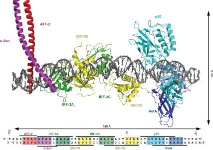
Figure 1.9. Structure of the interferon-b enhanceosome complex to demonstrate the relatively limited size of protein DNA binding sites, and protein-induced perturbations in the DNA double helix. Figure based on Figure 4 in Reference 37, and generated in PyMOL using a composite binding model obtained from the pdb file in the Supplemental Information.
Nuclear Localization of Polyamides:
goal, polyamides must be able to enter the cell and accumulate in the nucleus in sufficient quantities to bind to the appropriate DNA sequence and disrupt the protein-DNA interface. The mechanism by which polyamides enter the cell and translocate to the nucleus is not well understood. Previous work in the Dervan group has shown that polyamide cell uptake is an energy-dependent process, as it is impeded by lowered temperatures and in glucose-lacking cell culture media38. Further, internal data have demonstrated that the biological
activity of polyamides is enhanced in some cases by co-administration of verapamil, which inhibits function of drug-efflux pathways.39
Extensive attention has been devoted to determining the structural features of polyamides that play a role in nuclear accumulation. Initial investigations were inspired by the work of Bashkin et al. demonstrating that a C-terminal fluorescein (FITC) dye enabled demonstrable nuclear localization visualized by confocal microscopy of live, unfixed cells.39 Following
this seminal piece, confocal microscopy techniques were used to examine the nuclear and subcellular localization of polyamides as a function of fluorophore identity, polyamide heterocycle core sequence and composition, and polyamide structure.38, 40, 41 These
investigations revealed that a number of factors influence polyamide cell entry, but did not lead to a set of predictive structural requirements for polyamide nuclear accumulation.
Polyamides in Biological Systems:
Although polyamides had successfully inhibited protein-DNA binding and polyamide– small molecule activator domain conjugates had recruited proteins to DNA in vitro, gene regulation by polyamides in vivo was less established.42-48 The most notable example of
polyamide gene regulation in cell culture was disruption of HIV-1 replication in human T cells subsequent to dosing with two polyamides targeted to sites adjacent to three transcription-factor binding sites.49 A critical outgrowth of the extensive confocal microscopy studies
of polyamide cell entry and subcellular localization patterns was the revelation that a wide range of heterocycle core-dye combinations are able to cross into the cell.
In collaboration with the group of Dr. Kaelin, a polyamide was designed to bind to the hypoxia-response element in the promoter region of the vascular endothelial growth factor (VEGF) gene.50 VEGF is among a host of genes regulated by the hypoxia-inducible
factor/Aryl-hydrocarbon-receptor nuclear translocator (HIF-1$/ARNT (or HIF-1!))
transcription factor heterodimer.51 Under normal oxygen tension, the $-subunit of
HIF-1$ is hydroxylated at two proline residues for subsequent ubiquitination and degradation;
under hypoxic conditions, the hydroxylation event does not occur, and instead the HIF-1$/ARNT heterodimer forms and binds to the hypoxia response element (HRE).52
angiogenesis, glycolysis, cell survival, drug resistance and matrix remodeling.53-57 As
hypoxic conditions are frequently experienced by rapidly growing tissues, such as cancers, HIF-regulated genes and VEGF in particular are of great interest to the medical community for their involvement in cancer progression and survival.55, 58, 59
Summary:
Although it has been shown that a wide array of polyamide core-dye conjugates are able to localize to the nucleus of a range of cells, the widespread use of dye conjugates is problematic due to the high cost and instability of fluorescent dyes. The thrust of the work presented in this thesis deals with efforts to identify structural modifications to polyamides that facilitate their biological activity, are chemically stable, and do not interfere with binding affinities and specificities. As one of the aims of this work was to avoid pricey fluorescent dyes, such as FITC, detecting cell uptake and nuclear localization by confocal microscopy was not an option. The recently published result demonstrating VEGF mRNA regulation in HeLa cells was instead adopted as a biological readout of polyamide biological activity.
N N O
N H N O N H
N N O H N N O
H N N
N O H N N O H N N O H N N H O N O H N HN O O OH NH3
G H C
A T A A 5’ 3’ T T
C H G
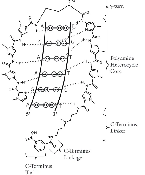
A T #-turn Polyamide Heterocycle Core C-Terminus Linker C-Terminus Linkage C-Terminus Tail
Figure 1.11. Schematic illustration of the hydrogen-bonding pattern mediating DNA recognition by a polyamide. The focus of work to identify structural modifications to improve polyamide biological activity discussed in this thesis, namely the C-Terminus linker, C-terminus linkage and C-terminus tail, are labeled.
[image:22.612.206.442.305.589.2]References:
1. Lander, E. S.; Linton, L. M.; et al., Initial sequencing and analysis of the human genome. Nature 2001, 409, (6822), 860-921.
2. Venter, J. C.; Adams, M. D., et al., The sequence of the human genome. Science
2001, 291, (5507), 1304-+.
3. Dickerson, R. E.; Drew, H. R.; Conner, B. N.; Wing, R. M.; Fratini, A. V.; Kopka, M. L., The Anatomy of A-DNA, B-DNA, and Z-DNA. Science 1982, 216, (4545), 475-485.
4. Mitra, S. N.; Wahl, M. C.; Sundaralingam, M., Structure of the side-by-side binding of distamycin to d(GTATATAC)(2). Acta Crystallographica Section D-Biological Crystallography 1999, 55, 602-609.
5. Kumar, R. A.; Ikemoto, N.; Patel, D. J., Solution structure of the calicheamicin gamma(I)(1)-DNA complex. Journal of Molecular Biology 1997, 265, (2), 187-201. 6. Quintana, J. R.; Lipanov, A. A.; Dickerson, R. E., Low-Temperature Crystallographic Analyses of the Binding of Hoechst-33258 to the Double-Helical DNA Dodecamer C-G-C-G-a-a-T-T-C-G-C-G. Biochemistry 1991, 30, (42), 10294-10306.
7. Arcamone, F.; Nicolell.V; Penco, S.; Orezzi, P.; Pirelli, A., Structure and Synthesis of Distamycin A. Nature 1964, 203, (494), 1064-&.
8. Mrksich, M.; Wade, W. S.; Dwyer, T. J.; Geierstanger, B. H.; Wemmer, D. E.; Dervan, P. B., Antiparallel Side-by-Side Dimeric Motif for Sequence-Specific Recognition in the Minor Groove of DNA by the Designed Peptide 1-Methylimidazole-2-Carboxamide Netropsin. Proceedings of the National Academy of Sciences of the United States of America
1992, 89, (16), 7586-7590.
9. Dervan, P. B., Molecular recognition of DNA by small molecules. Bioorganic & Medicinal Chemistry 2001, 9, (9), 2215-2235.
1998, 391, (6666), 468-471.
11. Briehn, C. A.; Weyermann, P.; Dervan, P. B., Alternative heterocycles for DNA recognition: The benzimidazole/imidazole pair. Chemistry-a European Journal 2003, 9, (9), 2110-2122.
12. Dervan, P. B.; Edelson, B. S., Recognition of the DNA minor groove by pyrrole-imidazole polyamides. Current Opinion in Structural Biology 2003, 13, (3), 284-299. 13. Marques, M. A.; Doss, R. M.; Foister, S.; Dervan, P. B., Expanding the repertoire of heterocycle ring pairs for programmable minor groove DNA recognition. Journal of the American Chemical Society 2004, 126, (33), 10339-10349.
14. Dervan, P. B.; Poulin-Kerstien, A. T.; Fechter, E. J.; Edelson, B. S., Regulation of gene expression by synthetic DNA-binding ligands. In DNA Binders and Related Subjects, 2005; Vol. 253, pp 1-31.
15. Viger, A.; Dervan, P. B., Exploring the limits of benzimidazole DNA-binding oligomers for the hypoxia inducible factor (HIF) site. Bioorganic & Medicinal Chemistry
2006, 14, (24), 8539-8549.
16. Chenoweth, D. M.; Poposki, J. A.; Marques, M. A.; Dervan, P. B., Programmable oligomers targeting 5’ -GGGG-3’ in the minor groove of DNA and NF-kappa B binding inhibition. Bioorganic & Medicinal Chemistry 2007, 15, (2), 759-770.
17. Kielkopf, C. L.; White, S.; Szewczyk, J. W.; Turner, J. M.; Baird, E. E.; Dervan, P. B.; Rees, D. C., A structural basis for recognition of A • T and T • A base pairs in the minor groove of B-DNA. Science 1998, 282, (5386), 111-115.
18. Kielkopf, C. L.; Baird, E. E.; Dervan, P. D.; Rees, D. C., Structural basis for G • C recognition in the DNA minor groove. Nature Structural Biology 1998, 5, (2), 104-109. 19. Kielkopf, C. L.; Bremer, R. E.; White, S.; Szewczyk, J. W.; Turner, J. M.; Baird, E. E.; Dervan, P. B.; Rees, D. C., Structural effects of DNA sequence on T • A recognition by hydroxypyrrole/pyrrole pairs in the minor groove. Journal of Molecular Biology 2000,
20. Foister, S.; Marques, M. A.; Doss, R. M.; Dervan, P. B., Shape selective recognition of T • A base pairs by hairpin polyamides containing N-terminal 3-methoxy (and 3-chloro) thiophene residues. Bioorg Med Chem 2003, 11, (20), 4333-40.
21. Mrksich, M.; Parks, M. E.; Dervan, P. B., Hairpin Peptide Motif – a New Class of Oligopeptides for Sequence-Specific Recognition in the Minor-Groove of Double-Helical DNA. J Am Chem Soc 1994, 116, (18), 7983-7988.
22. deClairac, R. P. L.; Geierstanger, B. H.; Mrksich, M.; Dervan, P. B.; Wemmer, D. E., NMR characterization of hairpin polyamide complexes with the minor groove of DNA. J Am Chem Soc 1997, 119, (34), 7909-7916.
23. Herman, D. M.; Baird, E. E.; Dervan, P. B., Stereochemical control of the DNA binding affinity, sequence specificity, and orientation preference of chiral hairpin polyamides in the minor groove. J Am Chem Soc 1998, 120, (7), 1382-1391.
24. Baird, E. E. D., P.B., Solid Phase Synthesis of polyamides containing imidazole and pyrrole amino acids. J Am Chem Soc 1996, 118, 6141-6146.
25. Belitsky, J. M.; Nguyen, D. H.; Wurtz, N. R.; Dervan, P. B., Solid-phase synthesis of DNA binding polyamides on oxime resin. Bioorg Med Chem 2002, 10, (8), 2767-74. 26. Trauger, J. W.; Baird, E. E.; Dervan, P. B., Recognition of DNA by designed ligands at subnanomolar concentrations. Nature 1996, 382, (6591), 559-561.
27. Dervan, P. B.; Edelson, B. S., Recognition of the DNA minor groove by pyrrole-imidazole polyamides. Curr Opin Struct Biol 2003, 13, (3), 284-99.
28. Oakley, M. G.; Mrksich, M.; Dervan, P. B., Evidence that a Minor Groove-Binding Peptide and a Major Groove-Binding Protein can simultaneously occupy a common site on DNA. Biochemistry 1992, 31, (45), 10969-10975.
29. Fechter, E. J.; Dervan, P. B., Allosteric inhibition of protein-DNA complexes by polyamide-intercalator conjugates. J Am Chem Soc 2003, 125, (28), 8476-8485.
minor-groove binding ligands. Biochemistry 2004, 43, (13), 3880-3890.
31. Nikolov, D. B.; Chen, H.; Halay, E. D.; Hoffmann, A.; Roeder, R. G.; Burley, S. K., Crystal structure of a human TATA box-binding protein/TATA element complex. Proc Natl Acad Sci U S A 1996, 93, (10), 4862-4867.
32. Leonard, G. A.; Hunter, W. N., Crystal and Molecular-Structure of D(CGTAGATCTACG) at 2-Center-Dot-25-Angstrom Resolution. J Mol Biol 1993, 234, (1), 198-208.
33. Kim, Y. C.; Geiger, J. H.; Hahn, S.; Sigler, P. B., Crystal-Structure of a Yeast Tbp Tata-Box Complex. Nature 1993, 365, (6446), 512-520.
34. Ellenberger, T. E.; Brandl, C. J.; Struhl, K.; Harrison, S. C., The Gcn4 Basic Region Leucine Zipper Binds DNA as a Dimer of Uninterrupted Alpha-Helices – Crystal-Structure of the Protein-DNA Complex. Cell 1992, 71, (7), 1223-1237.
35. Pabo, C. O.; Sauer, R. T., Transcription Factors – Structural Families and Principles of DNA Recognition. Ann Rev of Biochem 1992, 61, 1053-1095.
36. Pavletich, N. P.; Pabo, C. O., Zinc Finger DNA Recognition – Crystal-Structure of a Zif268-DNA Complex at 2.1-A. Science 1991, 252, (5007), 809-817.
37. Panne, D.; Maniatis, T.; Harrison, S. C., An atomic model of the interferon-beta enhanceosome. Cell 2007, 129, (6), 1111-23.
38. Best, T. P.; Edelson, B. S.; Nickols, N. G.; Dervan, P. B., Nuclear localization of pyrrole-imidazole polyamide-fluorescein conjugates in cell culture. Proc Natl Acad Sci U S A 2003, 100, (21), 12063-8.
39. Crowley, K. S.; Phillion, D. P.; Woodard, S. S.; Schweitzer, B. A.; Singh, M.; Shabany, H.; Burnette, B.; Hippenmeyer, P.; Heitmeier, M.; Bashkin, J. K., Controlling the intracellular localization of fluorescent polyamide analogues in cultured cells. Bioorg Med Chem Lett 2003, 13, (9), 1565-70.
Chem 2002, 10, (10), 3313-8.
41. Edelson, B. S.; Best, T. P.; Olenyuk, B.; Nickols, N. G.; Doss, R. M.; Foister, S.; Heckel, A.; Dervan, P. B., Influence of structural variation on nuclear localization of DNA-binding polyamide-fluorophore conjugates. Nucleic Acids Res 2004, 32, (9), 2802-18. 42. Chiang, S. Y.; Burli, R. W.; Benz, C. C.; Gawron, L.; Scott, G. K.; Dervan, P. B.; Beerman, T. A., Targeting the Ets binding site of the HER2/neu promoter with pyrrole-imidazole polyamides. J Biol Chem 2000, 275, (32), 24246-24254.
43. Yang, F.; Belitsky, J. M.; Villanueva, R. A.; Dervan, P. B.; Roth, M. J., Inhibition of Moloney murine leukemia virus integration using polyamides targeting the long-terminal repeat sequences. Biochem 2003, 42, (20), 6249-6258.
44. Simon, H.; Kittler, L.; Baird, E.; Dervan, P.; Zimmer, C., Selective inhibition of DNA gyrase in vitro by a GC-specific eight-ring hairpin polyamide at nanomolar concentration. Febs Letters 2000, 471, (2-3), 173-176.
45. Mapp, A. K.; Ansari, A. Z.; Ptashne, M.; Dervan, P. B., Activation of gene expression by small molecule transcription factors. Proc Natl Acad Sci U S A 2000, 97, (8), 3930-3935.
46. Ansari, A. Z.; Mapp, A. K.; Nguyen, D. H.; Dervan, P. B.; Ptashne, M., Towards a minimal motif for artificial transcriptional activators. Chemistry & Biology 2001, 8, (6), 583-592.
47. Arora, P. S.; Ansari, A. Z.; Best, T. P.; Ptashne, M.; Dervan, P. B., Design of artificial transcriptional activators with rigid poly-L-proline linkers. J Am Chem Soc 2002, 124, (44), 13067-13071.
48. Kwonj, Y.; Arndt, H. D.; Qian, M.; Choi, Y.; Kawazoe, Y.; Dervan, P. B.; Uesugi, M., Small molecule transcription factor mimic. J Am Chem Soc 2004, 126, (49), 15940-15941.
cells by synthetic DNA-binding ligands. Proc Natl Acad Sci U S A 1998, 95, (22), 12890-12895.
50. Olenyuk, B. Z.; Zhang, G. J.; Klco, J. M.; Nickols, N. G.; Kaelin, W. G., Jr.; Dervan, P. B., Inhibition of vascular endothelial growth factor with a sequence-specific hypoxia response element antagonist. Proc Natl Acad Sci U S A 2004, 101, (48), 16768-73.
51. Semenza, C. L., Hypoxia-inducible factor 1: Control of oxygen homeostasis in health and disease. Pediatric Research 2001, 49, (5), 614-617.
52. Semenza, G. L., Hydroxylation of HIF-1: Oxygen sensing at the molecular level. Physiology (Bethesda) 2004, 19, 176-82.
53. Kaelin, W. G., Jr., How oxygen makes its presence felt. Genes Dev 2002, 16, (12), 1441-5.
54. Ivan, M.; Kondo, K.; Yang, H. F.; Kim, W.; Valiando, J.; Ohh, M.; Salic, A.; Asara, J. M.; Lane, W. S.; Kaelin, W. G., HIF alpha targeted for VHL-mediated destruction by proline hydroxylation: Implications for O-2 sensing. Science 2001, 292, (5516), 464-468. 55. Semenza, G. L., Targeting HIF-1 for cancer therapy. Nat Rev Cancer 2003, 3, (10), 721-32.
56. Semenza, G. L., HIF-1 and human disease: One highly involved factor. Genes Dev
2000, 14, (16), 1983-91.
57. Melillo, G., Inhibiting hypoxia-inducible factor 1 for cancer therapy. Mol Cancer Res 2006, 4, (9), 601-5.
58. Melillo, G., Targeting hypoxia cell signaling for cancer therapy. Cancer Metastasis Rev 2007, 26, (2), 341-52.
59. Yancopoulos, G. D.; Davis, S.; Gale, N. W.; Rudge, J. S.; Wiegand, S. J.; Holash, J., Vascular-specific growth factors and blood vessel formation. Nature 2000, 407, (6801), 242-8.
Chapter 2
Improved Nuclear Localization of DNA-Binding Polyamides
2.1 Abstract
2.2 Introduction
Polyamides containing N-methylimidazole (Im) and N-methylpyrrole (Py) comprise a class of programmable DNA-binding ligands capable of binding to a broad repertoire of DNA sequences with affinities and specificities comparable to those of natural DNA-binding proteins (1, 2). Sequence specificity is programmed by side-by-side pairings of the heterocyclic amino acids in the minor groove of DNA: Im/Py distinguishes G•C from C•G; Py/Py binds both A•T and T•A; and 3-chlorothiophene/N-methylpyrrole (Ct/Py) prefers T•A over A•T at the amino terminus position (3–5). The use of polyamides to modulate the expression of selected genes through interaction with transcriptional machinery could have applications in biology and human medicine (1, 2). Regulation of endogenous genes by DNA-binding small molecules requires cellular uptake and nuclear localization (6– 9), chromatin accessibility (10, 11), and site-specific interactions with gene promoters sufficient to interfere with specific transcription factor–DNA interfaces (12–15).
The use of confocal microscopy to visualize subcellular localization of fluorophore-labeled molecules is a convenient method to study uptake and trafficking of polyamides in living cells (6–9). Nuclear localization of more than one hundred hairpin polyamide-fluorophore conjugates in several human cell lines has been examined using this method (8, 9). Py/Im content, number and location of positive and negative charges, presence of a
!-alanine residue at the carboxy terminus, choice of fluorophore, linker composition, and
studied in model systems of greater biological complexity, molecules that can access the nuclei of cells without the use of fluorescent dyes are desirable.
NH3 NH HN N H N N N H N N O O O H N O H N NH O S N H N N H N N O O O H N N H N O N NH S -O 2C O O OH Cl
1 O + +
NH3 N N H N N H N N O O O H N O H N NH O N N N H N N H N O O O N H N N O N NH HN H N NH S -O 2C O O OH 2 + O + NH3 NH HN N H N N N H N N O O O H N O H N NH O S N H N N H N N O O O H N N H N O N -O 2C O O OH Cl H N O O 3 + O +
Figure 2.1. Structures of polyamide-fluorescein conjugates 1–3. Polyamides 1 and 3 target the sequence 5’-WTWCGW-3’ , while polyamide 2 targets the sequence 5’-WGGWCW-3’. Polyamide 3 differs from 1 and 2 in the linker region between the polyamide core sequence and the fluorescein moiety. Open circles designate N-methylpyrrole, closed circles designate N-methylimidazole, squares designate 3-chlorothiophene, and diamonds designate !-alanine.
Polyamide-fluorescein conjugate 1 (Figure 2.1) was designed to bind the sequence 5’-ATACGT-3’ within the hypoxia response element (HRE) in the vascular endothelial growth factor (VEGF) enhancer. It was found to bind its target site with an affinity of 6.3 ! 109 M-1, disrupt hypoxia-inducible factor 1 (HIF-1) binding to the HRE in vitro,
The current study utilizes the inhibition of hypoxia-induced VEGF expression as a biological readout for nuclear localization of polyamides targeted to the HRE. As a control, we show that a polyamide-fluorescein conjugate 3 that targets the HRE but exhibits poor nuclear localization in HeLa cells due to a non-optimal linker (8) has a limited effect on hypoxia-induced VEGF expression. We have synthesized a focused library of polyamides with an identical core sequence that targets the HRE while varying carboxy terminus (tail) moieties. The carboxy terminus was chosen as the site of chemical variation in our library, as subtle modifications to this area were previously found to influence nuclear localization of polyamide-fluorophore conjugates (8, 9). The polyamides generated in this study retain high binding affinity and specificity to the target site. Synthesized as a control group was a library of “mismatch” polyamides with identical carboxy terminus modifications but a different core targeted to the DNA sequence 5’-WGGWCW-3’, which is not found at the VEGF HRE. The majority of polyamides from both libraries failed to inhibit hypoxia induced VEGF expression. We identified, however, two non-fluorescent polyamides with biological activity and binding affinities and specificities comparable to those of polyamide-fluorescein conjugate 1.
2.3 Materials and Methods Synthesis of Polyamides
Polyamides were synthesized by solid phase methods on Kaiser oxime resin (Nova Biochem) except polyamide 3, which was synthesized on Boc-!-alanine-PAM-resin (Peptides
yield the final product after reverse-phase HPLC purification. The purity and identity of all polyamides were verified by analytical HPLC and MALDI-TOF MS.
Confocal Microscopy
Microscopy of live, unfixed cells was performed as previously described (8, 9). Briefly, cells were plated on glass-bottom culture dishes for 24 hours prior to overnight incubation with 2 µM polyamide. Imaging was performed on a Zeiss LSM 5 Pascal inverted laser
scanning microscope.
Determination of DNA-Binding Affinities and Sequence Specificities
Quantitative DNase I footprint titration experiments were used to determine polyamide binding affinities (Ka) to the 5’-ATACGT-3’ sequence within the HRE of the VEGF promoter. These experiments were performed using the 5’ 32P-labeled 197 basepair
PCR amplification product of the plasmid pGL2-VEGF-Luc isolated by nondenaturing gel electrophoresis (16, 21). Quantitative DNase I footprint titration experiments were conducted as previously reported (21).
Measurement of Hypoxia–Induced Relative VEGF mRNA
Cells were plated in 24-well plates at a density of 15–20 ! 103 cells per well (30–40 !
103 cells/mL). Polyamides were added to adhered cells in solutions of cell media at the
appropriate concentration and allowed to incubate with the cells for 48 hours. Then, hypoxic induction of gene expression was chemically induced by adding deferoxamine (DFO) to 300 µM for an additional 16 hours (22, 23). Cells were harvested, RNA isolated,
cDNA synthesized, and quantitative real-time RT-PCR conducted as previously described (16). Briefly, RNA was isolated using the RNeasy kit (Qiagen), cDNA synthesized using Powerscript (BD Biosciences), and quantitative real-time RT-PCR performed using SYBR Green PCR Master Mix (Applied Biosystems) on an ABI 7300 instrument. VEGF expression was measured relative to !-glucuronidase (GUSB) as an endogenous control. Statistical
purchased from ATCC, and U251 cells were received as a generous gift from Dr. Giovanni Melillo of the National Cancer Institute.
Chromatin Immunoprecipitation
Cells were plated on 15 cm diameter culture dishes and left to attach overnight. Polyamides were incubated with the cells for 48 hours, and the cells were incubated for an additional 16 hours after addition of DFO to a final concentration of 300 µM. Cells were then treated
with 1% formaldehyde for ten minutes. Chromatin was isolated and sheared. HIF-1$
antibodies (Novus Biologicals) were used to immunoprecipitate HIF-1 bound DNA fragments. After crosslink reversal, PCR reactions using primers flanking the HRE of VEGF were used to assess enrichment of bound fragments as compared to mock-precipitated (no antibody) controls. PCR reactions were monitored either using SYBR Green PCR Master Mix (Applied Biosystems) on an ABI 7300 instrument, or directly visualized using gel electrophoresis.
2.4 Results
Confocal Microscopy of Polyamide-Fluorophore Conjugates 1–3
Cellular uptake of polyamide-fluorescein conjugates 1–3 (Figure 2.1) was examined by confocal microscopy in live, unfixed HeLa cells (Figure 2.2B). These data demonstrate that polyamide-FITC conjugates 1 and 2, targeted to 5’-WTWCGW-3’ and 5’-WGGWCW-3’, respectively, both localize in the nucleus of HeLa cells. In contrast, polyamide conjugate
3, also targeted to 5’-WTWCGW-3’ but with a !-alanine residue at the carboxy terminus,
is largely extracellular.
Suppression of Hypoxia-Inducible Transcription in Cell Culture by Polyamide-Fluorophore Conjugates 1–3
approximately 50%. Polyamides 2 and 3 show diminished biological activity compared to
1. Together, the uptake and gene expression data for 1–3 suggest that a polyamide targeted to the HRE of VEGF must have efficient nuclear uptake in order to inhibit hypoxia–induced expression of VEGF. Chromatin immunoprecipitation experiments in cultured HeLa cells were performed to assess whether changes in VEGF promoter occupancy by HIF-1" were consistent with displacement of protein by polyamide 1. Chromatin immunoprecipitation assays with anti-HIF1" or mock antibody treatment are consistent with inhibition of HIF-1 binding to the HRE in the presence of polyamide 1, and polyamide 2 to a modest degree (Figure 2.2D). In the absence of either polyamide 1 or 2, HIF-1 occupies the VEGF HRE following DFO-induction. HIF-1 occupancy at the VEGF HRE is reduced in cells treated with 1 prior to DFO-induction. Polyamide 2 has a more limited effect.
0 0.2 0.4 0.6 0.8 1.0 1.2
1 2 3
- + + + +
Relative
VEGF mRN
A
Levels
-
-DFO Polyamide
C.
D.
Polyamide DFO
$
-HIF-1
$
-+
-+ 1
+ 2 +
-B.
1 2 3
A.
O +
+ HRE
5’- C A G T G C A T A C G T G G G C T C -3’
vegf HIF-1
X
$ !
1
2
3’- G T C A C G T A T G C A C C C G A G -5’
O +
[image:38.612.115.530.344.626.2]+
Non-fluorescent HRE-Targeted Polyamides
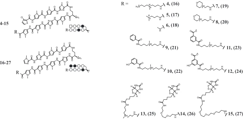
We set out to synthesize a series of non-fluorescent HRE-targeted polyamides (4–15) and their mismatch analogues (16–27; Figure 2.3). Polyamides 4–15 can be considered in three groups: 4–8 were designed with minimized tail motifs; 9–12 were designed to probe the functional groups present in fluorescein dyes potentially responsible for the favorable nuclear uptake profile of 1; and 13–15 were biotin conjugates.
NH3 N N H N N H N N O O O H N O H N NH O N N N H N N H N O O O N H N N O N R NH3 N H N N N H N N O O O H N O H N NH O S N H N N H N N O O O N H N O N Cl R 4-15 16-27 R + R + R = HN NH S O H H H NH O NH H N HN NH S O H H H NH O O O N H H N HN NH O HO O O H N HN NH O O O H N HN NH O HO H N HN NH O H N N H N H O NH N H O
NH HN H3N HN N
H HN NH S O H H H NH O O
O O NH
4, (16) 5, (17) 7, (19) 8, (20) 6, (18) 9, (21) 10, (22) 11, (23) 12, (24)
[image:39.612.116.523.227.428.2]13, (25) 14, (26) 15, (27)
Figure 2.3. Structures of polyamides 4–27. Polyamides 4-15 target 5’-WTWCGW-3’ DNA sequences; 16– 27 target 5’-WGGWCW-3’.
10 and 12 each have a hydroxyl group meta- to the attachment site. Polyamides 13–15 are polyamide-biotin conjugates. The high affinity of the biotin moiety to streptavidin may be useful for biochemical pull-down experiments. Polyamide 13 retains the linker found in 1–3 while 14 and 15 contain 2- and 3-oxygen PEG linkers, respectively. Polyamides
16–27 were synthesized as mismatch control polyamides for 4–15, respectively.
Binding Affinities and Specificities
Polyamides 4–15, designed to bind the 5’-ATACGT-3’ HRE binding site,share the same hairpin polyamide core as polyamides 1 and 3 but have different carboxy terminus tail groups. The DNA binding affinities of 4–15 for the HRE of VEGF were measured by quantitative DNase I footprint titrations using a 5’ 32P-labeled PCR amplification product
of the plasmid pGL2-VEGF-Luc, which contains the VEGF HRE. Polyamide conjugate 1
was previously found to have Ka = 6.3 ! 109 M-1 at this site (16). Polyamides 4–15 were
footprinted (Figure 2.4, Figure 2.5), and the Ka values obtained range from 4.4 (± 1.7) ! 109
for 15 to 7.7 (± 0.9) ! 1010 for 4 (Table 2.1). With the exception of 15, the values obtained
are all higher than that measured for 1. Polyamides 4–15 retain good specificity for the 5’-ATACGT-3’ HRE binding site, although a modest degree of non-specific binding is observed at high concentrations for polyamides 4, 5, 7, 8 and 10. Modifications to the tail moiety did not significantly abrogate high binding affinity and specificity.
-1 Table 2.1: Equilibrium Association Constants: K (M )a
7.7 (± 0.9) x 1010
4
5 6.2 (± 1.2) x 1010
2.5 (± 0.3) x 10 10
6
7 4.0 (± 0.9) x 1010
8 3.8 (± 0.5) x 1010
9 1.5 (± 0.6) x 1010
10 2.1 (± 0.4) x 1010
11 2.6 (± 0.4) x 1010
12 1.1 (± 0.2) x 1010
13 7.2 (± 1.1) x 109
14 4.8 (± 1.9) x 109
15 4.4 (± 1.7) x 109
A
5'-AGACTCCACAGTGCA TACGTG GGCTCCAACAGGTCC-3' 3'-TCTGAGGTGTCACGT ATGCAC CCGAGGTTGTCCAGG-5'
32 P
pGL2-VEGF-Luc 197 bp
M
HRE
C
[12] -0.2
0 0.2 0.4
0.6
0.8 1 1.2
norm
!
10-13 10-12
10-11 10-10
10-9 10-8
10-7 10-6
M
Intact
DNase I A G
1 pM
100 nM
12
1 2 3 4 5 6 7 8 9 10 11 12 13 14 15
B
M
[11] 10-13
10-12 10-11
10-10 10-9
10-8 10-7
10-6 -0.2
0 0.2 0.4
0.6
0.8 1 1.2
norm
!
1 2 3 4 5 6 7 8 9 10 11 12 13 14 15
Intact
DNase I A G
1 pM
100 nM
[image:41.612.173.451.77.596.2]11
A
Intact
DNase I G
100 nM
A
1 pM
4
1 2 3 4 5 6 7 8 9 10 11 12 13 14 15
M
[4]
norm
!
10-1310-1210-1110-1010-9 10-8 10-7 10-6
-0.2 0 0.2 0.4 0.6 0.8 1 1.2 B Intact
DNase I G
100 nM
A
1 pM
M
5
1 2 3 4 5 6 7 8 9 10 11 12 13 14 15
[5]
10-1310-1210-1110-1010-910-810-710-6
norm ! -0.2 0 0.2 0.4 0.6 0.8 1 1.2 C Intact
DNase I A G
1 pM
1 2 3 4 5 6 7 8 9 10 11 12 13 14 15
100 nM
6
M
[6]
-0.2 0 0.2 0.4 0.6 0.8 1 1.2 norm ! 10-13 10-12 10-11 10-10 10-9 10-8 10-7 10-6 D Intact
DNase I G
100 nM
A
1 pM
M
7
1 2 3 4 5 6 7 8 9 10 11 12 13 14 15
[7]
-0.2 0 0.2 0.4 0.6 0.8 1 1.2 norm !
10-1310-1210-1110-1010-9 10-810-710-6
G
G
100 nM
10 1 pM DNase I Intact A
1 2 3 4 5 6 7 8 9 10 11 12 13 14 15
M
[10]
-0.2 0 0.2 0.4 0.6 0.8 1 1.2 norm !
10-1310-1210-1110-1010-910-810-710-6
G Intact DNase I 100 nM A 1 pM 9
1 2 3 4 5 6 7 8 9 10 11 12 13 14 15
M
[9]
norm
!
10-1310-1210-1110-1010-9 10-810-710-6
-0.2 0 0.2 0.4 0.6 0.8 1 1.2 F E Intact
DNase I G
100 nM
A
1 pM
8
1 2 3 4 5 6 7 8 9 10 11 12 13 14 15
M
[8]
norm ! -0.2 0 0.2 0.4 0.6 0.8 1 1.2
10-1310-1210-1110-1010-9 10-810-710-6
H
Intact
DNase I G
100 nM
A
1 pM
M
13
1 2 3 4 5 6 7 8 9 10 11 12 13 14 15
[13]
-0.2 0 0.2 0.4 0.6 0.8 1 1.2 norm !
10-1310-1210-1110-1010-910-810-710-6
I Intact DNase I 100 nM A 1 pM 14 M
1 2 3 4 5 6 7 8 9 10 11 12 13 14 15
G
[14]
10-1310-1210-1110-1010-910-8 10-7 10-6
-0.2 0 0.2 0.4 0.6 0.8 1 1.2 norm ! J Intact
DNase I G
100 nM
A
1 pM
15
1 2 3 4 5 6 7 8 9 10 11 12 13 14 15
M
[15]
10-1310-1210-1110-1010-910-810-710-6
[image:42.612.114.537.89.608.2]norm ! -0.2 0 0.2 0.4 0.6 0.8 1 1.2
Screening for Suppression of Hypoxia-Inducible Transcription by 4–27 in HeLa cells
Induction of VEGF mRNA by the hypoxia mimetic DFO in HeLa cells in the presence of polyamides 4–15 was measured by quantitative real-time RT-PCR and compared to that of polyamide 1 (Figure 2.6). Polyamides 5, 6,and 8 showed modest inhibition of induced VEGF expression and polyamides 4 and 7 showed no difference relative to the untreated, induced control. Mismatch polyamides 16–19 showed no difference in VEGF mRNA relative to the untreated, induced control. Polyamide 20 showed a modest effect.
1 4 5 6 7 8 9 10 11 12 13 14 15
DFO - + + + + + + + + + + + + + +
Relative
VEGF mRNA
Fold Change -1.9 1.0 -1.2-1.2 1.0 -1.4-1.6-1.5-2.1-2.1-1.4-1.3 -1.3
Polyamide - -1.4
0.0 0.2 0.4 0.6 0.8 1.0 1.2 A
0 0.2 0.4 0.6 0.8 1.0 1.2 1.4
Relative
VEGF mRNA
4 16 5 17 6 18 7 19 8 20
DFO - + + + ++ + + ++ ++
Polyamide -
-B C
9 21 10 22 11 23 12 24
- + + + + + + + + +
-
-0 0.2 0.4 0.6 0.8 1.0 1.2 1.4
Relative
VEGF mRNA
DFO Polyamide
Polyamides 9 and 10 had a modest effect on induced VEGF expression, whereas 11 and 12
inhibited induced VEGF expression comparably to polyamide 1. Control polyamides 23
and 24 had a reduced effect as compared to their match congeners 11 and 12 (Figure 2.7A). HeLa cell growth was not inhibited by 1 µM 11, 12, 23 or 24 (Figure 2.7B). Polyamides
13–15 had a modest effect on induced VEGF expression. An appreciable difference in relative VEGF mRNA levels was seen in cells treated with biotin conjugates 13, 14 and mismatch congeners 25, 26 (Figure 2.7C). It is noted that cells treated with polyamides 25
and 26 had VEGF expression slightly higher than that of the untreated, induced control.
0.2 0.4 0.6 0.8 1.0 1.2 1.4 1.6
0
11 23
DFO - + + + + +
12 24
+ + + + Polyamide -
-Relative
VEGF mRNA
A
B
13 25 14 26 15 27
+ + + + + +
DFO - +
Polyamide -
-0.2 0.4 0.6 0.8 1.0 1.2 1.4
0
Relative
VEGF mRNA
no polyamide 11 12 23 24
25 x 103 50 x 103 75 x 103 100 x 103 125 x 103 150 x 103
0 24 48 72
Time (hours)
Number of cells
C
Suppression of Hypoxia-Inducible Transcription by Polyamides in U251 Cells
The effects of HRE targeted polyamides 1 and 11 and control polyamides 2 and 23 were tested in U251 cells, which express a higher level of VEGF than HeLa cells. Uptake of optically tagged polyamides 1 and 2 was examined by confocal microscopy in live, unfixed U251 cells dosed with 2 µM of 1 or 2 (Figure 2.8).
1 2
Figure 2.8. Uptake of polyamides 1 (left) and 2 (right)in U251 cells.
Induction of VEGF mRNA in the presence of polyamides 1 and 11 was inhibited dose dependently (Figure 2.9A). Polyamide 2 at had minimal effect on VEGF expression at 0.2
#M. However, at 1 #M, polyamide 2 had a moderate effect, though still less than that of its match congener 1. Polyamide 23 had a more modest effect than its match congener 11
at both 0.2 and 1 #M. We also undertook chromatin immunoprecipitation experiments in U251 cells to assess changes in VEGF promoter occupancy by HIF-1" in the presence of
1, 2, 11, and 23 (Figure 2.9B). Chromatin immunoprecipitation assays with anti-HIF1" or mock antibody treatment are consistent with decreased occupancy of HIF-1" at the VEGF HRE in the presence of polyamides 1 and 11 at 1 #M. Polyamide 23 has a more modest effect compared to that of its match congener 11. At the concentration tested (1 µ%),
A Relative VEGF mRNA 0.2 0.4 0.6 0.8 1.0 1.2 0 1
DFO - + + + + +
2 Polyamide
-Relative VEGF mRNA 0.2 0.4 0.6 0.8 1.0 1.2 0 DFO Polyamide 11 - + + + + + 23 -0 1.0 2.0 3.0 4.0 5.0 6.0 7.0 Fold enrichment 1
DFO - + + +
2
Polyamide -
-B 0 1.0 2.0 3.0 4.0 5.0 6.0 7.0 Fold enrichment 11
DFO - + + +
23
Polyamide -
-Figure 2.9. (A) Induction of VEGF mRNA by the hypoxia mimetic deferoxamine (DFO) in the presence of polyamides 1, 2, 11, and 23 in U251 cells measured by quantitative real-time PCR. Concentrations of polyamides are 0.2 and 1 µM. (B) Chromatin immunoprecipitation assays with anti-HIF1$ or mock antibody treatment expressed as fold-enrichment (specific/mock) of a 120 base pair sequence at the vegf HRE measured by real-time PCR. Concentrations of polyamides are 1 µM.
2.5 Discussion
Previous studies on fluorophore-labeled polyamides have revealed several parameters that affect cellular trafficking of these molecules, including number of heterocyclic ring pairs, relative Py/Im content and sequence, and choice of fluorophore and attachment site (8, 9). Among fluorophores considered, the protonation state of fluorescein is pH sensitive, while those of tetramethylrhodamine and Bodipy are not. In previous work, nuclear localization of polyamide 2 in HeLa cells was ablated when the cells were grown in glucose- and sodium pyruvate-free media, but subsequently restored when normal growth media was added (8). This suggests that for some polyamides, nuclear localization is energy dependent. Many fluorophore-polyamide conjugates that exhibit a punctate, cytoplasmic staining pattern co-localize with LysoTracker, a lysosomal stain. Verapamil, a calcium channel and P-glycoprotein efflux pump inhibitor, was shown to facilitate nuclear uptake of a Bodipy-polyamide conjugate otherwise sequestered within cytoplasmic vesicles (7). Some polyamides with minimal nuclear localization, e.g. 3, localize mostly in the extracellular space. Nevertheless, the fact that many eight-ring hairpin polyamide-fluorophore conjugates do localize in the nuclei of live cells is encouraging.
of designed DNA-binding ligands to modulate the expression of specific sets of genes. Moving ahead, anticipated experiments include studies in mammalian model systems to account for biodistribution, availability, and metabolism of polyamides. Towards this end, we have attempted to identify polyamides capable of nuclear localization but of simpler structures and lower molecular weights than dye conjugates. The inhibition of induced VEGF expression with HRE-targeted polyamide 1, but not 2, which targets a different DNA sequence or 3, which exhibits considerably less nuclear localization, demonstrates the viability of VEGF inhibition as a proxy for nuclear localization of polyamides that bind the HRE.
The ability to interrogate polyamide cellular uptake and nuclear localization by affecting expression of an endogenous inducible gene enables exploration of the nuclear uptake profiles of non-fluorescent tail moieties. The quantitative real-time RT-PCR data presented show that removal of the fluorescein dye (polyamide 4) and step-wise ablation of the linker moiety (polyamides 5 and 6) diminish the biological activity of these compounds as compared to polyamide 1 in this system. The Ka values measured for 4–6 are between four– and 12–fold higher than that measured for the parent polyamide 1 on the same DNA sequence. Similarly, the binding affinities of N-alkylaminemorpholino tail polyamides 7
and 8 are ~six-fold higher than that of the parent polyamide 1 for the same binding site, but only modest biological activities are observed.
biological activity, but those conjugated to either isophthalic acid or 5-hydroxy isophthalic acid, 11 and 12, respectively, demonstrate a biological effect similar to that observed for the parent polyamide 1. These results suggest that the presence of the carboxylic acid group on the benzene ring is a positive determinant of nuclear localization in this system.
Biotin conjugates 13–15 differ in the identity of the linker moiety and were of interest due to the potential utility of these conjugates for pull-down experiments. The binding affinities measured by quantitative DNase I footprint titration experiments for these conjugates were similar to that of the parent polyamide 1. By quantitative real-time RT-PCR, polyamides 13–15 all demonstrated modest biological activities, and the results do not suggest strong differences in activities based on linker identity. The modest activities of the biotin conjugates, which might reflect a moderate degree of nuclear uptake, are neither encouraging nor dismissive of potential applications of these conjugates.
Our working hypothesis regarding VEGF inhibition by polyamides is through direct interactions with the DNA-HIF-1 interface at the HRE that prevents HIF-1 binding, most likely by an allosteric mechanism. To explore this model further, we used chromatin immunoprecipitation to assess HIF-1 occupancy at the VEGF HRE under hypoxia induced conditions in HeLa cells in the presence and absence of match (and mismatch) polyamides with variable C-terminus tails. Antibodies against HIF-1$ were found to enrich fragments
of DNA containing the VEGF HRE after DFO-induction. This enrichment was reduced in samples treated with polyamide 1 and, to a lesser extent, polyamide 2. We also undertook experiments in U251 cells, which express VEGF more robustly and have previously been used as a model cell line for studying hypoxia-induced VEGF expression (17).
Polyamide-fluorescein conjugates 1 and 2 localize in the nucleus of U251 cells. Induction of VEGF mRNA in the presence of polyamides 1, 2, 11, and 23 at 0.2 and 1 µM in U251 cells was measured by quantitative real-time RT-PCR. Polyamide 1 showed
significant inhibition at both concentrations. Interestingly, at 1 µM polyamide 2 showed
Polyamide 11 exhibits dose-dependent inhibition of VEGF similar to that measured in HeLa cells, while 23 showed modest activity at both concentrations comparable to that measured in HeLa cells. Chromatin immunoprecipitation of HIF-1$ from U251 cells
treated with HRE-targeted polyamides 1 and 11 and mismatch polyamides 2 and 23 shows that enrichment of HIF-1$ bound DNA fragments containing the VEGF HRE after DFO
induction is inhibited by pre-treatment with polyamides 1 and 11, and to a lesser extent 2
and 23. This is generally consistent with a mechanism whereby polyamides 1 and 11 exert their effect by preventing HIF-1 binding to the cognate HRE. This is also consistent with DNase I footprint titrations and gel shift assays in vitro (16).
Table 2.2: MALDI-ToF MS Data for Polyamide Conjugates 3–27
MALDI-TOF MS Data for Polyamide Conjugates (3–27):
(3) CtPyPyIm-(R)-!(NH2)-PyImPyPy-"-NH(CH2)3N(CH3)(CH2)3NH(FAM): MALDI-TOF-MS Calculated [M+H+]: 1676.17; Found: 1676.9.
(4) CtPyPyIm-(R)-!(NH2)-PyImPyPy-NH(CH2)3N(CH3)(CH2)3NH2: MALDI-TOF-MS Calculated [M+H+]: 1246.50; Found: 1246.5.
(5) CtPyPyIm-(R)-!(NH2)-PyImPyPy-NH(CH2)3N(CH3)2: MALDI-TOF-MS Calculated [M+H+]:1203.46 ; Found: 1203.4.
(6) CtPyPyIm-(R)-!(NH2)-PyImPyPy-NH(CH3): MALDI-TOF-MS Calculated [M+H+]: 1132.38; Found: 1132.4.
(7) CtPyPyIm-(R)-!(NH2)-PyImPyPy-NH(CH2)2(Morpholine): MALDI-TOF-MS Calculated [M+H+]:1231.45; Found:1231.7.
(8) CtPyPyIm-(R)-!(NH2)-PyImPyPy-NH(CH2)3(Morpholine): MALDI-TOF-MS Calculated [M+H+]:1245.47 ; Found: 1245.9.
(9) CtPyPyIm-(R)-!(NH2)-PyImPyPy-NH(CH2)3N(CH3)(CH2)3NH(Benzoic acid): MALDI-TOF-MS Calculated [M+H+]: 1350.53; Found: 1350.5.
(10) CtPyPyIm-(R)-!(NH2)-PyImPyPy-NH(CH2)3N(CH3)(CH2)3NH(3-Hydroxy benzoic acid): MALDI-TOF-MS Calculated [M+H+]: 1366.52; Found: 1366.4. (11) CtPyPyIm-(R)-!(NH2)-PyImPyPy-NH(CH2)3N(CH3)(CH2)3NH(Isophthalic acid):. MALDI-TOF-MS Calculated [M+H+]:1394.52; Found: 1394.5.
(12) CtPyPyIm-(R)-!(NH2)-PyImPyPy-NH(CH2)3N(CH3)(CH2)3
NH(5-Hydroxyisophthalic acid): MALDI-TOF-MS Calculated [M+H+]:1410.51; Found:
1410.5.
(13) CtPyPyIm-(R)-!(NH2)-PyImPyPy-NH(CH2)3N(CH3)(CH2)3NH(Biotin): MALDI-TOF-MS Calculated [M+H+]: 1474.10; Found: 1474.5.
(14) CtPyPyIm-(R)-!(NH2)-PyImPyPy-NH(CH2)2(O(CH2)2)2NH(Biotin): MALDI-TOF-MS Calculated [M+H+]: 1477.05; Found: 1476.8.
(15) CtPyPyIm-(R)-!(NH2)-PyImPyPy-NH(CH2)3(O(CH2)2)3CH2NH(Biotin):
MALDI-TOF-MS Calculated [M+H+]: 1549.16; Found: 1549.5.
(16) ImImPyPy-(R)-!(NH2)-ImPyPyPy-NH(CH2)3N(CH3)(CH2)3NH2: MALDI-TOF-MS Calculated [M+H+]:1210.59; Found:1210.7.
(17) ImImPyPy-(R)-!(NH2)-ImPyPyPy-NH(CH2)3N(CH3)2: MALDI-TOF-MS Calculated [M+H+]:1267.55; Found:1267.2.