© 2001 OsteoArthritis Research Society International 1063–4584/01/030203+12 $35.00/0 doi:10.1053/joca.2000.0377, available online at http://www.idealibrary.com on
Studies on YKL-40 in knee joints of patients with rheumatoid arthritis
and osteoarthritis. Involvement of YKL-40 in the joint pathology
B. Volck*§, J. S. Johansen*, M. Stoltenberg*, C. Garbarsch§, P. A. Price¶, M. Østergaard*,
K. Ostergaard
\
, P. Løvgreen-Nielsen†, S. Sonne-Holm‡ and I. Lorenzen*
Departments of *Rheumatology, †Pathology, and ‡Orthopedic Surgery, Hvidovre Hospital,
University of Copenhagen, Denmark; §Institute of Medical Anatomy Section A, The Panum Institute, University of Copenhagen, Denmark;\Osteoarthritis Research Unit, Institute for Inflammation Research, Rigshospitalet, Copenhagen, Denmark; and ¶Department of Biology, University of California, San Diego, La Jolla, California, U.S.A.
Summary
Objective: The presence of YKL-40 (human cartilage glycoprotein 39) in synovium, cartilage and synovial fluid (SF) from knee joints of patients with rheumatoid arthritis and osteoarthritis (OA) were related to histopathological changes in synovium and cartilage and to serum YKL-40 and other biochemical markers.
Methods: The localization of YKL-40 in synovium and cartilage was determined by immunohistochemistry. Synovial inflammation was estimated histologically and by magnetic resonance imaging (MRI). Biochemical markers of inflammation, neutrophil activation and cartilage metabolism were analysed. YKL-40 concentrations in serum and SF were determined by RIA and ELISA.
Results: In the synovium YKL-40 positive cells were found in lining and stromal cells (macrophages) and the number of YKL-40 positive cells was related to the degree of synovitis. In arthritic cartilage, YKL-40 was located to chondrocytes. YKL-40 levels in SF were higher in RA patients with moderate/severe or none/slight synovitis of the knee joint compared to OA patients with moderate/severe or none/slight synovitis. SF YKL-40 correlated with the synovial membrane and the joint effusion volumes determined by magnetic resonance imaging (MRI) and with other biochemical markers of intercellular matrix metabolism. SF YKL-40 was higher than serum YKL-40, and a relationship existed between the YKL-40 levels in SF and serum. Intraarticular glucocorticoid injection was followed by clinical remission and a decrease in serum YKL-40, which increased again at clinical relapse.
Conclusions: YKL-40 in SF is derived from cells in the inflamed synovium, chondrocytes and SF neutrophils. Joint derived YKL-40 influences serum YKL-40. YKL-40 may be involved in the pathophysiology of the arthritic processes and reflect local disease activity. © 2001 OsteoArthritis Research Society International
Key words: YKL-40, Human cartilage glycoprotein 39, Rheumatoid arthritis, Osteoarthritis.
Introduction
YKL-40*, also called human cartilage glycoprotein-39 (HC gp-39)1, is a mammalian member of family 18 glycosyl
hydrolases1–8and has a known gene sequence7. YKL-40
is a heparin and chitin-binding lectin4,8 without chitinase
activity1,5,8. The physiological function of YKL-40 is
unknown, but the protein contains several DR4 peptide binding motifs and may be a target of the immune response in rheumatoid arthritis (RA)9. Several cells present in the
arthritic joint can secrete YKL-40. It is one of the most abundant proteins secreted in vitro by chondrocytes1,5and
synovial cells2 from patients with RA. We have recently
reported a high number of YKL-40 positive chondrocytes in articular cartilage from the hip joint of patients with osteo-arthritis (OA)10. YKL-40 positive chondrocytes were in
particular located in the superficial and middle layer of the cartilage and especially in areas of the joint with a consider-able biomechanical load. Chondrocytes from normal carti-lage were mainly YKL-40 negative10. Another recent
study11 demonstrated by in-situ hybridization the same
zonal distribution of YKL-40 (HC gp-39) in osteoarthritic cartilage, as well as the absence of the protein in normal cartilage. YKL-40 expression by osteophytic tissue in end stage osteoblasts and by primary osteocytes from osteoar-thritic patients was also reported11. YKL-40 is not produced
by normal monocytes but is secreted by differentiated and activated human macrophages in many different
Received 24 November 1999; revision requested 7 April 2000; revision received 22 June 2000; accepted 27 July 2000.
Address correspondence to: Birgitte Volck, MD, Division of Medicine, Department of Rheumatology 232, Hvidovre Hospital, University of Copenhagen, Kettega˚rd Alle´ 30, DK-2650 Hvidovre, Denmark. Tel: +45 36 32 35 19; Fax: +45 36 47 14 10; E-mail: [email protected]
The study was supported by grants from ‘The Danish Rheuma-tism Association’, ‘Clara Hansens Mindelegat’, ‘Henny og Helge Holgersens Mindelegat’, ‘Lægeforeningens Forskningsfond’, ‘Michaelsen Fonden’, ‘Puljen til fremme af klinisk forskning’, ‘Hjørdis og Jørgen Peter Christiansens Familiefond’, ‘Ingeniør af Frederikssund, Søren Alfred Andersens Legat’ and ‘Thomas og Elisabeth Frølund Nielsens Foundation’.
*YKL-40 is also named human cartilage glycoprotein-39 (HC gp-39) (Ref. 1), 38-kDa heparin binding glycoprotein (Ref. 4), and CHI3L1 (Ref. 7). The protein had been termed YKL-40 from its molecular weight (40 kDa) and the one letter code for its three NH2-terminal amino acids (tyrosine, lysine, and leucine) (Ref. 18).
tissues6–8,12,13, including inflamed synovium from patients
with active RA6. Furthermore, YKL-40 is present in the
specific granules of neutrophils and is exocytosed by activation14.
YKL-40 in serum and synovial fluid can be measured by radioimmunoassay (RIA)3 or enzyme linked
immuno-sorbent assay (ELISA)15. Increased levels of YKL-40 in
serum and synovial fluid are found in patients with active RA3,15–16or severe knee OA17compared to normal
sub-jects17. A correlation exits between the level of YKL-40 in
serum and synovial fluid with approximately 10-fold higher values in synovial fluid3,17.
The object of the present study was to elucidate a possible involvement of YKL-40 in the pathophysiology of RA and OA as well as the role of YKL-40 as a marker of disease activity. The presence of YKL-40 in the synovial membrane and articular cartilage was examined by immunohistochemical methods, and the YKL-40 concen-trations in synovial fluid and serum were examined by immunoassays (RIA and ELISA) in patients with RA and OA. We related the YKL-40 findings with histopathological changes in the synovial membrane and articular cartilage, with the synovial membrane and the joint effusion volumes [determined by magnetic resonance imaging (MRI)] and with biochemical markers of inflammation, neutrophil activation and cartilage metabolism.
Patients and methods
The investigation included a cross-sectional and a longi-tudinal study. The knee joint was used as a model and dominating knee joint involvement was an inclusion criteria. The research protocols were approved by the Ethics Committee for Medical Research in Copenhagen. In accordance with the Helsinki Declaration II each patient was informed about the study verbally and in writing, and all gave their written consent.
CROSS SECTIONAL STUDY
Twenty patients with RA and 39 patients with OA were included. The patients were scheduled to undergo total knee joint replacement [RA (N=13) and OA (N=39)] or arthroscopic synovectomy of the knee joints [RA (N=7)]. The patients fulfilled the American College of Rheumatol-ogy 1987 classification criteria for RA19or the ACR 1986
classification criteria for idiopathic OA of the knee20.
Patients with malignant disease or elevated liver enzymes were not included. Seven of the patients with RA were treated with slow-acting antirheumatic drugs (DMARDs) [methotrexate (N=2), sulfasalazine (N=4), penicillamine (N=1)], either as the only treatment (N=4), in combination with non-steroidal antiinflammatory drugs (NSAIDs) (N=1), or in combination with low dose oral prednisolone (N=2). Low dose oral prednisolone (1.25–10.0 mg/day) was given as the only treatment in six patients, and given in combi-nation with NSAID in three patients. Twelve patients were treated with NSAID alone [RA (N=1) and OA (N=11)]. Three patients with RA did not receive any kind of antirheu-matic medicine. Just prior to surgery a clinical examination and blood samples were performed. In case of joint effusion synovial fluid was aspirated by the surgeon when the operation was initiated. Variable amounts of synovial fluid were collected dependent upon the amount available in the joint and the degree of waste in connection with the
collection. Thirty-one of these patients underwent a MRI determination of the knee joint and were included in a project investigating MRI determination of synovial mem-brane and joint effusion volumes in relation to signs of synovial inflammation21.
LONGITUDINAL STUDY
Eighteen patients with RA according to the ACR 1987 classification criteria19 and clinical signs of knee joint
synovitis (15 females and three males) were included. Median disease duration was 5.5 years (range 0.3–36 years) and duration of knee symptoms was 0.6 years (range 0.1–7 years). The patients had a clinical indication for arthrocentesis and intraarticular corticosteroid injection. During arthrocentesis, as much synovial fluid as possible was aspirated, and 80 mg methylprednisolone acetate (2 ml, 40 mg/ml) plus 6 ml lidocaine 0.5% (lidocaine hydro-chloride 5 mg/ml) were injected into the knee joint. Serum samples were collected just prior to corticosteroid injection and after 1, 7, 14, 30, 60, 90 and 180 days. Seven patients received continuous therapy with low-dose oral pred-nisolone (2.5–8.75 mg/day) in combination with DMARD (sulfasalazine (N=2) and methotrexate (N=1)) and NSAID (N=3), or with NSAID alone (N=2). Three of the patients were treated with DMARD [penicillamine (N=2) and sulfasalazine (N=1)] in combination with NSAID, eight patients received NSAID alone. No patients had received intraarticular glucocorticoid therapy within the last 4 weeks.
SYNOVIAL MEMBRANE BIOPSIES
From all patients included in the cross-sectional study, synovial biopsy specimens were obtained during surgery. In patients who had a MRI determination of the synovial membrane and joint effusion volume performed, four pre-selected sites of the synovial membrane of the knee joint21
were chosen. Patients, who did not have a MRI determi-nation, had synovial membrane biopsies obtained from three different sites of the knee joint: (1) the lateral; (2) the medial; and (3) the suprapatellar recess. The surgeon selected these three biopsies to represent the overall degree of synovitis of the knee joint [assessed by edema, volume and vascularity (redness) of the synovial mem-brane]. The synovial biopsy specimens were immediately fixed in 10% neutral buffered formalin, pH 7.0 until the next day, dehydrated in graded mixtures of ethanol and water, immersed in xylene, paraffin embedded, cut at 5m and stained with hematoxylin and eosin.
SYNOVIAL MEMBRANE AND JOINT EFFUSION VOLUMES DETERMINED BY MRI
MRI was performed using a 1.5-Tesla Magnetom (Siemens, Erlangen, Germany) equipped with a dedicated knee coil. Synovial membrane and joint effusion volumes were calculated from continuous pre- and post-gadolinium-DPTA (Shering, Berlin, Germany) T1-weighted transversal MR-images, by means of image processing software. The method is described in detail elsewhere21.
HISTOLOGICAL EVALUATION OF SYNOVIAL SPECIMENS
Synovial inflammatory activity was graded under blinded conditions by an experienced histopathologist. The grading
of the inflammatory reaction was done as a general aver-age of all of the synovial tissue from all the biopsies and was based upon a grading of the following nine parameters and described in details elsewhere21:
(1) subsynovial infiltration of polymorphonuclear leuko-cytes;
(2) subsynovial infiltration of mononuclear leukocytes; (3) surface fibrin deposition;
(4) multiplication of the synovial lining; (5) villous hypertrophy of the synovial surface; (6) proliferation of blood vessels;
(7) perivascular edema;
(8) formation of granulation tissue; and (9) fibrosis.
Each of the nine parameters was graded as 0 (none), 1 (mild), 2 (moderate), or 3 (severe), and an ‘average grade’ for each parameter was calculated as the average of the grading of the collected biopsies from each knee. Based upon a total score for each knee joint achieved from the average score for all of the nine parameters considered, the knees were divided into two groups: ‘None/slight’ syno-vial inflammation (N=40) included knees with an average grade ≤1 for all signs of inflammatory activity considered; and ‘moderate/severe’ synovial inflammation (N=19) included knees with an average grade >1 for all signs of inflammatory activity considered.
IMMUNOHISTOCHEMICAL STAINING FOR YKL-40 ANTIGEN IN SYNOVIAL MEMBRANE
Alkaline phosphatase staining technique for polyclonal antibodies was used and included the following steps (all performed at room temperature and separated by washes in TBS). Non-specific binding was inhibited by incubation for 20 min with 3% bovine serum albumin (BSA) (Sigma A-4503) in Tris buffered saline (TBS, 5 mM Tris-HCl, 146 mM NaCl, pH 7.4). This buffer was used for dilution of the different antibodies. Incubation for 30 min with an affinity-purified rabbit polyclonal immunoglobulin (IgG) against human YKL-40 (IgG concentration of the YKL-40 antibody was 33g/mL). The human YKL-40 used for immunization of rabbits and for affinity purification of anti-bodies is described elsewhere3. The YKL-40 antibody used
in these studies was purified from rabbit antiserum by affinity chromatography using a Sepharose support with covalently attached purified human YKL-40. The antibodies were eluted by 100 mM glycine (pH 2.5). Non-immune rabbit IgG (DAKO X0936, Copenhagen, Denmark) was used as negative control in the same IgG concentration (33g/mL). Thereafter incubation for 30 min with alkaline phosphatase-conjugated swine antibodies to rabbit IgG (DAKO D0306, diluted 1:20). Sigma FASTY BCIP/NBT tablets (Sigma B-5655; 5-Bromo-4-chloro-3-indolylphosphate/Nitro Blue Tetrazolium) with 0.25 mg/mL Levamisole in 0.1 M Tris-HCl, pH 9.5 was used as color substrate (30 min incubation time). Positive staining was recognized as a dark blue/violet color.
DOUBLE STAINING FOR YKL-40 AND CD68 (MACROPHAGE PROTEIN) IN THE SYNOVIAL MEMBRANE
Peroxidase/DAB (3.3–diaminobenzidine tetrahydrochlor-ide (DAB; KemEnTec, Copenhagen, Denmark) was used as immunostain for the second immunostaining step in the
double staining procedure, which included the following sequences (all performed at room temperature and sep-arated by washes in TBS/Tween20). The sections were pre-incubated with 0.6% H2O2 (Perhydrol, Merc, Darmstadt, Germany) in 0.005 M TBS for 30 min, and staining for YKL-40 antigen was performed as described in the section above. After incubation with Sigma FASTY BCIP/NBT the color reaction was stopped in distilled water at 4°C. Nonspecific binding was inhibited by incubation for 30 min with 3% BSA in TBS (this buffer was also used for dilution of the various antibodies). Then incubation was performed for 30 min with mouse antihuman CD68 (DAKO M814), diluted 1:50, followed by incubation with a rabbit antimouse IgG (DAKO Z259), diluted 1:20. Non-immune mouse serum (DAKO X0931) was used as a negative control in the same IgG concentration. Then incubation was carried out for 30 min with peroxidase labeled mouse monoclonal PAP [horseradish peroxidase labeled mouse monoclonal antihorseradish peroxidase (DAKO P850)]. Pre-incubation for 10 min with DAB (KemEnTec 4170), 0.5 mg/mL buffer, pH 7.0 was followed by incubation for 10 min in the same DAB solution but with the addition of 0.018% H2O2. The reaction was stopped with distilled water at 4°C for 15 min. Positive staining was recognized as a dark blue/violet color for YKL-40 antigen and a brown color for CD68 antigen.
MICROSCOPIC EVALUATION OF IMMUNOHISTOCHEMICAL STAINING OF SYNOVIAL SPECIMENS
The synovial sections were examined blindly two times by the same observer and scored for the presence of YKL-40 positive cells in a scale as follows: score 0=all cells negative; score 1=few cells positive (approximately 1–25%); score 2=a considerable number of cells positive (approximately 25–75%); score 3=most/all cells positive (approximately 75–100%).
ARTICULAR CARTILAGE BIOPSIES
Standardized samples of cartilage representing all aspects of the joint surfaces (femoral, tibial and patellar surface) of the knee were collected from up to 16 topo-graphically different areas (Fig. 1) from four of the patients with RA (mean age 63 years, range 34–74) and from four of the patients with OA (mean age 77 years, range 74–79) from the cross-sectional study. Only 58 biopsies from the patients with RA and 59 biopsies from the patients with OA were collected, because some areas of the joint surfaces contained only denuded bone. The cartilage samples were stored in cryovials at −80°C until immunohistochemical analysis.
HISTOLOGICAL EVALUATION OF CARTILAGE SPECIMENS
The cartilage sections were evaluated for the presence or absence of histopathological cartilage changes, includ-ing surface fibrillation/fissures and clusters/clones22.
IMMUNOHISTOCHEMICAL STAINING FOR YKL-40 ANTIGEN IN CARTILAGE
Four micrometer cryostat cartilage sections were fixed with acetone at room temperature for 15 min. Avidin/
biotinylated horseradish peroxidase staining technique was used as described recently10. To assess if the accessibility
of the YKL-40 epitope of the antibody was hindered by extracellular matrix interactions in the cartilage some of the sections were treated with either protease, 0.05% (type 14, Sigma P5147), trypsin, 0.1% (Sigma T8128) or hyaluroni-dase, 0.1% (Fluka 531712) for 10 min at 37°C prior to the immunohistochemical staining with an affinity-purified rabbit polyclonal antiserum against human YKL-40 (IgG concentration of 33g/mL) followed by incubation with biotinylated swine antirabbit IgG (DAKO E0353, diluted 1:400). Antibody binding was visualized by a complex of avidin-biotinylated horseradish peroxidase (ABComplex/ HRP, DAKO K0355) and AEC (3-amino-9-ethylcarbazole) staining kit (SIGMA AEC101). Positive staining was a red color associated with the cytoplasm of the cell.
MICROSCOPIC EVALUATION OF IMMUNOHISTOCHEMICAL STAINING OF CARTILAGE SPECIMENS
Two of the sections [RA (N=1) and OA (N=1)] were not evaluated due to inadequate quality (tissue folds). The cartilage sections were examined blindly by two observers and scored for the presence of YKL-40 positive chondro-cytes in a scale as follows: score 0=all chondrochondro-cytes negative; score 1=few chondrocytes positive (1–25% tive); score 2=considerable number of chondrocytes posi-tive (25–75%); and score 3=most/all chondrocytes posiposi-tive (75–100%).
BIOCHEMICAL ANALYSIS OF SERUM AND SYNOVIAL FLUID
Blood samples were allowed to clot at room temperature and then centrifuged at 2000 g for 10 min. Serum was either analysed immediately or stored at −80°C until analy-sis was performed. The synovial fluid samples were centri-fuged at 2000 g for 10 min and the cell-free supernatants were stored at −80°C until analysis was performed. ESR in blood was determined by the Westergren method and serum CRP was determined with nephelometry. YKL-40
concentrations in synovial fluid and serum were determined by two different methods; an in-house RIA using polyclonal rabbit antibodies3 and a commercial ELISA using both
monoclonal and polyclonal antibodies (Metra Biosys-tems)15. The two assays and the normal YKL-40 levels in
serum and synovial fluid are described in detail else-where3,15,17. The source of the YKL-40 antigen used for
standards and production of antibodies in both methods were purified from the conditioned medium of the human osteosarcoma cell line MG63. However, the methods used to calibrate the standards were different. The concentration of the amino-terminal propeptide of type III procollagen was determined by RIA23. The concentrations in synovial fluid
and serum of cartilage oligomeric matrix protein (COMP) and sulfated glucosaminoglycans (GAG) were determined by Wieslab (Lund, Sweden)24,25. The concentrations in
synovial fluid of myeloperoxidase26, lactoferrin27,
neu-trophil gelatinase associated lipocalin (NGAL)28 and
human cathelicidin (hCAP-18)29 were determined by
ELISA. Due to restricted amounts of serum and synovial fluid available, incomplete number of measurements occurred in some of the variables analysed.
CLINICAL JOINT ASSESSMENT
Clinical assessment of disease activity in the joints included assessment of joint swelling and tenderness. The knee joints as well as the total number of joints involved were registered. Clinical indication for arthrocentesis of the knee joint and subsequent intraarticular corticosteroid injection was determined by clinical signs of intraarticular fluid and of synovitis. Remission was defined by absence of synovial swelling and tenderness of the knee joint. Clinical relapse was defined by recurrence of joint swelling and tenderness.
STATISTICAL ANALYSIS
The statistical analysis was performed with SPSS®
(Stat-istical Package for the Social Science) Software. Results are given as median, range and ratios. Comparison between groups was performed by the non-parametric Mann–Whitney test or the Kruskal–Wallis test (and subse-quent Dunn’s method). Comparison between two related samples was performed by the Wilcoxon signed ranks test. Subsequent comparison between the consecutive changes over time was performed by regression analysis (random coefficient regression model) using the ‘Ime’ software by Pinheiro and Bates. Correlation analysis between the dif-ferent parameters was calculated by the Spearman rho test. P-values less than 0.05 were considered significant.
Results
The clinical characteristics of the patients are shown in
Table I. Patients with RA had more swollen joints, a higher histological score of synovial inflammation and a larger volume of synovial membrane of the knee joint (determined by MRI) than patients with OA. ESR, serum CRP, serum and synovial fluid YKL-40 and PIIINP were highest in patients with RA. Serum YKL-40 levels were higher in RA patients compared to healthy subjects (median 296g/L vs 102g/L, P<0.01) (Table I). No differences between the two groups of patients were found in the synovial fluid concentrations of COMP and GAG. However, serum
Fig. 1. Standardized samples of cartilage were collected from up to 16 topographically different areas of the arthritc knee joint; Tibia: condylus medialis tibiae: 1=anterior, 2=intermediate, 3=posterior, 4=central; condylus lateralis tibiae: 5=anterior, 6=intermediate, 7=posterior, 8=central; Femur: condylus lateralis femoris: 9=anterior, 10=intermediate, 11=posterior; condylus medialis femoris: 12=anterior, 13=intermediate, 14=posterior; 15=facies
COMP was significant higher in OA patients compared to RA patients (P<0.01) (Table I).
SYNOVIAL FLUID YKL-40 LEVELS IN RELATION TO HISTOLOGICAL ASSESSMENT OF SYNOVIAL INFLAMMATION
Highest levels of YKL-40 in synovial fluid were found in knee joints from patients with RA and moderate to severe degree of synovial inflammation (median 6300g/L) and the levels were higher (P=0.054) compared to patients with OA and moderate to severe synovial inflammation (3400g/L) (Table IIandFig. 2). Patients with RA and none to slight synovial inflammation had higher levels of YKL-40 in synovial fluid compared to OA patients with none to slight synovitis (median 4200g/L vs 2450g/L, P=0.026). Simi-lar results were found when the ELISA method was used to measure YKL-40. There was a trend towards higher SF YKL-40 levels in RA patients with moderate to severe synovial inflammation compared to those with none to slight synovitis; however, the difference was not significant (P=0.096).
IMMUNOHISTOCHEMICAL STAINING OF YKL-40 IN SYNOVIAL MEMBRANE IN RELATION TO SYNOVIAL INFLAMMATION AND SYNOVIAL FLUID YKL-40 LEVELS
Positive staining for CD68 antigen was observed in cells of the synovial lining and in mononuclear cells in the stroma of the synovial membrane in patients with RA and OA, and some of these cells were also positive for YKL-40 antigen
[Fig. 3(A)]. The number of YKL-40 positive cells was related with the degree of synovial inflammation. A severely inflamed synovial membrane had a higher number of YKL-40 positive cells compared to a less inflamed synovial membrane. No difference was observed in the staining pattern of synovial membranes of RA and OA patients. The score of YKL-40 positive cells in the synovium gained from 0 (no positive cells) to 2 (25–75% positive cells) (Table III). No sections had positive YKL-40 staining in all cells. YKL-40 positive cells were found in 29% of the sections with none to slight synovial inflammation and in 70% of the sections with moderate to severe synovial inflammation. The level of YKL-40 in synovial fluid was related to the score of YKL-40 positive cells in the synovial membrane (Fig. 4) (Kruskal–Wallis test, P=0.0156, N=22) with a significant difference (Dunn’s method, P<0.05) between synovial membranes with the higher (score=2) and the lower (score=0) number of YKL-40 positive cells.
SYNOVIAL FLUID YKL-40 LEVELS IN RELATION TO THE SYNOVIAL MEMBRANE AND JOINT EFFUSION VOLUMES DETERMINED BY MRI
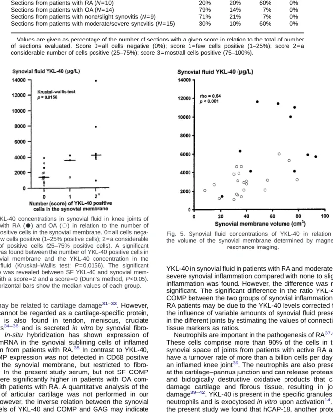
The concentration of YKL-40 in synovial fluid was signifi-cantly correlated with the MRI-determined volumes of the synovial membrane (rho=0.64, P<0.001, N=31) (Fig. 5) as well as the joint effusion volumes (rho=0.59; P<0.001,
N=31) in patients with RA and OA. A significant correlation
was found between serum PIIINP and the volume of the synovial membrane (rho=0.39, P<0.05) and of the joint effusion determined by MRI (rho=0.33; P=0.07). Synovial fluid levels of COMP, GAG and the markers of different
Table I
Patient characteristics in the cross-sectional and the longitudinal study of arthritic knee joints Cross-sectional study Longitudinal study
RA Normal values
RA OA
Age 66 (29–77)20*** 74 (50–85)39 58 (19–78)18
Number of swollen joints—total 3 (0–18)19*** 0 (0–3)35 3 (1–30) Number of swollen joints—large 2 (0–6)19*** 0 (0–3)35 2.5 (1–10)
Number of swollen joints—small 0 (0–13)19*** 0 (0)35 0 (0–25)
ESR, mm/h 44 (3–95)20*** 12 (2–92)35 47 (2–105) <20
Serum CRP, nmol/L 246 (30–1413)20*** 89 (30–419)37 327 (29–777) <90
Serum YKL-40 (RIA),g/L 296 (55–920)19* 130 (56–1290)31 276 (104–540) <208 (Ref. 17)
Serum YKL-40 (ELISA),g/L 87 (20–218)12 73 (26–565)31 <95 (Ref. 15)
Serum COMP, mg/L 0.9 (0.6–1.3)14** 1.2 (0.7–1.7)22
Serum PIIINP,g/L 4.5 (1.5–8.4)19** 3.4 (1.4–6.0)31 <3.5
Synovial inflammation, 0–1 1 (0–1)20*** 0 (0–1)39 MRI/synovial membrane volume, cm3 69 (21–93)10* 36 (8–79)21 MRI/joint effusion volume, cm3 40 (4–232)10 21 (0–119)21
SF YKL-40 (RIA),g/L 4890(910–13800)20*** 2700 (351–6000)39 <1350 (Ref. 17) SF YKL-40 (ELISA),g/L 2120 (485–6850)14** 1190 (274–2600)29 <402 (Ref. 15) SF PIIINP,g/L 3230 (600–6700)19*** 960 (240–3310)37
SF COMP, mg/L 7.2 (3.1–22.1)18 9.2 (3.9–20.5)34
SF GAG, mg/L 42 (10–132)16 42 (3–79)33
Ratio SF/serum YKL-40 (RIA) 20 (2–67)19 13 (3–55)31 Ratio SF/serum YKL-40 (ELISA) 16 (8–112)39 15 (3–56)33 Ratio SF YKL-40 (RIA)/SF COMP 548 (122–2721)17** 247 (38–939)34 Ratio SF YKL-40 (RIA)/SF GAG 156 (15–540)16* 63 (8–530)33 Ratio SF YKL-40 (ELISA)/SF COMP 295 (34–991)14* 144 (19.7–440)29 Ratio SF YKL-40 (ELISA)/SF GAG 75 (11–234)13 34 (10–200)28
Values are given as median and rangesnumber of patients. CRP=C-reactive protein; ESR=erythrocyte sedimentation rate; PIIINP=amino terminale propeptide of type III procollagen; COMP=cartilage oligomeric matrix protein; GAG=glycosaminoglycans; MRI=magnetic resonance imaging. SF=synovial fluid; synovial inflammation (0=none/slight, 1=moderate/severe). *=P<0.05; **=P<0.01; ***=P<0.001 vs patients with OA tested by the Mann–Whitney U-test.
granules in neutrophils did not correlate statistically with the volume of the synovial membrane and joint effusion.
IMMUNOHISTOCHEMICAL STAINING OF YKL-40 IN ARTICULAR CARTILAGE IN RELATION TO HISTOPATHOLOGICAL CARTILAGE CHANGES
All the arthritic cartilage sections had considerable his-topathological changes, including surface fibrillation or fis-sures and/or clones or clusters. In the pannus-invaded cartilage [Fig. 3(B)] a prominent YKL-40 staining was shown in the cytoplasm of the different cells types present in the pannus, and the extracellular matrix also exhibited some staining [Fig. 3(C)]. We did not attempt to differentiate between the YKL-40 staining in the different cell types. The extracellular matrix in the underlying cartilage was YKL-40 negative around both YKL-40 positive and negative chondrocytes [Fig. 3(C)]. The cartilage from patients with RA exhibited frequently prominent fibrous pannus and clusters/clones [Fig. 3(C)] compared with cartilage from OA patients [Fig. 3(D)]. YKL-40 was found in the cytoplasm of the chondrocytes [Fig. 3(E)] but not in the extracellular matrix, except in areas of pannus-invaded cartilage. Treat-ment with protease, trypsin or hyaluronidase prior to the immunohistochemical staining did not reveal any overt additional YKL-40 staining of the extracellular cartilage matrix or in the chondrocytes. YKL-40 positive chondro-cytes were observed in all layers of the cartilage. Most of the sections included non-intact residual cartilage with only the middle and/or the deep layer left, and no differences in the staining pattern between the different layers could be identified. Chondrocytes located in clusters/clones and in areas of excessive surface fibrillation showed a similar pattern of YKL-40 staining as found in isolated chondro-cytes and chondrochondro-cytes in other areas of the same section. No difference was found between the YKL-40 staining pattern in articular cartilage in RA and OA patients. Most cartilage sections had a score of 1 (1–25% positive chondrocytes). Seven sections had a YKL-40 score of 2
(25–75% positive chondrocytes) (three sections from RA patients and four sections from OA patients), and no sections had a score of 3. Arthritic knees without YKL-40 staining in chondrocytes were not found. There was no difference in respect of topographic site in the knee joint and the YKL-40 staining (Table IV).
IMMUNOHISTOCHEMICAL STAINING OF YKL-40 IN ARTICULAR CARTILAGE IN RELATION TO SYNOVIAL INFLAMMATION AND SYNOVIAL FLUID YKL-40 LEVELS
YKL-40 staining in chondrocytes from knees with mod-erate to severe synovial inflammation was similar to the YKL-40 staining in chondrocytes from knees with none to slight synovial inflammation. Two of the knees from which cartilage specimens were obtained were graded with mod-erate to severe synovial inflammation (both knees were from RA patients) and these knees had high SF YKL-40 levels (7830 and 13 800g/L). Two knees were graded with no signs of synovial inflammation (both from OA patients) but the YKL-40 concentration in the synovial fluid was higher (1991 and 4200g/L) than the level in normal knees (<1350g/L).17
SYNOVIAL FLUID YKL-40 LEVELS IN RELATION TO BIOCHEMICAL MARKERS OF SYNOVIAL INFLAMMATION, CARTILAGE
DEGRADATION AND ACTIVATED NEUTROPHILS
In patients with RA or OA, no significant correlation existed between SF levels of YKL-40 as compared with PIIINP, COMP, GAG or the markers of the specific granules in neutrophils. The concentrations of PIIINP, COMP, GAG, lactoferrin, NGAL and hCAP-18 (located to the specific granules as YKL-40) were not different in the synovial fluid from patients with moderate to severe synovial inflamma-tion and patients with none to slight synovial inflammainflamma-tion (Table II), neither in patients with RA nor OA.
Table II
The synovial fluid concentration of the biochemical markers in relation to the degree of synovial inflammation of the knee joint in patients with RA and OA
RA OA
Synovial inflammation None/slight Moderate/severe None/slight Moderate/severe
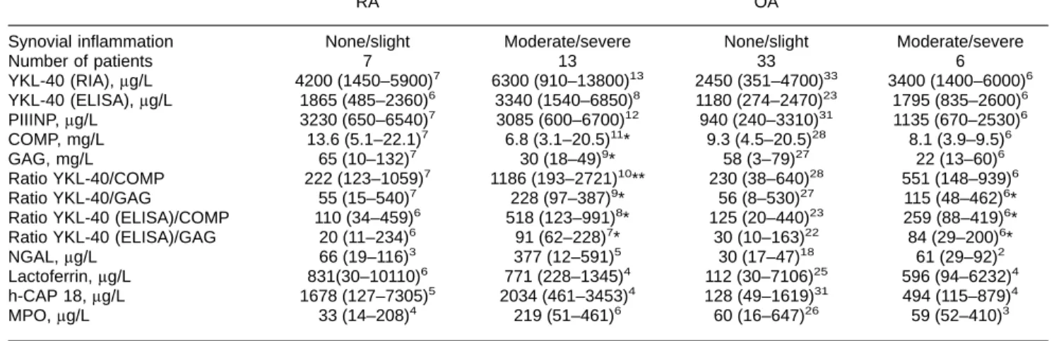
Number of patients 7 13 33 6 YKL-40 (RIA),g/L 4200 (1450–5900)7 6300 (910–13800)13 2450 (351–4700)33 3400 (1400–6000)6 YKL-40 (ELISA),g/L 1865 (485–2360)6 3340 (1540–6850)8 1180 (274–2470)23 1795 (835–2600)6 PIIINP,g/L 3230 (650–6540)7 3085 (600–6700)12 940 (240–3310)31 1135 (670–2530)6 COMP, mg/L 13.6 (5.1–22.1)7 6.8 (3.1–20.5)11* 9.3 (4.5–20.5)28 8.1 (3.9–9.5)6 GAG, mg/L 65 (10–132)7 30 (18–49)9* 58 (3–79)27 22 (13–60)6 Ratio YKL-40/COMP 222 (123–1059)7 1186 (193–2721)10** 230 (38–640)28 551 (148–939)6 Ratio YKL-40/GAG 55 (15–540)7 228 (97–387)9* 56 (8–530)27 115 (48–462)6*
Ratio YKL-40 (ELISA)/COMP 110 (34–459)6 518 (123–991)8* 125 (20–440)23 259 (88–419)6* Ratio YKL-40 (ELISA)/GAG 20 (11–234)6 91 (62–228)7* 30 (10–163)22 84 (29–200)6*
NGAL,g/L 66 (19–116)3 377 (12–591)5 30 (17–47)18 61 (29–92)2
Lactoferrin,g/L 831(30–10110)6 771 (228–1345)4 112 (30–7106)25 596 (94–6232)4 h-CAP 18,g/L 1678 (127–7305)5 2034 (461–3453)4 128 (49–1619)31 494 (115–879)4
MPO,g/L 33 (14–208)4 219 (51–461)6 60 (16–647)26 59 (52–410)3
Values are given as medians and rangesnumber of patients. RIA=radioimmunoassay; ELISA=enzyme linked immunosorbent assay; PIIINP=amino terminal propeptide of type III procollagen; COMP=cartilage oligomeric matrix protein; GAG=glycosaminoglycans; markers of neutrophil activation: NGAL=neutrophil gelatinase associated lipocalin; lactoferrin; cathelicidin h-CAP 18; MPO=myeloperoxidase. *=P<0.05; **=P<0.01; ***=P<0.001 vs none/slight synovial inflammation tested by the Mann–Whitney U-test.
To minimize the influence of variable amounts of synovial fluid present in the knee joints we also calculated the synovial fluid results as ratios. The median values of the ratios of YKL-40/COMP and YKL-40/GAG were signifi-cantly higher in RA knees with moderate to severe synovial inflammation (Table II) compared with RA knees with none to slight synovial inflammation (P=0.024 and P=0.025, respectively), whereas significant differences between the groups in patients with OA were only found for the ratio of YKL-40/GAG (P=0.045) and not for the ratio YKL-40/ COMP.
RELATIONSHIP BETWEEN YKL-40 LEVELS IN SYNOVIAL FLUID AND SERUM
YKL-40 in synovial fluid correlated significantly with serum YKL-40 (rho=0.46, P=0.05, N=19) in patients with RA as well as in patients with OA (rho=0.50, P=0.004,
N=31). The median ratio of YKL-40 in synovial fluid and
serum was 19.8 in patients with RA and 12.8 in patients with OA (Table I). Furthermore, significant correlations were found between SF YKL-40 and the ESR (rho=0.68,
P=0.008) and serum CRP (rho=0.62, P=0.003) in patients
with RA, but not in patients with OA. Serum YKL-40 correlated with serum CRP (rho=0.56, P=0.02) in patients with RA. The serum and synovial fluid levels of YKL-40 were highest using the in-house RIA. Highly significant correlations were found between both the serum values and the synovial fluid levels of YKL-40 when the results of the two methods were compared (synovial fluid: rho=0.77,
P<0.001 and serum: rho=0.90, P<0.001).
CHANGES IN SERUM YKL-40 AFTER INTRAARTICULAR CORTICOSTEROID
Serum YKL-40 was initially 276g/L and significantly higher in the RA patients (P<0.001) compared with the value of healthy controls17. Following intraarticular
corticos-teroid injection clinical remission occurred. Serum YKL-40 decreased significantly after only 1 day (median 180g/L,
P=0.03). After 7 and 14 days serum YKL-40 was
decreased by 15.7% (P=0.017) and 18.4% (P=0.025) (Table V). However, when a separate test (random coef-ficient regression model) for an initial effect of glucocorti-coid injection and for a trend in the subsequent development (taking into account a possible variation of these effects between persons), the initial effect of gluco-corticoid was found to be significant (P=0.0018), but the subsequent trend with sustained decrease at day 7 and 14 with return to baseline at day 30 was non-significant (P=0.127). Eight patients had a clinical relapse during the next 6 months and these patients had an increase in serum YKL-40 at the time of relapse (350g/L) compared with the previous value (224g/L, P<0.05). Serum PIIINP was also decreased 1 day after glucocorticoid injection (Table V). After 7 days the serum PIIINP concentrations were also significantly decreased, but after 14 days the values were not different from the baseline values. Significant decreases in serum CRP were seen at day 1, 7, and 14 and in ESR at day 7 and 14 after glucocorticoid injection (Table V).
Discussion
The present study demonstrated high concentrations of YKL-40 in synovial fluid of the knee joint of patients with RA and OA. By immunohistochemical methods we demon-strated the presence of YKL-40 in synovial cells of the inflamed synovial membrane with a positive relation between the number of YKL-40 positive cells and the severity of the synovial inflammation. In addition, the MRI-determined volume of the synovial membrane and joint effusion, an estimation of the degree of synovial inflamma-tion, was also related to the levels of YKL-40 in synovial fluid. Others have reported that activated macrophages secrete YKL-40 in vitro6–8,12,13 and in-situ hybridization
studies have demonstrated YKL-40 expression in macro-phages located in the synovial membrane of patients with RA6. Our demonstration of macrophage CD68 positive
cells in the inflamed synovial membrane, also positive for YKL-40 antigen, is consistent with the macrophage as a source of secretion of YKL-40 into the synovial fluid. Furthermore, in RA patients a positive relation between the degree of synovial inflammation and the ratios of YKL-40/ COMP in synovial fluid was shown, with highest synovial fluid levels of YKL-40 in knees with the highest degree of synovial inflammation. The findings provide evidence that YKL-40 is released from the synovial membrane under
Fig. 2. Synovial fluid concentrations of YKL-40 (determined by radioimmunoassay (RIA) and ELISA, respectively), COMP, and GAG in relation to the degree of synovial inflammation determined by histological criteria. =patients with RA and =patients with OA. COMP=cartilage oligomeric matrix protein; GAG= glycosaminoglycans. The hatched areas represent the normal values of the different parameters. Normal values of COMP and
pathophysiological conditions. However, the high amounts of YKL-40 detectable in the synovial fluid may also origi-nate from other sources, including chondrocytes and activated neutrophils.
In the articular cartilage from the arthritic knee joints YKL-40 was discovered in the cytoplasm of the chondro-cytes, in accordance with two recent studies on cartilage from osteoarthritic hip joints10,11. It is known that
chondro-cytes secrete YKL-40 in vitro1,5, and one would expect to
find YKL-40 in the articular extracellular matrix. However, YKL-40 staining in the extracellular matrix of cartilage was only found in areas of pannus-invaded cartilage. Enzyme digestion of the cartilage sections prior to the immunohis-tochemical staining did not expose YKL-40 staining in the extracellular matrix. The presence of YKL-40 in the pericel-lular matrix or extracelpericel-lular matrix can not be excluded, since YKL-40 may be bound in a way that prevents its detection or the protein may be present in too low a concentration to be detected by our antibody.
Unfortu-nately, we did not have the opportunity to obtain ‘normal’ cartilage from non-arthritic knee joints. In a recent study of arthritic and normal cartilage of hip joints10we showed that
chondrocytes from normal cartilage were, in general, YKL-40 negative. The finding is in accordance with the studies of Hakala et al.1and Connor et al.11, demonstrating
no YKL-40 mRNA expression by normal human chondro-cytes. A few of the studied arthritic knees showed no signs of synovial inflammation; nevertheless, the synovial fluid YKL-40 level was higher compared to normal knee joints. The observations may indicate that chondrocytes of the arthritic cartilage contributes to the high level of YKL-40 in synovial fluid of the knee joints from patients with RA and OA. The findings are in accordance with our previous reported hypothesis that YKL-40 may play a role in cartilage remodeling in arthritic joints10.
COMP, a non-collagenous glycoprotein belonging to the heterogeneous family of thrombospondin30was originally
isolated from cartilage. COMP levels in synovial fluid and
Fig. 3. Representative light micrographs of immunohistochemical staining of YKL-40 in synovial membranes and cartilage from knee joints of patients with RA and OA. (a) A moderately to severely inflamed synovial membrane from a patient with OA. Some of the macrophage CD68 positive cells (brown/red color) were also positive for YKL-40 antigen (dark blue/violet color) as illustrated in the double stained section from a patient with OA and moderate to severe synovial inflammation. The arrow indicates a double stained cell (bar=40m). (b) A pannus from a patient with RA illustrating YKL-40 positive staining of the cells. The arrowhead indicates an YKL-40 positive mononuclear cell. The arrow indicates disintegrated cartilage (bar=100m). (c) Pannus-covered cartilage section rich in clones (arrow) from location no. 10 (see Fig. 1) of the knee joint of a patient with RA, illustrating YKL-40 staining in the cytoplasm of the chondrocytes. No staining is detectable in the cartilage matrix (bar=100m). However, the pannus as well as the pannus-invaded cartilage shows diffuse YKL-40 staining in the extracellular matrix. (d) Osteoarthritic cartilage section from location no. 7 (seeFig. 1), illustrating YKL-40 positive chondrocytes through the superficial zone (sf) and middle zone (mz) of the cartilage. YKL-40 positive chondrocytes are also found in the deep zone of the cartilage (not shown) (bar=100m). (e) A high magnification of a chondrocyte from (D) showing the granular distribution of YKL-40 in the cytoplasm of the
serum may be related to cartilage damage31–33. However,
COMP cannot be regarded as a cartilage-specific protein, since it is also found in tendon, meniscus, cruciate ligaments34–36 and is secreted in vitro by synovial
fibro-blasts35. In-situ hybridization has shown expression of
COMP mRNA in the synovial sublining cells of inflamed synovium from patients with RA.35In contrast to YKL-40,
the COMP expression was not detected in CD68 positive cells of the synovial membrane, but restricted to fibro-blasts.35 In the present study serum, but not SF COMP
levels were significantly higher in patients with OA com-pared with patients with RA. A quantitative analysis of the amount of articular cartilage was not performed in our study. However, the inverse relation between the synovial fluid levels of YKL-40 and COMP and GAG may indicate that the release of YKL-40 from the cartilage is inferior to the release of YKL-40 from the synovium. We found a significant difference between the SF ratios YKL-40/COMP in RA patients with moderate to severe synovial inflamma-tion compared to RA patients with none to slight synovial inflammation. A trend towards higher concentrations of
YKL-40 in synovial fluid in patients with RA and moderate to severe synovial inflammation compared with none to slight inflammation was found. However, the difference was not significant. The significant difference in the ratio YKL-40/ COMP between the two groups of synovial inflammation in RA patients may be due to the YKL-40 levels corrected for the influence of variable amounts of synovial fluid present in the different joints by estimating the values of connective tissue markers as ratios.
Neutrophils are important in the pathogenesis of RA37,38.
These cells comprise more than 90% of the cells in the synovial space of joints from patients with active RA and have a turnover rate of more than a billion cells per day in an inflamed knee joint39. The neutrophils are also present
at the cartilage–pannus junction and can release proteases and biologically destructive oxidative products that can damage cartilage and fibrous tissue, resulting in joint damage39–42. YKL-40 is present in the specific granules of
neutrophils and is exocytosed in vitro upon activation14. In
the present study we found that hCAP-18, another protein exocytosed from the specific granules of neutrophils, showed a trend towards highest levels in the patients with moderate to severe synovial inflammation of the knee joint, but there were only a few patients in each group (N=4 and
N=5) and no statistical difference was achieved. These
observations and the fact that the different organelles of the
Table III
Number of YKL-40 positive cells in the synovial membrane (evaluated as a score) from knee joints of patients with rheumatoid arthritis and osteoarthritis
Score 0 1 2 3
Sections from patients with RA (N=10) 20% 20% 60% 0%
Sections from patients with OA (N=14) 79% 14% 7% 0%
Sections from patients with none/slight synovitis (N=9) 71% 21% 7% 0% Sections from patients with moderate/severe synovitis (N=15) 30% 10% 60% 0% Values are given as percentage of the number of sections with a given score in relation to the total of number of sections evaluated. Score 0=all cells negative (0%); score 1=few cells positive (1–25%); score 2=a considerable number of cells positive (25–75%); score 3=most/all cells positive (75–100%).
Fig. 4. YKL-40 concentrations in synovial fluid in knee joints of patients with RA () and OA () in relation to the number of YKL-40 positive cells in the synovial membrane. 0=all cells nega-tive; 1=few cells positive (1–25% positive cells); 2=a considerable number of positive cells (25–75% positive cells). A significant relation was found between the number of YKL-40 positive cells in the synovial membrane and the YKL-40 concentration in the synovial fluid (Kruskal–Wallis test: P=0.0156). The significant difference was revealed between SF YKL-40 and synovial mem-branes with a score=2 and a score=0 (Dunn’s method, P<0.05).
The horizontal bars show the median values of each group.
Fig. 5. Synovial fluid concentrations of YKL-40 in relation to the volume of the synovial membrane determined by magnetic
neutrophils are exocytosed in a strict hierarchy41, suggest
that some of the YKL-40 present in the synovial fluid from patients with moderate to severe synovitis originates from neutrophils in the joint. However, the ratios between YKL-40 and the other granule proteins of neutrophils in the synovial fluid were all higher as compared with the ratios that were calculated by results from a recent study where the granule proteins were mobilized from neutrophils in suspension in response to stimulation14. This finding
indi-cates that synovial fluid YKL-40 also originates from sources other than neutrophils.
In the present study we used two different methods3,15to
determine the YKL-40 levels in synovial fluid and serum. We do not know if the two antibodies used in the ELISA method recognize the same epitope on the YKL-40 antigen as recognized by the polyclonal antibody used in our in-house YKL-40 RIA. We found a good correlation between YKL-40 determined by the two methods. There was, however, a difference between the exact levels of YKL-40. This may be explained by differences in the calculation of the standard used in the two assays.
RA is a polyarticular disease. Correlations between con-centrations of components in synovial fluid and serum therefore reflect changes in the global amount of synovial fluid and synovial tissue. The positive correlations between serum YKL-40 and YKL-40 in the synovial fluid of the knee joints accords with the dominating knee joint synovitis in the present study and the quantitative dominance of synovial tissue in the knee joint. The ratios of YKL-40 in synovial fluid to serum did not differ between patients with RA and OA reflecting the non-specific nature of joint inflammation. The synovial fluid concentrations of YKL-40 are determined by the release of YKL-40 to the synovial fluid, as well as by the clearance of YKL-40 from the joint cavity, which is unknown so far.
Intraarticular glucocorticoid injection was followed by a decrease in clinical signs of synovitis and a decrease of serum YKL-40. Similarly, a clinical relapse of the knee synovitis was accompanied by an increase in serum YKL-40. The findings demonstrate that local
synovitis-derived YKL-40 influences serum YKL-40. Similar results were found for serum PIIINP though the values returned to baseline earlier than the values of YKL-40 did. The mech-anism by which glucocorticoid influences YKL-40 produc-tion is unknown. The changes in serum YKL-40 were positively correlated with changes in the serum levels of PIIINP, CRP and ESR, though following a somewhat differ-ent course. When the synovial membrane is inflamed, the synthesis as well as degradation rate of type III collagen is increased43. PIIINP is a marker of type III collagen
syn-thesis and is synthesized and secreted by synovial cells44.
We found high concentrations of SF PIIINP in patients with RA, which probably reflect an increased turnover of type III collagen in the inflamed synovium. The positive correlation between YKL-40 and PIIINP supports the assumption that YKL-40 in part originates from the inflamed synovial mem-brane. CRP and ESR, the gold standards of biochemical assessment of the disease activity in RA, are produced in the liver by the hepatocytes and are thus an indirect measure of joint inflammation. By contrast, serum YKL-40 is a more direct measure of joint inflammation. Future studies may reveal the potential of serum YKL-40 to provide new information on disease activity and pathophysiology of the synovitis in RA.
In conclusion, our study showed that YKL-40 was detected in the inflamed synovial membrane and the number of YKL-40 positive cells was related with the degree of synovial inflammation. Apart from YKL-40 derived from the synovial membrane, YKL-40 derived from articular cartilage and neutrophils in the synovial fluid may contribute to SF YKL-40. A relationship exists between YKL-40 in serum and synovial fluid with approximately 15-fold higher levels in synovial fluid. Intraarticular gluco-corticoid injection, inducing remission in joint inflammation, was followed by a decrease in serum YKL-40. The findings are consistent with a local release of YKL-40 in the arthritic joint influencing serum YKL-40. Assessment of serum YKL-40 may provide new and direct information on the local disease activity as well as on the pathophysiological processes in the arthritic joint.
Table IV
Topographical distribution of YKL-40 positive chondrocytes in cartilage from arthritic knee joints (RA and OA)
Location no. 1 2 3 4 5 6 7 8 9 10 11 12 13 14 15 16
Median score 16 0.58 07 0.58 08 17 0.58 17 17 18 17 17 17 08 0.58 0.56
Range 0–2 0–1 0–1 0–1 0–1 0–1 0–2 0–2 0–2 0–2 0–1 0–2 0–1 0–1 0–2 0–1
Values are given as median scoresnumber of sections evaluatedand ranges. The location numbers are presenting the topographical locations of the knee joint for the cartilage samples (seeFig. 1).
Table V
The changes in four different serological variables of synovial inflammation following intra-articular glucocorticoid injection (GCC) in knee joints of patients with RA
Time of intraarticular GGC
Day 1 Day 7 Day 14 Day 30
Serum YKL-40 (g/L) 276 (104–480)18 180 (108–485)17* 164 (84–414)15* 172 (54–470)15** 179 (56–500)14 Serum PIIINP (g/L) 4.2 (2.2–8.6)14 2.9 (1.1–5.1)13*** 2.9 (1.7–5.0)13** 3.7 (2.1–8.3)11 3.6 (2.3–11.9)10 Serum CRP (nmol/L) 329 (29–1016)17 210 (29–1547)15* 64 (29–327)15*** 166 (29–520)14*** 126 (29–1616)15 ESR (mm/h) 46 (2–105)17 46 (2–130)15 30 (2–84)13** 28 (8–100)14* 41 (4–109)15
Values are given as medians and rangesnumber of patients. The data shown are only from patients who did not suffer from relapse of knee joint synovitis in the 30 days time period. *=P<0.05; **=P<0.01; and ***=P<0.001 vs value at time of intraarticular glucocorticoid injection by Wilcoxon signed rank test.
Acknowledgments
We appreciate the excellent technical assistance offered by Inger Aakaard and Susanne Munch, Department of Rheu-matology, Hvidovre Hospital, Denmark, for the determi-nation of YKL-40 and PIIINP in serum and synovial fluid. We wish to thank study nurse Brigitta Pedersen-Zbinden for her valued contributions. We also appreciate the excel-lent technical assistance of Birgitte Olsen and Sussi Forchammer, Institute of Medical Anatomy, Section A, The Panum Institute, Copenhagen, Denmark, for the immuno-histochemical procedures of YKL-40 in synovial tissue and cartilage. We wish to thank Professor Niels Borregaard and Dr Ole Sørensen, The Granulocyte Research Laboratory, Rigshospitalet, Copenhagen, Denmark, for providing the ELISAs and the laboratory facilities for the determination of markers of neutrophil activation in serum and synovial fluid and for expert advice and discussion concerning the neu-trophils. The helpful co-operation of Dr Jesper Hvolris and the staff of the Department of Orthopedic Surgery, Hvidovre Hospital, Denmark are gratefully appreciated. Niels Johansen and Michel Normark, technicians at the Depart-ment of Pathology, Hvidovre Hospital, Denmark, are thanked for their helpfulness and for the sectioning of the articular cartilage. Wieslab are acknowledged for analyzing COMP levels in serum and synovial fluid and GAG in synovial fluid.
References
1. Hakala BE, White C, Recklies AD. Human cartilage gp-39, a major secretory product of articular chondro-cytes and synovial cells, is a mammalian member of a chitinase protein family. J Biol Chem 1993;268:25803–10.
2. Nyirkos P, Golds EE. Human synovial cells secrete a 39 kDa protein similar to a bovine mammary protein expressed during the non-lactating period. Biochem J 1990;68:265–8.
3. Johansen JS, Jensen HS, Price PA. A new biochemical marker for joint injury. Analysis of YKL-40 in serum and synovial fluid. Br J Rheumatol 1993;32:949–55. 4. Shackelton LM, Mann D, Millis ATJ. Identification of a 38-kDa heparin-binding glycoprotein (gp38k) in dif-ferentiating vascular smooth muscle cells as a mem-ber of a group of proteins associated with tissue remodelling. J Biol Chem 1995;270:13076–83. 5. Hu B, Trinh K, Figueira WF, Price PA. Isolation and
sequence of a novel human chondrocyte protein related to mammalian members of the chitinase protein family. J Biol Chem 1996;271:19415–20. 6. Kirkpatrik RB, Emery JG, Connor JR, Dodds R, Lysko
PG, Rosenberg M. Induction and expression of human cartilage glycoprotein 39 in rheumatoid inflammatory and peripheral blood monocyte-derived macrophages. Exp Cell Res 1997;237:46–54. 7. Rehli M, Krause SW, Andreesen R. Molecular
charac-terization of the gene for human cartilage gp-39 (CHI3L1), a member of the chitinase protein family and a marker for late stages of macrophage differen-tiation. Genomics 1997;43:221–5.
8. Renkema GH, Boot GR, Au FL, Donker-Koopman WE, Strijland A, Muijsers AO, et al. Chitotriosidase, a chitinase, and the 39 kDa human cartilage glycopro-tein, a chitin-binding lectin, are homologues of family 18 glycosyl hydrolases secreted by human macro-phages. Eur J Biochem 1998;251:504–9.
9. Verheijden GFM, Rijnders AWM, Bos E, Coenen-de Roo CJJ, van Staveren CJ, Miltenburg AMM, et al. Human cartilage glycoprotein-39 as a candidate autoantigen in rheumatoid arthritis. Arthritis Rheum 1997;40:1115–25.
10. Volck B, Ostergaard K, Johansen JS, Garbarsch C, Price PA. The distribution of YKL-40 in osteoarthritic and normal human articular cartilage. Scand J Rheum 1999;28:171–9.
11. Connor JR, Dodds RA, Emery JG, Kirkpatrick RB, Rosenberg M, Gowen M. Human cartilage glycopro-tein 39 (HC gp-39) mRNA expression in adult and fetal chondrocytes, osteoblasts and osteocytes by in-situ hybridization. Osteoarthritis Cart 2000;8:87– 95.
12. Krause SW, Rehli M, Kreutz M, Schwarzfischer L, Paulauskis JD, Andreesen R. Differential screen-ing identifies genetic markers of monocyte to macrophage maturation. J Leukoc Biol 1996; 60:540–5.
13. Boot RG, van Achterberg TAE, van Aken BE, Renkema GH, Jacobs MJHM, Aerts JMFG, et al. Strong induc-tion of members of the chitinase family of proteins in atherosclerosis. Chitotriosidase and human cartilage gp-39 expressed in lesion macrophages. Arterioscler Thromb Vasc Biol 1999;19:687–94.
14. Volck B, Price PA, Johansen JS, Sørensen O, Benfield TL, Nielsen HJ, et al. YKL-40, a mammalian member of the chitinase family, is a matrix protein of specific granules in human neutrophils. Proc Assoc Am Phys 1998;110:351–60.
15. Harvey S, Weisman M, O’dell J, Scott T, Krusemeier M, Visor J, et al. Chondrex: new marker of joint disease. Clin Chem 1998;44:509–16.
16. Johansen JS, Stoltenberg M, Hansen M, Florescu A, Hørslev-Petersen K, Lorenzen I, et al. Serum YKL-40 concentrations in patients with rheumatoid arthritis: relation to disease activity. Rheumatology 1999; 38:618–26.
17. Johansen JS, Hvolris J, Hansen M, Backer V, Lorenzen I, Price PA. Serum YKL-40 levels in healthy children and adults. Comparison with serum and synovial fluid levels of YKL-40 in patients with osteo-arthritis or trauma of the knee joint. Br J Rheumatol 1996;35:553–9.
18. Johansen JS, Williamson MK, Rice JS, Price PA. Identification of proteins secreted by human osteo-blastic cells in culture. J Bone Miner Res 1992;7:501–12.
19. Arnett FC, Edworthy SM, Bloch DA, McShane DJ, Fries JF, Cooper NS, et al. The American Rheuma-tism Association 1987 revised criteria for the classifi-cation of rheumatoid arthritis. Arthritis Rheum 1988;31:315–24.
20. Altman R, Asch E, Bloch D, Bole G, Borenstein D, Brandt K, et al. Development of criteria for the classification and reporting of osteoarthritis: classifi-cation of osteoarthritis of the knee. Arthritis Rheum 1986;29:1039–49.
21. Østergaard M, Stoltenberg M, Løvgreen-Nielsen P, Volck B, Hjorth-Jensen C, Lorenzen I. Magnetic reso-nance imaging-determined synovial membrane and joint effusion volumes in rheumatoid arthritis and osteoarthritis: Comparison with the macroscopic and microscopic appearance of the synovium. Arthritis Rheum 1997;40:1856–67.
22. Ostergaard K, Salter DM. Immunohistochemistry in the study of normal and osteoarthritic articular cartilage. Prog Histochem Cytochem 1998;33:93–168. 23. Risteli J, Niemi S, Trivedi P, Ma¨entausta O, Mowat AP,
Risteli L. Rapid equilibrium radioimmunoassay for the amino-terminal propeptide of human type III procollagen. Clin Chem 1988;34:715–18.
24. DiCesare PE, Mo¨rgelin M, Carlson CS, Pasumarti S, Paulsson M. Cartilage oligomeric matrix protein; isolation and characterization from human articular cartilage. J Orthop Res 1995;3:422–8.
25. Bjo¨rnsson S. Quantitation of proteoglycans as gly-coaminoglycans in biological fluids using an Alcian blue dot analysis. Anal Biocham 1998;256:229–37. 26. Kjeldsen L, Bainton DF, Sengeløv H, Borregaard N.
Identification of neutrophil gelatinase-associated lipocalin as a novel matrix protein of specific granules in human neutrophils. Blood 1994;83:799–807. 27. Kjeldsen L, Bjerrum OW, Hovgaard D, Johnsen AH,
Sehested M, Borregaard N. Human neutrophil gela-tinase: a marker for circulating blood neutrophils. Purification and quantitation by enzyme linked immu-nosorbent assay. Eur J Haematol 1992;49:180–91. 28. Kjeldsen L, Koch C, Arnljots K, Borregaard N.
Charac-terization of two ELISAs for NGAL, a newly described lipocalin in human neutrophils. J Immunol Methods 1996;198:155–64.
29. Sørensen O, Cowland JB, Askaa J, Borregaard N. An ELISA for hCAP-18, the cathelicidin present in human neutrophils and plasma. J Immunol Methods 1997;206:53–9.
30. Oldberg Ar, Antonsson P, Lindblom K, Heinega˚rd D. COMP (cartilage oligometric matrix protein) is struc-turally related to the thrombospondins. J Biol Chem 1992;267:22346–50.
31. Lohmander LS, Saxne T, Heinega˚rd D. Release of cartilage oligomeric matrix protein (COMP) into joint fluid after knee injury and in osteoarthritis. Ann Rheum Dis 1994;53:8–13.
32. Sharif M, Saxne T, Shepstone L, Kirwan JR, Elson CJ, Dieppe PA. Relationship between serum cartilage oligomeric matrix protein (COMP) into joint fluid after knee injury and in osteoarthritis. Ann Rheum Dis 1992;53:8–13.
33. Petterson IF, Boega˚rd T, Svensson B, Heinega˚rd D, Saxne T. Changes in bone and cartilage metabolism
identified by serum markers in early osteoarthritis of the knee joint. Br J Rheum 1998;37:46–50.
34. DiCesare PE, Hauser N, Lehman D, Pasumarti S, Paulsson M. Cartilage oligomeric matrix protein (COMP) is an abundant component of tendon. FEBS lett 1994;354:237–40.
35. Hummel KM, Neidhart M, Vilim V, Hauser N, Aicher WK, Gay RE, et al. Analysis of cartilage oligomeric matrix protein (COMP) in synovial fibroblasts and synovial fluids. Br J Rheum 1998;37:721–8.
36. Neidhart M, Hauser N, Paulsson M, DiCesara PE, Michel BA, Hauselmann HJ. Small fragment of carti-lage oligomeric matrix protien (COMP) in synovial fluid and serum as markers for cartilage degradation. Br J Rheumatol 1997;36:1151–60.
37. Smith JA. Neutrophils, host defense, and inflamma-tion: a double-edged sword. J Leukocyte Biol 1994;56:672–86.
38. Phillinger MH, Abramson SB. The neutrophil in rheu-matoid arthritis. Rheum Dis Clin North Am 1995;21:691–714.
39. Hollingsworth JW, Siegel ER, Creasy WA. Granulocyte survival in synovial exudate of patients with rheuma-toid arthritis and other inflammatory joint disease. Yale J Biol Med 1967;39:289–96.
40. Chatham WW, Swain R, Frohsin H Jr, Heck LW, Miller EJ, Blackburn WD Jr. Degradation of human articular cartilage by neutrophils in synovial fluid. Arthritis Rheum 1993;36:51–8.
41. Borregaard N, Cowland J. Granules of the human neutrophilic polymorphonuclear leukocyte. Blood 1997;89:3503–21.
42. Weiss SJ. Tissue destruction by neutrophils. N Engl J Med 1989;320:365–74.
43. Hørslev-Petersen K, Bendtsen KD, Junker P, Mathiesen FK, Hansen TM, Lorenzen I. Serum aminoterminal type III procollagen peptide in inflam-matory and degenerative rheumatic disorders. Clin Rheumatology 1988;7:61–8.
44. Hørslev-Petersen K. Circulating extracellular matrix components as markers for connective tissue response to inflammation. A clinical and experimental study with special emphasis on serum aminoterminal type III procollagen peptide in rheumatic diseases. Dan Med Bull 1990;37:308–29.
![catena Poly[[trimethyltin(IV)] μ 2 p tolylacetato κ2O:O′]](data:image/gif;base64,R0lGODlhAQABAIAAAP///wAAACH5BAEAAAAALAAAAAABAAEAAAICRAEAOw==)