Original Article
CREB-binding protein expression and the correlation
with clinical aspects of oral squamous cell carcinoma
Chia-Ming Yeh1, Shun-Fa Yang1,2, Mu-Kuan Chen1,3, Kun-Tu Yeh1,4, Shu-Hui Lin1,4,5, Chiao-Wen Lin6,7
Institutes of 1Medicine and 6Oral Sciences, Chung Shan Medical University, Taiwan; Departments of 2Medical
Research and 7Dentistry, Chung Shan Medical University Hospital, Taichung, Taiwan; 3Department of
Otorhinolaryngology-Head and Neck Surgery, Changhua Christian Hospital, Changhua, Taiwan; 4Department of
Surgical Pathology, Changhua Christian Hospital, Changhua, Taiwan; 5Department of Medical Technology, Jen-Teh
Junior College of Medicine, Nursing and Management, Miaoli, Taiwan
Received January 14, 2016; Accepted March 24, 2016; Epub May 1, 2016; Published May 15, 2016
Abstract: Background: The CREB-binding protein (CBP) is a member of the histone acetyltransferase family of tran-scriptional coactivators that regulate the chromosomal infrastructure and mediate the combination of transcription factors and DNA. However, the clinical significance of CBP expression in oral squamous cell carcinoma (OSCC) and
its correlation with patients’ prognoses remain unclear. Materials and methods: CBP expression in 276 patients
with OSCC was detected using tissue microarrays, and the associations between nuclear CBP expression and the clinical parameters of OSCC patients were evaluated. Results: Nuclear CBP expression was observed in 182 pa -tients (68.2%), and high nuclear CBP expression was associated with a more receded clinical stage (P = 0.031) and smaller tumor size (P = 0.007) but not associated with positive lymph node metastasis or distal metastasis.
Conclusion: Our results revealed that the loss of CBP nuclear expression in OSCC samples can predict the progres
-sion of OSCC patients in Taiwan.
Keywords:CREB-binding protein, OSCC, tissue microarrays, clinical stage
Introduction
Oral squamous cell carcinoma (OSCC) is a com
-mon cancer worldwide. Its incidence is increas-ing rapidly every year in Asian countries, includ-ing in Taiwan. In addition to alcohol and tobacco
consumption, chewing betel quid is the main
reason for the increased prevalence of OSCC in this region [1]. OSCC is characterized by a high
degree of local invasiveness and a high rate of metastasis, which increase the death rate among these patients [2, 3]. Despite extensive research, the 5-year mortality rate of oral
can-cer remains approximately 50%, with no signifi
-cant decrease worldwide [4]. Therefore, identi-fying a reliable biomarker for prediction of
metastasis and prognosis of OSCC is crucial.
Abnormal regulation of the chromatin structure may lead to aberrant gene expression and can-cer development [5]. CREB-binding protein (CBP) and its paralog, E1A-binding protein (p300), are members of the histone acetyl-transferase (HAT) family of transcriptional
coactivators [6, 7]. They play a crucial role in the mechanisms of transcriptional activation. Relaxation of chromatin through their intrinsic HAT can regulate multiple biological activities such as cell proliferation, differentiation, metas-tasis, and apoptosis [8]. Several studies have suggested that CBP and p300 have tumor rele-vance because they regulate the activities of
tumor-related proteins such as p53 and NF-κB
[9, 10] and control the modification of nucleo
-somal histones that govern epigenetic expres-sion and silence several tumor-related genes [11, 12]. CBP and p300 expression is associat-ed with a poor prognosis in small-cell lung can-cer; Gao et al. reported that the overall survival rate of patients with CBP- and p300-positive
tumors was significantly lower than that of
of CBP expression in OSCC remains unclear. Therefore, the present study investigated CBP expression and clinicopathologic features in
surgically resected OSCC patients for identify
-ing patients with increased risks of cancer recurrence and providing a theoretical basis for
further clinical prevention of OSCC.
Materials and methods
Patients and tissue microarray
In this study, we collected 276 OSCC patients
who underwent treatment at Changhua Ch-
ristian Hospital, (Changhua, Taiwan) between 2000 and 2006 as previously described [17]. Before commencement of this study, approval was obtained from the Institutional Review Board of Changhua Christian Hospital and informed written consent to participate in the study was obtained from each person.
Immunohistochemical staining
OSCC TMA block slides were deparaffinized in
xylene, rehydrated through a series of decreas-ing dilutions of alcohol and distilled water, and washed with phosphate-buffered saline as
pre-viously described [18]. Then, the slides were
incubated with CREB-binding protein anti-body (Abcam; ab10490) in a dilution of 1:100.
Expression of CBP was assessed semi-quanti
-tatively based on the staining intensity by two pathologists, who blinded to clinical outcome, scoring coded sections under a light micro-scope independently. The intensity of staining was scored as negative (score 0), weak (score 1 +), and strong (score 2 +), respectively.
Statistical analysis
Statistical analyses were performed with the SPSS statistical software 17.0 (SPSS Inc., Chicago, IL, USA). Demographic data including
age, six, clinical stage, T classification, N clas
-sification, M clas-sification, differentiation, the
continuous variables were presented by mean ± standard deviation; the categorical variables
were presented by numbers (%). A P-value of less than 0.05 was regarded as statistically
significant.
[image:2.612.92.286.86.543.2]Results
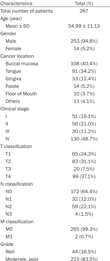
Table 1 lists the clinicopathologic
characteris-tics of patients with OSCC. Weenrolled 267
patients (253 male patients; mean age = 54.99 ± 11.13 y, range = 44-66 y) and analyzed their conditions. The cancers were located at the buccal mucosa (n = 108), tongue (n = 91), gin-giva (n = 33), palate (n = 14), floor of the mouth
(n = 10), and other locations (n = 11). According to the American Joint Committee on Cancer system, the tumors were classified into TNM
stages I (n = 51), II (n = 56), III (n = 30), and IV (n = 130).
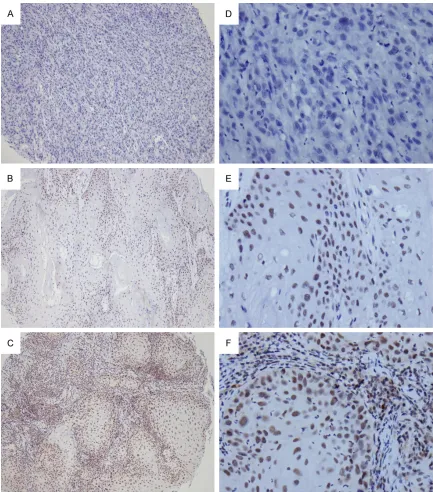
Nuclear CBP expression was detected by immu-nohistochemistry. Representative examples of tumors demonstrating overall negative (score: Table 1. Patient characteristics
Characteristics Total (%)
Total number of patients 267 Age (year)
Mean ± SD 54.99 ± 11.13 Gender
Male 253 (94.8%)
Female 14 (5.2%)
Cancer location
Buccal mucosa 108 (40.4%)
Tongue 91 (34.2%)
Gingiva 33 (12.4%)
Palate 14 (5.2%)
Floor of Mouth 10 (3.7%)
Others 11 (4.1%)
Clinical stage
I 51 (19.1%)
II 56 (21.0%)
III 30 (11.2%)
IV 130 (48.7%) T classification
T1 65 (24.3%)
T2 83 (31.1%)
T3 20 (7.5%)
T4 99 (37.1%) N classification
N0 172 (64.4%)
N1 32 (12.0%)
N2 59 (22.1%)
N3 4 (1.5%) M classification
M0 265 (99.3%)
M1 2 (0.7%)
Grade
Well 44 (16.5%)
5637 Int J Clin Exp Pathol 2016;9(5):5635-5641 0), low (score: 1), and high CBP (score: 2)
expression are illustrated in Figure 1.
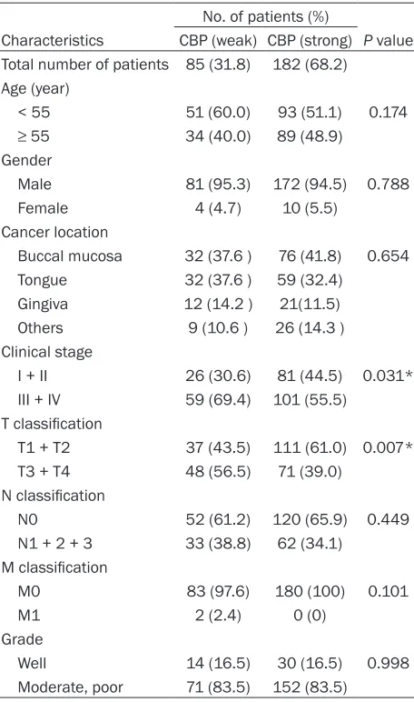
Table 2 depicts the relationships between selected clinicopathologic factors and CBP expression. CBP immunohistological stains
were classified according to the level of nuclear CBP expression into 2 groups, weak (score: 0)
and strong (score: 1 and 2). A higher CBP expression was significantly associated with a
more receded clinical stage (P = 0.031) and smaller tumor size (P = 0.007). However, the results revealed no significant association
[image:3.612.89.522.69.561.2]between CBP expression and patient age, sex, lymph node metastasis, or distal metastasis (Table 2).
Figure 1. CREB-binding protein (CBP) in primary oral cancer. Tissue microarrays of primary oral squamous cell carci
Table 3 illustrates the relationships between selected clinicopathologic factors and CBP expression in patients aged < 55 years. The results revealed that higher CBP expression
was significantly associated with a more reced
-ed clinical stage (P = 0.020) and smaller tumor size (P = 0.021); no significant associations
between CBP expression and patient age, sex, lymph node metastasis, or distal metastasis were observed. Table 4 illustrates that CBP
expression had no significant correlation with clinicopathologic factors in patients aged ≥ 55 years.
Discussion
Oral cancer is a fatal disease and has the
four-th highest mortality of malignancy among
ous malignancies including colorectal, breast, hepatocellular, and non-small cell lung carcino-mas [24-27]. Ianculescu et al. reported that CBP and p300 promoted prostate cancer pro-gression, which could be blocked by siRNA
[28]. CBP and p300 also promoted cancer pro
-gression in colon cancer cell lines with micro-satellite instability [29]. However, our results revealed that nuclear CBP expression was associated with an obstructed clinical stage and tumor size in patients with OSCC (Table 2).
These findings suggest that CBP may act as a
tumor suppressor in patients of OSCC. CBP and
p300 can promote cell proliferation and cancer
development under specific conditions; howev
-er, they are also crucial for the transactivation
function of p53, BRCA1, and FOXO3, which are
[image:4.612.92.320.95.481.2]all critical tumor suppressors [30-32]. In the Table 2. Patient characteristics regarding CBP
expres-sion
No. of patients (%)
Characteristics CBP (weak) CBP (strong) P value Total number of patients 85 (31.8) 182 (68.2)
Age (year)
< 55 51 (60.0) 93 (51.1) 0.174
≥ 55 34 (40.0) 89 (48.9)
Gender
Male 81 (95.3) 172 (94.5) 0.788
Female 4 (4.7) 10 (5.5) Cancer location
Buccal mucosa 32 (37.6 ) 76 (41.8) 0.654 Tongue 32 (37.6 ) 59 (32.4)
Gingiva 12 (14.2 ) 21(11.5)
Others 9 (10.6 ) 26 (14.3 ) Clinical stage
I + II 26 (30.6) 81 (44.5) 0.031* III + IV 59 (69.4) 101 (55.5)
T classification
T1 + T2 37 (43.5) 111 (61.0) 0.007* T3 + T4 48 (56.5) 71 (39.0)
N classification
N0 52 (61.2) 120 (65.9) 0.449 N1 + 2 + 3 33 (38.8) 62 (34.1)
M classification
M0 83 (97.6) 180 (100) 0.101 M1 2 (2.4) 0 (0)
Grade
Well 14 (16.5) 30 (16.5) 0.998
Moderate, poor 71 (83.5) 152 (83.5)
*P < 0.05.
Taiwanese males. The relatively high prev-alence of oral cancer in Taiwan is due mainly to the prevalence of chewing betel
quid [19]. Oral cancer arises from several
anatomic sites within the oral cavity, with the tongue and buccal mucosa being the most common sites in Taiwanese patients [20]. Despite advances in cancer diagnosis and treatment, the prognosis of OSCC
remains dismal. Most OSCC patients die of
recurrence or metastasis. The present study detected CBP expression in tumor tissues by using immunohistochemistry, revealing that high CBP expression in
patients with OSCC is significantly associ
vari-5639 Int J Clin Exp Pathol 2016;9(5):5635-5641 present study, the tumor-suppressive function of CBP both in vitro and in vivo supported our clinical data: (1) CBP pos-sibly induces an imbalance in the expres-sion of oncogenes and tumor-suppressor genes by the deregulation of histone acet-ylation [33], (2) CBP dysfunction alters the normal acetylation pattern of several can-cer-related nonhistone substrates caus-ing malignant transformation [34-36], (3) p53 acetylated and activated by CBP can inhibit cell growth [37], and (4) CBP may strengthen DNA damage signals by inhib-iting tumor cell growth [38]. p53 is as a tumor suppressor and plays a crucial role in regulating cell-cycle progression and apoptosis. Inactivation of the p53 path-way accounts for the most common molecular defects in human cancers [39]. Previous studies have reported that p53 downregulation through CBP enhances tumorigenesis [40]. Jin et al. also demon-strated that a histone deacetylase inhibi-tor, DWP0016, induced p53 acetylation,
which benefited from the upregulation of
the coactivators, CBP and p300, to inhibit cell growth in glioblastoma cells [37]. Therefore, CBP has a dual functional prop-erty, depending on the cellular environ-ment. Previous studies have reported that p53 is associated with tumorigenesis; activation of p53 expression is a potential strategy for anticancer treatment in he-
ad and neck squamous cell carcinoma.
Therefore, the correlation between CBP and p53 may provide a useful prognostic marker in OSCC diagnostics.
We conducted an immunohistochemical analysis and observed that nuclear CBP expression was associated with an ob- structed clinical stage and tumor size in
patients with OSCC. In addition, CBP
expression was not associated with poor overall survival. We could not study the relationship between CBP expression and poor overall survival. Therefore, further research with additional patient informa-tion is warranted to conclusively deter-mine the usefulness of nuclear CBP in
OSCC diagnostics and to clarify the down
-stream mechanisms involved in
control-ling the biological behavior of OSCC via
[image:5.612.92.324.97.383.2]histone-modifying molecules. Table 3. Patient characteristics regarding CBP
expres-sion in the young patient (age < 55) No. of patients (%)
Characteristics CBP (weak) CBP (strong) P value Total number of patients 51 (35.4) 93 (64.6)
Gender
Male 49 (96.1) 88 (94.6) 0.698
Female 2 (3.9) 5 (5.4) Cancer location
Buccal mucosa 21 (41.2) 35 (37.6) 0.417 Tongue 18 (35.3) 34 (36.6)
Gingiva 8 (15.7) 9 (9.7)
Others 4 (7.8) 15 (16.1)
Clinical stage
I + II 14 (27.5) 44 (47.3) 0.020* III + IV 37 (72.5) 49 (52.7)
T classification
T1 + T2 21 (41.2) 57 (61.3) 0.021* T3 + T4 30 (58.8) 36 (38.7)
N classification
N0 29 (56.9) 62 (66.7) 0.243 N1 + 2 + 3 22 (43.1) 31 (33.3)
Grade
Well 10 (19.6) 18 (19.4) 0.971 Moderate, poor 41 (80.4) 75 (80.6)
*P < 0.05.
Table 4. Patient characteristics regarding CBP
expres-sion in the older patient (age ≥ 55)
No. of patients (%)
Characteristics CBP (weak) CBP (strong) P value Total number of patients 34 (27.6) 89 (72.4)
Gender
Male 32 (94.1) 84 (94.4) 0.955 Female 2 (5.9) 5 (5.6)
Cancer location
Buccal mucosa 11 (32.4) 41 (46.1) 0.460 Tongue 14 (41.1) 25 (28.1)
Gingiva 4 (11.8) 12 (13.5)
Others 5 (14.7) 11 (12.3)
Clinical stage
I + II 12 (35.3) 37 (41.6) 0.525 III + IV 22 (64.7) 52 (58.4)
T classification
T1 + T2 16 (47.1) 54 (60.7) 0.173 T3 + T4 18 (52.9) 35 (39.3)
N classification
N0 23 (67.6) 58 (65.2) 0.795 N1 + 2 + 3 11 (32.4) 31 (34.3)
Grade
Well 4 (11.8) 12 (13.5) 0.800
[image:5.612.92.323.433.724.2]Acknowledgements
This study was supported by a research grant from National Science Council, Taiwan
(NSC96-2628-B-040-021-MY3 and NSC102-2314-B-040-008-MY3) and Chung Shan Medical
Uni-versity Hospital, Taiwan (CSH-2015-C-015; CSH-2015-E-001-Y2).
Disclosure of conflict of interest
None.
Address correspondence to: Dr. Chiao-Wen Lin,
Institute of Oral Sciences, Chung Shan Medical
University, 110 Chien-Kuo N. Road, Section 1,
Taichung 402, Taiwan. Tel: +886-4-24739595 Ext. 34255; Fax: +886-4-24723229; E-mail: cwlin@
csmu.edu.tw
References
[1] Chiba I. Prevention of Betel Quid Chewers’ Oral Cancer in the Asian-Pacific Area. Asian Pac J
Cancer Prev 2001; 2: 263-269.
[2] Tanaka T. Chemoprevention of oral
carcino-genesis. Eur J Cancer B Oral Oncol 1995; 31B:
3-15.
[3] Petersen PE. Oral cancer prevention and con
-trol--the approach of the World Health Or-ganization. Oral Oncol 2009; 45: 454-460.
[4] Kantola S, Parikka M, Jokinen K, Hyrynkangs
K, Soini Y, Alho OP and Salo T. Prognostic fac -tors in tongue cancer-relative importance of demographic, clinical and histopathological
factors. Br J Cancer 2000; 83: 614-619.
[5] Popovic R and Licht JD. Emerging epigene- tic targets and therapies in cancer medicine. Cancer Discov 2012; 2: 405-413.
[6] Bannister AJ and Kouzarides T. The CBP co-activator is a histone acetyltransferase. Nature
1996; 384: 641-643.
[7] Ogryzko VV, Schiltz RL, Russanova V, Howard
BH and Nakatani Y. The transcriptional coacti-vators p300 and CBP are histone
acetyltrans-ferases. Cell 1996; 87: 953-959.
[8] Goodman RH and Smolik S. CBP/p300 in cell growth, transformation, and development. Genes Dev 2000; 14: 1553-1577.
[9] Avantaggiati ML, Ogryzko V, Gardner K,
Giordano A, Levine AS and Kelly K. Recruitment of p300/CBP in p53-dependent signal
path-ways. Cell 1997; 89: 1175-1184.
[10] Cras A, Politis B, Balitrand N, Darsin-Bettinger D, Boelle PY, Cassinat B, Toubert ME and Chomienne C. Bexarotene via CBP/p300 in-duces suppression of NF-kappaB-dependent cell growth and invasion in thyroid cancer. Clin
Cancer Res 2012; 18: 442-453.
[11] Zhao X and Benveniste EN. Transcriptional ac -tivation of human matrix metalloproteinase-9 gene expression by multiple co-activators. J
Mol Biol 2008; 383: 945-956.
[12] Wang F, Zhang R, Wu X and Hankinson O.
Roles of coactivators in hypoxic induction of
the erythropoietin gene. PLoS One 2010; 5:
e10002.
[13] Gao Y, Geng J, Hong X, Qi J, Teng Y, Yang Y, Qu
D and Chen G. Expression of p300 and CBP is associated with poor prognosis in small cell lung cancer. Int J Clin Exp Pathol 2014; 7: 760-767.
[14] Tang Z, Yu W, Zhang C, Zhao S, Yu Z, Xiao X, Tang R, Xuan Y, Yang W, Hao J, Xu T, Zhang Q,
Huang W, Deng W and Guo W. CREB-binding protein regulates lung cancer growth by target-ing MAPK and CPSF4 signaltarget-ing pathway. Mol
Oncol 2016; 10: 317-29.
[15] Santer FR, Hoschele PP, Oh SJ, Erb HH,
Bouchal J, Cavarretta IT, Parson W, Meyers DJ, Cole PA and Culig Z. Inhibition of the acetyl-transferases p300 and CBP reveals a targeta-ble function for p300 in the survival and inva-sion pathways of prostate cancer cell lines. Mol Cancer Ther 2011; 10: 1644-1655. [16] Xu LX, Li ZH, Tao YF, Li RH, Fang F, Zhao H, Li G,
Li YH, Wang J, Feng X and Pan J. Histone acet -yltransferase inhibitor II induces apoptosis in glioma cell lines via the p53 signaling pathway.
J Exp Clin Cancer Res 2014; 33: 108.
[17] Lin SH, Lin YM, Yeh CM, Chen CJ, Chen MW, Hung HF, Yeh KT and Yang SF. Casein kinase 1 epsilon expression predicts poorer prognosis in low T-stage oral cancer patients. Int J Mol Sci
2014; 15: 2876-2891.
[18] Ko CP, Yang LC, Chen CJ, Yeh KT, Lin SH, Yang SF, Chen MK and Lin CW. Expression of
my-eloid zinc finger 1 and the correlation to clini
-cal aspects of oral squamous cell carcinoma.
Tumour Biol 2015; 36: 7099-7105.
[19] Kao SY and Lim E. An overview of detection and screening of oral cancer in Taiwan. Chin J
Dent Res 2015; 18: 7-12.
[20] Ho PS, Ko YC, Yang YH, Shieh TY and Tsai CC. The incidence of oropharyngeal cancer in
Taiwan: an endemic betel quid chewing area. J Oral Pathol Med 2002; 31: 213-219.
[21] Wolffe AP. Chromatin remodeling: why it is
im-portant in cancer. Oncogene 2001; 20:
2988-2990.
[22] Brasacchio D, Noori T, House C, Brennan AJ,
Simpson KJ, Susanto O, Bird PI, Johnstone RW
and Trapani JA. A functional genomics screen
identifies PCAF and ADA3 as regulators of hu -man granzyme B-mediated apoptosis and Bid
cleavage. Cell Death Differ 2014; 21: 748-760.
[23] Guo W, Lu J, Dai M, Wu T, Yu Z, Wang J, Chen W,
Shi D, Yu W, Xiao Y, Yi C, Tang Z, Xu T, Xiao X,
5641 Int J Clin Exp Pathol 2016;9(5):5635-5641
coactivator CBP upregulates hTERT expression and tumor growth and predicts poor prognosis
in human lung cancers. Oncotarget 2014; 5:
9349-9361.
[24] Chen LC, Kurisu W, Ljung BM, Goldman ES, Moore D 2nd and Smith HS. Heterogeneity for allelic loss in human breast cancer. J Natl
Cancer Inst 1992; 84: 506-510.
[25] Takahashi K, Kudo J, Ishibashi H, Hirata Y and
Niho Y. Frequent loss of heterozygosity on chro -mosome 22 in hepatocellular carcinoma. Hepatology 1993; 17: 794-799.
[26] Duriez C, Schmitz A, Fouchet P, Buecher B, Thuille B, Lerebours F, Leger R, Boman F, Flejou JF, Monges G, Paraf F, Bedossa P, Sabourin JC, Salmon RJ, Laurent-Puig P,
Thomas G and Olschwang S. [Localization of a
tumor suppressor gene distal to D22S270 in colorectal cancers]. Gastroenterol Clin Biol
1997; 21: 358-364.
[27] Hou X, Li Y, Luo RZ, Fu JH, He JH, Zhang LJ and Yang HX. High expression of the transcriptional
co-activator p300 predicts poor survival in re-sectable non-small cell lung cancers. Eur J
Surg Oncol 2012; 38: 523-530.
[28] Ianculescu I, Wu DY, Siegmund KD and Stallcup MR. Selective roles for cAMP response ele-ment-binding protein binding protein and p300 protein as coregulators for androgen-regulated gene expression in advanced prostate cancer
cells. J Biol Chem 2012; 287: 4000-4013.
[29] Ionov Y, Matsui S and Cowell JK. A role for p300/CREB binding protein genes in promot-ing cancer progression in colon cancer cell lines with microsatellite instability. Proc Natl
Acad Sci U S A 2004; 101: 1273-1278.
[30] Watts GS, Oshiro MM, Junk DJ, Wozniak RJ,
Watterson S, Domann FE and Futscher BW. The acetyltransferase p300/CBP-associated factor is a p53 target gene in breast tumor
cells. Neoplasia 2004; 6: 187-194.
[31] Ogiwara H and Kohno T. CBP and p300 histone
acetyltransferases contribute to homologous recombination by transcriptionally activating
the BRCA1 and RAD51 genes. PLoS One 2012; 7: e52810.
[32] Wang F, Marshall CB, Li GY, Yamamoto K, Mak TW and Ikura M. Synergistic interplay between promoter recognition and CBP/p300
coactiva-tor recruitment by FOXO3a. ACS Chem Biol
2009; 4: 1017-1027.
[33] Nagy Z and Tora L. Distinct GCN5/PCAF-containing complexes function as co-activators and are involved in transcription factor and
global histone acetylation. Oncogene 2007;
26: 5341-5357.
[34] Gu W and Roeder RG. Activation of p53
se-quence-specific DNA binding by acetylation of
the p53 C-terminal domain. Cell 1997; 90: 595-606.
[35] Zhao LY, Liu Y, Bertos NR, Yang XJ and Liao D.
PCAF is a coactivator for p73-mediated
trans-activation. Oncogene 2003; 22: 8316-8329.
[36] Cohen HY, Lavu S, Bitterman KJ, Hekking B, Imahiyerobo TA, Miller C, Frye R, Ploegh H, Kessler BM and Sinclair DA. Acetylation of the C terminus of Ku70 by CBP and PCAF controls Bax-mediated apoptosis. Mol Cell 2004; 13:
627-638.
[37] Jin H, Liang L, Liu L, Deng W and Liu J. HDAC inhibitor DWP0016 activates p53 transcription and acetylation to inhibit cell growth in U251 glioblastoma cells. J Cell Biochem 2013; 114:
1498-1509.
[38] Yan G, Eller MS, Elm C, Larocca CA, Ryu B, Panova IP, Dancy BM, Bowers EM, Meyers D, Lareau L, Cole PA, Taverna SD and Alani RM. Selective inhibition of p300 HAT blocks cell cycle progression, induces cellular senescen- ce, and inhibits the DNA damage response in melanoma cells. J Invest Dermatol 2013; 133: 2444-2452.
[39] Brown CJ, Lain S, Verma CS, Fersht AR and Lane DP. Awakening guardian angels: drugging the p53 pathway. Nat Rev Cancer 2009; 9:
862-873.
[40] Mazza D, Infante P, Colicchia V, Greco A, Alfonsi R, Siler M, Antonucci L, Po A, De Smaele E, Ferretti E, Capalbo C, Bellavia D, Canettieri G, Giannini G, Screpanti I, Gulino A and Di
Marcotullio L. PCAF ubiquitin ligase activity in -hibits Hedgehog/Gli1 signaling in p53-depen-dent response to genotoxic stress. Cell Death