ORIGINAL RESEARCH
Forensic Application of Postmortem
Diffusion-Weighted and Diffusion Tensor MR Imaging of
the Human Brain in Situ
E. Scheurer K.-O. Lovblad R. Kreis S.E. Maier C. Boesch R. Dirnhofer K. Yen
BACKGROUND AND PURPOSE: DWI and DTI of the brain have proved to be useful in many neurologic disorders and in traumatic brain injury. This prospective study aimed at the evaluation of the influence of the PMI and the cause of death on the ADC and FA for the application of DWI and DTI in forensic radiology.
MATERIALS AND METHODS: DWI and DTI of the brain were performed in situ in 20 deceased subjects with mapping of the ADC and FA. Evaluation was performed in different ROIs, and the influence of PMI and cause of death was assessed.
RESULTS: Postmortem ADC values of the brain were decreased by 49%–72% compared with healthy living controls. With increasing PMI, ADCs were significantly reduced when considering all ROIs together and, particularly, GM regions (all regions,P⬍.05; GM,P⬍.01), whereas there was no significant effect in WM. Concerning the cause of death, ADCs were significantly lower in mechanical and hypoxic brain injury than in brains from subjects having died from heart failure (traumatic brain injury,P⬍.005; hypoxia,P⬍.001). Postmortem FA was not significantly different from FA in living persons and showed no significant influence of PMI or cause of death.
CONCLUSIONS:Performing postmortem DWI and DTI of the brain in situ can provide valuable infor-mation for application in forensic medicine. ADC could be used as an indicator of PMI and could help in the assessment of the cause of death.
ABBREVIATIONS:ADC⫽apparent diffusion coefficient; ADCTc⫽temperature corrected apparent diffusion coefficient; CC⫽corpus callosum; DWI⫽diffusion-weighted imaging, DTI⫽diffusion tensor imaging; FA⫽fractional anisotropy; FSE⫽fast spin-echo; GM⫽gray matter, HF⫽cardiac failure; MRI⫽MR imaging; n.a.⫽measurements not performed; PMI⫽postmortem interval; ROI⫽region of interest; SEM⫽standard error of the mean; STIR⫽short tau inversion recovery; STR⫽strangulation; Tapp⫽approximate body core temperature at the time of MR imaging; TBI⫽ traumatic brain injury; WM⫽white matter
I
n DWI, signal intensity strongly depends on the rate of water diffusion, which can be used quantitatively to determine ADC.1The scalar FA derived from DTI adds detailed data onthe average directionality of diffusion, which allows investigat-ing connectivity and microstructural integrity of internal fibrous structures, such as neuronal tracts in the brain.2-5In clinical
med-icine, both DWI and DTI of the brain have proved to be useful particularly to assess ischemia, edema, and structural integrity in many neurologic disorders and in traumatic brain injury.6-12
In the past 10 years, radiologic methods such as CT and MR imaging have been increasingly used in forensic medicine to address, among others, neurotraumatologic and neuropatho-logic issues.13-15The application of DWI and DTI to the post-mortem brain is expected to support the diagnosis of brain
parenchyma damage due to traumatic incidents on a micro-structural level by noninvasively revealing edema and rupture of fiber tracts.16,17
Numerous postmortem DWI and DTI studies were per-formed in the animal and human brain; however, most eval-uated the effect of formalin fixation and PMI on diffusion properties18-23or investigated isolated fixed human brains for
the diagnosis of disease.24,25To date, there are only very few
studies on DWI or DTI in the postmortem unfixed human brain in situ that have concentrated on general characteristic changes in postmortem MR imaging and CT, including DWI on one hand and postmortem DTI in a single case with a brain stem trauma on the other.17,26
In this study, we aimed at the evaluation of a potential application of DWI and DTI and particularly ADC and FA in different regions of the postmortem brain in situ for the ap-plication in forensic diagnostics. The main goals were to assess the following: 1) whether there was an influence of the time since death on ADC and FA, which could be used for estima-tion of the PMI, and 2) whether there was a correlaestima-tion with the cause of death.
Materials and Methods
Subjects
A consecutive sample of 20 deceased subjects with a forensic autopsy request by the legal authorities was included in this study (15 males, 5 Received October 22, 2010; accepted after revision December 17.
From the Ludwig Boltzmann Institute for Clinical-Forensic Imaging (E.S., K.Y.), Graz, Austria; Medical University Graz (E.S., K.Y.), Graz, Austria; Institute of Forensic Medicine (E.S., R.D., K.Y.), University of Bern, Bern, Switzerland; Department of Radiology (K.-O.L.), University of Geneva, Geneva, Switzerland; Department of Neuroradiology (K.-O.L.), Inselspital, Bern, Switzerland; Department of Clinical Research (R.K., C.B.), MR-Spectroscopy and -Method-ology, University of Bern, Bern, Switzerland; and Department of Radiology (S.E.M.), Brigham and Women’s Hospital, Harvard Medical School, Boston, Massachusetts.
Please address correspondence to Eva Scheurer, MD, MSc, Ludwig Boltzmann Institute for Clinical-Forensic Imaging, Universita¨tsplatz 4/II, A-8010 Graz, Austria; e-mail: eva.scheurer@ cfi.lbg.ac.at
http://dx.doi.org/10.3174/ajnr.A2508
females; mean age, 45 years; median, 46 years; age range, 3–94 years). Two healthy living volunteers (a man, aged 26 years; a woman, aged 31 years) were examined as controls. Before scanning, the corpses were examined externally by a forensic pathologist to ensure compli-ance with the inclusion criteria (ie, PMI at the time of inclusion⬍120 hours, no signs of decomposition, no history of neurologic disorder). PMI was determined either by witnessed death (eg, when death oc-curred in hospital) or by standard forensic methods (body tempera-ture, livor and rigor mortis). For the evaluation of the quantitative effect of PMI, subjects were divided into 3 groups: PMI group 1, with PMIⱕ24 hours (n⫽7; mean age⫾SEM, 38⫾22.8 years), PMI group 2 with PMI 25– 48 hours (n⫽7, age, 52⫾20.3 years), PMI group 3 with PMI⬎48 hours (n⫽6, age, 45⫾13.7 years). Mean ages in the groups were not significantly different from each other. In 12 subjects, the cause of death, as diagnosed at forensic autopsy after scanning, was traumatic; 3 had died from intoxication or medical maltreatment, and 5 from natural causes (Table 1). For the analysis of the influence of the cause of death, subject groups were defined as the following: 1) mechanical brain trauma (n⫽4), with all subjects show-ing signs of a direct blunt force impact and intracranial hemorrhage and varying additional findings, such as skull fractures, cerebral con-tusions, and edema; 2) hypoxic brain injury caused by strangulation (n⫽5); and 3) heart failure (n⫽4). The study was approved by the local ethics committee, and informed consent was obtained from the living volunteers.
MR Imaging
MR imaging of the brain in situ was performed within 141 hours after death (mean, 40 hours; median, 38 hours; range, 13–141 hours) at 1.5T (Signa EchoSpeed Horizon, Version 5.8; GE Healthcare, Mil-waukee, Wisconsin) by using a quadrature head coil. Depending on cooling time at 4°C, body core temperature at the beginning of the acquisition was between 5°C and 30°C (median, 11°C). The measure-ment was performed with a digital thermometer in the rectum. For MR imaging, the bodies were wrapped in 2 artifact-free body bags to prevent contamination and to guarantee anonymity.
Data were acquired in the axial orientation with a multisection line-scan sequence; for each section, 6 images with high b-values (bmax⫽1000 s/mm2) in 6 noncollinear and noncoplanar directions [relative amplitudes: (Gx,Gy,Gz)⫽{(1,1,0),(0,1,1),(1,0,1)(⫺1,1,0), (0,⫺1,1)(1,0,⫺1)}] and 2 images with low b-values (5 s/mm2) were obtained. The imaging parameters were the following: TR/TE, 3520/96 ms; matrix, 128⫻128; FOV, 24⫻24 cm; NEX, 1; sections, 22; section thickness, 5 mm; section gap, 1 mm. Additionally, a stan-dard protocol, including an axial T1-weighted (TR/TE, 400/14 ms), T2-weighted FSE (TR/TE, 4000/15 ms), STIR (TR/TE, 11 002/217 ms), and a fast multiplanar spoiled gradient-recalled acquisition in the steady state (TR/TE, 270/4.2 ms) sequence, was performed. Total scanning times ranged from 35 to 75 minutes.
For the living volunteers, the same protocol with parameters typ-ical for in vivo examinations was used (DTI: TR/TE, 3392/84.5 ms; matrix, 128⫻128; FOV, 22⫻22 cm; NEX, 1; sections, 12; section thickness, 5 mm; section gap, 6 mm; and bmax, 1000 s/mm2).
Data analysis was performed on an Advantage Windows worksta-tion (Version 9.1, GE Healthcare).
Evaluation of ADC and FA
Maps of ADC and FA were calculated on a pixel-by-pixel basis after zero-filling to a matrix size of 256⫻256. Quantitative measurements were performed by 2 independent examiners (neuroradiologist, 11 years of experience; forensic expert, 10 years of experience) section by section in anatomically specified ROIs (circular ROIs for ADC, 10 mm2; for FA, 20 mm2, respectively). ADC was measured bilaterally in 20 ROIs in GM and WM as well as regions with mixed proportions (ie, fractions of both white and gray matter, as listed in the On-line Table) and 1 ROI in the cerebellar vermis (Fig 1); FA was measured bilaterally in 9 ROIs in WM and mixed regions only (Table 2). For statistical evaluation, the ROIs were grouped in WM, GM, and all ROIs (ie, including ROIs with mixed tissue). Because diffusion is temperature-dependent, ADCs of the postmortem cases were tem-perature-corrected to 38°C by using a correction factor of 2% per degree Celsius according to the equation27: ADC
[image:2.594.53.532.57.295.2]Tc⫽ADC(100%⫹ Table 1: Case data
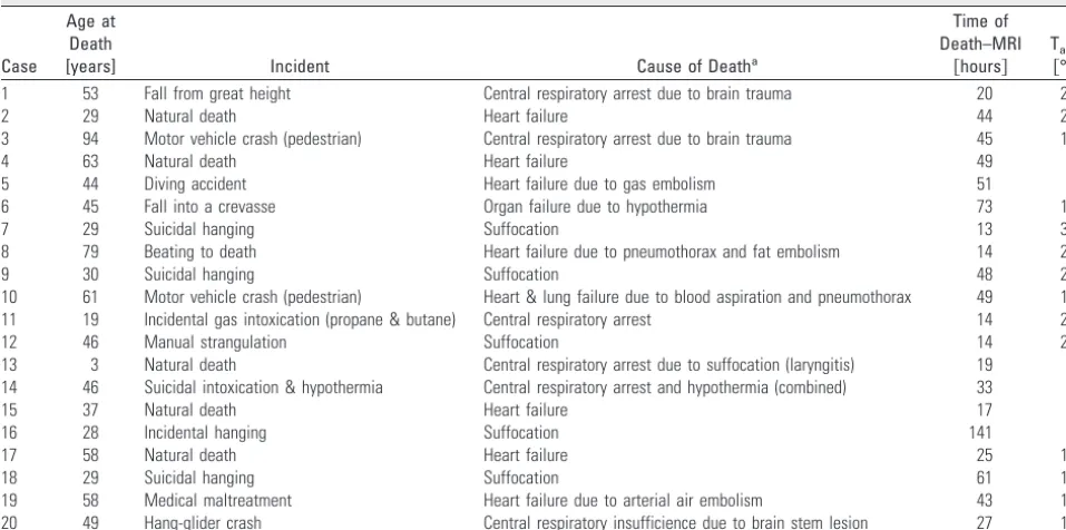
Case
Age at Death
[years] Incident Cause of Deatha
Time of Death–MRI
关hours兴
Tappb 关°C兴
1 53 Fall from great height Central respiratory arrest due to brain trauma 20 20
2 29 Natural death Heart failure 44 20
3 94 Motor vehicle crash (pedestrian) Central respiratory arrest due to brain trauma 45 10
4 63 Natural death Heart failure 49 5
5 44 Diving accident Heart failure due to gas embolism 51 5
6 45 Fall into a crevasse Organ failure due to hypothermia 73 12
7 29 Suicidal hanging Suffocation 13 30
8 79 Beating to death Heart failure due to pneumothorax and fat embolism 14 28
9 30 Suicidal hanging Suffocation 48 23
10 61 Motor vehicle crash (pedestrian) Heart & lung failure due to blood aspiration and pneumothorax 49 10
11 19 Incidental gas intoxication (propane & butane) Central respiratory arrest 14 25
12 46 Manual strangulation Suffocation 14 22
13 3 Natural death Central respiratory arrest due to suffocation (laryngitis) 19 8
14 46 Suicidal intoxication & hypothermia Central respiratory arrest and hypothermia (combined) 33 5
15 37 Natural death Heart failure 17 8
16 28 Incidental hanging Suffocation 141 5
17 58 Natural death Heart failure 25 10
18 29 Suicidal hanging Suffocation 61 15
19 58 Medical maltreatment Heart failure due to arterial air embolism 43 10
20 49 Hang-glider crash Central respiratory insufficience due to brain stem lesion 27 19
aIn cases with combined or concurring causes of death only the most relevant are mentioned. b
Approximate body core temperature at the time of MR imaging
BRAIN
ORIGINAL
2%)(38°C-Tapp). The body core temperature measured at the start of the scan (Tapp) was used for the correction.
Forensic Autopsy and Histology
After MR imaging, autopsy was performed by 2 forensic pathologists within 24 hours, in 2 cases a few hours later (34 and 44 hours, respec-tively). Autopsy included detailed neuropathologic evaluation in all cases; histologic examination was performed from visible lesions. Vi-sual comparison of MR images with autopsy findings and, if available, with their histologic correlates was performed.
Statistical Analysis
For analysis, ADC and FA measurements of both examiners were pooled for each ROI. The evaluation of effects of PMI and cause of
death was performed with the Statistical Package for the Social Sci-ences, Version 17.0 (SPSS, Chicago, Illinois) by using a univariate analysis of variance with a Bonferroni post hoc correction for multi-ple tests (pair-wise comparisons). APvalue of⬍.05 was considered significant. The brain regions were assigned to 3 groups (GM, WM, mixed) as shown in the On-line Table. The effect of PMI on ADC was evaluated once for all brain regions with PMI group and brain region as factors. A second analysis was calculated separately for GM and WM regions, respectively, with the PMI group as a factor. Addition-ally, the Pearson correlation of PMI and ADC was evaluated (2-sided). For the assessment of the effect of PMI on FA, only the PMI group was used as a factor because only WM and mixed tissue ROIs had been measured. The influence of a cause of death involving me-chanical or hypoxic brain injury on ADC was assessed with cause of
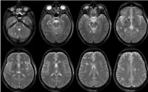
Fig 1.Trace-weighted image (b-value⫽5 s/mm2
[image:3.594.52.533.44.343.2]) of a deceased subject showing the placement of ROIs for the evaluation of ADC values. ROIs are only shown unilaterally; the ROI in the medulla is not shown. The ROIs are the following: 1) pons, 2) cerebellum, 3) hippocampus, 4) mesencephalon, 5) vermis, 6) putamen, 7) pallidum, 8) temporal cortex, 9) internal capsule anterior, 10) internal capsule posterior, 11) thalamus, 12) occipital cortex, 13) corpus callosum genu, 14) corpus callosum splenium, 15) frontal WM, 16) caudate nucleus, 17) frontal cortex, 18) motor cortex, 19) centrum semiovale, and 20) parietal cortex.
Table 2: FA in different ROIs
ROI
FA
Postmortem Casesa Living Controls
Mean SD Median Min Max Mean SD
Frontal white matter 0.25 0.05 0.25 0.15 0.34 0.22 0.01
Centrum semiovale 0.26 0.04 0.25 0.18 0.34 0.32 0.03
Internal capsule anterior 0.31 0.07 0.31 0.18 0.41 0.32 0.02
Internal capsule posterior 0.41 0.04 0.41 0.33 0.51 0.36 0.01
Corpus callosum genu 0.36 0.08 0.37 0.19 0.48 0.36 0.04
Corpus callosum splenium 0.49 0.08 0.51 0.32 0.59 0.56 0.04
Mesencephalon 0.31 0.05 0.30 0.24 0.49 0.43 0.03
Pons 0.31 0.05 0.31 0.23 0.39 0.31 0.04
Medulla 0.22 0.04 0.22 0.16 0.28 n.a. n.a.
[image:3.594.52.539.398.546.2]death and brain region as factors, while for the effect on FA, only cause of death was used as a factor.
Results
ADC
Good-quality DWI and DTI were acquired in deceased sub-jects as well as in healthy living volunteers. Figure 2 shows an example of postmortem and in vivo DWI and the ADC and FA maps in the respective subjects.
The On-line Table summarizes mean, SD, median, and minimal and maximal values for the ADCTcin different brain
regions in comparison with ADCs of the living controls. Mean postmortem ADCTcranged from 21⫻10⫺5mm2/s (vermis of
the cerebellum) to 41⫻10⫺5mm2/s (medulla). Postmortem ADCTcof GM with a mean ADCTcbetween 34⫻10⫺5mm2/s
(frontal cortex) and 38⫻10⫺5mm2/s (occipital cortex) was
significantly higher than ADCTcvalues in WM (P⬍.01) with
a mean ADCTc between 29⫻ 10⫺5mm2/s (centrum
semi-ovale) and 41⫻10⫺5mm2/s (medulla) and in mixed regions
(P⬍.05). Compared with the ADCs of the living volunteers, which ranged from 67⫻10⫺5mm2/s (cerebellum) to 81⫻
10⫺5mm2/s (hippocampus), postmortem ADCTcwas
consid-erably reduced. The greatest decrease was observed in the ver-mis, with a reduction of 72% compared with the ADC of the healthy living controls, while the least reduction was 49% in the occipital cortex.
Figure 3 compares the ADCs of the living controls with the postmortem values and shows the great decrease in all exam-ined ROIs. In contrast to this huge effect, approximate tem-perature correction had only a minor influence.
To evaluate the effect of the PMI on the ADCTcof different
brain regions, we compared 3 groups. Group 1 had a mean PMI of 16 hours (SD, 3 hours; median, 14 hours; range, 13–20 hours), group 2 a mean PMI of 38 hours (SD, 9 hours; median, 43 hours; range, 25– 48), and group 3 a mean PMI of 71 hours since death (SD, 36 hours; median, 56 hours; range, 49 –141 hours). Within the 3 groups, the distribution of the causes of death (brain trauma, strangulation, and heart failure) was not significantly different. Figure 4 shows that with increasing PMI, postmortem ADCTcin the overall brain and in GM
re-gions decreased significantly between group 1 and group 3 (all ROIs,P⬍.05; GM,P⬍.01), while there were no significant
Fig 2.Example of a DWI image,b0image, ADC map, and FA map of a healthy living volunteer (woman, 31 years) (top row) and a deceased subject (woman; age, 45 years; PMI, 73 hours;
body temperature at acquisition, 12°C) (bottom row).A, DWI (b⫽1000 s/mm2).B, Trace-weighted image with b-value⫽5 s/mm2.C, ADC map.D) FA map.
[image:4.594.52.533.43.342.2] [image:4.594.303.532.380.537.2]differences between groups 1 and 2, or between groups 2 and 3. In WM regions, no significant effect was observable. Corre-lation of ADC with PMI showed a decreasing linear trend; however, with a moderate and not statistically significant cor-relation coefficient ofr⫽0.42 for GM (P⫽.06).
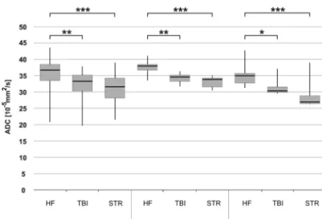
The influence of the cause of death on the ADCTcin all
ROIs and in GM and WM separately is demonstrated in Fig 5. The subjects having died from a natural cause due to cardiac failure had significantly higher ADCTcvalues in all brain
re-gions than those who died from mechanical brain trauma (all ROIs,P⬍.005; GM,P⬍.005; WM,P⬍.05) or the sub-jects with hypoxic brain injury due to congestion resulting from strangulation (all ROIs,P⬍.001; GM,P⬍.001; WM, P⬍.001). There was no significant difference between the 2 types of brain injury, and no correlation between the cause of death and PMI.
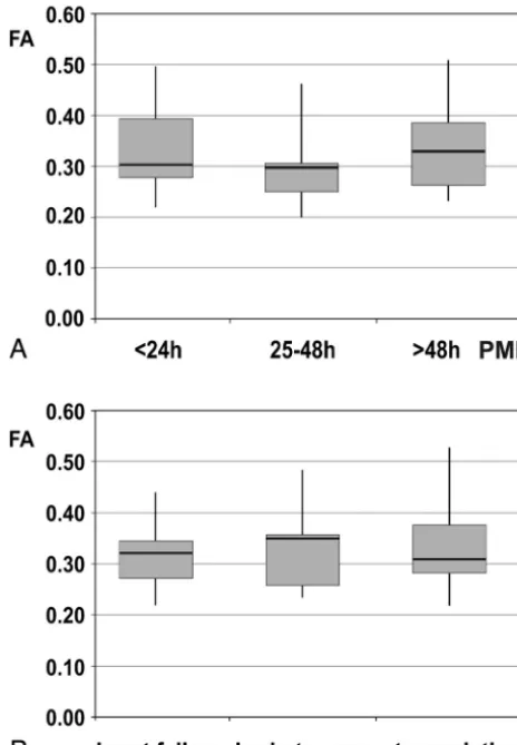
FA
Between the deceased subjects and the living controls, there was no significant difference concerning FA of all brain re-gions (Fig 6). Mean values were between 0.22 and 0.49 in the
postmortem cases and between 0.22 and 0.56 in the living controls (Table 2). The highest FA was measured in the splenium of the corpus callosum in both postmortem and liv-ing subjects, while the lowest value was found in the medulla and frontal WM. The PMI did not have any significant effect on FA (ie, the pair-wise comparisons among the 3 groups of subjects did not show any significant difference [Fig 7A]). FA was also stable regarding the influence of different types of brain injury (ie, mechanical brain trauma and hypoxic injury due to strangulation). None of the pair-wise comparisons reached significance when comparing the subjects with brain injury with those having died from cardiac failure (Fig 7B).
Traumatologic Changes
In 6 cases, traumatic changes of the brain were observed at autopsy, which were mainly intracerebral hematoma (n⫽1), lacerations (n⫽3), and subarachnoid (n⫽8) and subdural hematomas (n⫽3). Of these, 4 subarachnoid hemorrhages and 1 thin subdural blood layer were not seen on MR imaging. All other findings were detected with MR imaging with a good correlation to autopsy.
Discussion
ADC and FA were measured in the postmortem brain in situ to evaluate DWI and DTI for forensic application. Visual com-parison of postmortem ADC maps with those in the living controls already showed clearly reduced diffusivity. Although differences in body temperature could well be one reason for this, postmortem ADCTcproved to be significantly decreased
compared with the values of living subjects in all brain regions, confirming previous studies,26,28agreeing also with clinical
findings in acute ischemia due to cerebral occlusion.29-31 Post-mortem ADCTcvalues were significantly higher in GM than in
WM. Because this observation was also reported in living sub-jects,32this difference between gray and white matter seems to
be preserved postmortem.
In forensic medicine, PMI (ie, the time span between death and either examination or fixation) is essential for criminal investigations. Particularly beyond approximately 30 hours after death, when the body reaches ambient temperature,
Fig 4.Influence of PMI on ADCTcin all ROIs, GM, and WM, respectively. Whiskers show
the maximum and the minimum values. A single asterisk indicatesP⬍.05; double asterisks,P⬍.01.
Fig 5.Influence of mechanical brain trauma and hypoxic brain injury due to strangulation on ADCTcof all ROIs, GM, and WM, respectively. A group of brain trauma cases (n⫽4)
and a group of strangulation cases (n⫽5) are compared with a group of subjects with natural death due to cardiac failure (n⫽4). Whiskers show the maximum and the minimum values. A single asterisk indicatesP⬍.05; double asterisks,P⬍.005; and triple asterisks, P⬍.001.
[image:5.594.55.283.42.210.2] [image:5.594.302.530.42.215.2] [image:5.594.53.285.257.415.2]objective methods are hardly developed.33,34A significant
dif-ference of ADCTcwas found mainly in the GM between the
group with PMIsⱕ1 day and that with PMIs of⬎2 days. This is in agreement with previous studies that investigated the ef-fect of PMI in tissue samples of animal brain fixed at different time points after death.20,22 However, in contrast to these
studies, no significant effect of PMI on ADC could be observed for WM. Linear correlation of ADC with PMI was moderate, which can be attributed to several reasons such as an overlay by the differences in the cause of death, incomplete data with higher PMIs, as well as an influence of noise and imperfect temperature correction. Additionally, the decrease may be nonlinear. However, the measurement of postmortem ADCTc
in GM could emerge as a valuable method for a noninvasive and objective estimation of PMI.
Regarding the cause of death, ADCTc was significantly
lower in all brain regions in 2 groups of subjects, with a cause of death associated with brain injury compared with subjects with cardiac failure. No significant difference between brain injury caused by mechanical trauma and hypoxic injury due to strangulation was found. The higher ADCTcin subjects with
heart failure could be an indicator of the length of the process of dying (ie, agony). This would be in good agreement with the ADCTcvalues in the groups with brain injury, because
accord-ing to forensic experience, agony is usually prolonged to sev-eral minutes in death due to cardiac failure, while in strangu-lation agony is expected to be very short, in some cases even shorter than in death due to mechanical brain trauma. In fo-rensic medicine, the assessment of the cause of death can be challenging, particularly in cases without any exteriorly visible injuries. Thus, postmortem ADCTccould help in
noninva-sively differentiating natural death from death caused by oc-cult brain injury mechanisms. Because ADC in WM seems to be less influenced by PMI, the evaluation of WM regions would be preferential to address this question.
Postmortem FA did not change significantly compared with the values of the healthy living volunteers. Additionally, PMI and the presence of brain injury did not show any signif-icant change of FA. This observation disagrees with those in other studies, which found decreasing anisotropy following brain death.22,23,35-37However, direct comparison is difficult
because in some of these studies, fixed brain or tissue samples were used while we examined entire unfixed brains in situ. An effect of fixation on most diffusivity indices has been shown but obviously does not affect all indices and locations of the brain equally.18,20,21,23Small effects of PMI on FA may have
been hidden by using all ROIs containing WM and mixed tissue; however, the obviously inconsistent effect of death on FA as seen in Fig 4 does not support such speculation. The FA values found in the healthy living volunteers were lower than those reported previously.11,38This was probably caused by
averaging over large ROIs with contamination by GM and WM regions with fiber crossings.
While most traumatic findings were seen in MR imaging, the detection of thin blood layers seems to be difficult. This is in agreement with previous results.39
Regarding limitations of this study, the influence of age, sex, and hemispheric differences was not evaluated because there were no significant differences between the investigated groups and, additionally, because they seem to have no or limited impact.28,32,40The sample size of the investigated
sub-jects was limited, and there is a need for corroboration of the results, particularly for the evaluation of the cause of death and a possible influence of agony. To minimize the effect of vary-ing brain temperatures as a consequence of PMI, approximate temperature correction was performed. However, only body core temperature was available, which might differ from brain temperature. The cortical ADC measurements may have been influenced by partial volume effects with the surrounding CSF. Additionally, given the strongly reduced postmortem ADCs found in this study, the definition of ADC and FA would be more accurate if a much higher maximum b-value could be chosen than is normally used in vivo. For example, with the current finding of ADCTcbeing about half the in vivo value, a
3.5 times higher maximum b-value should be used at 10°C to reach a diffusion weighting comparable with in vivo. Whether this is possible with the same TE depends on the technically achievable gradient strengths of a particular MR imaging scanner.
Conclusions
Postmortem DWI of the brain in situ has the potential to im-prove forensic diagnostics, specifically regarding PMI estima-tion and definiestima-tion of the cause of death. Addiestima-tionally, the fact
Fig 7.A, Influence of the PMI on FA in 3 groups of subjects (PMI⬍24 hours,n⫽7; PMI⫽
[image:6.594.52.284.47.381.2]that the brain seems to remain structurally intact in the early postmortem period is relevant for future postmortem imaging studies.
References
1. Le Bihan D, Breton E, Lallemand D, et al.MR imaging of intravoxel incoherent motions: application to diffusion and perfusion in neurologic disorders. Ra-diology1986;161:401– 07
2. Basser PJ, Pierpaoli C.Microstructural and physiological features of tissues elucidated by quantitative-diffusion-tensor MRI. J Magn Reson B
1996;111:209 –19
3. Moseley ME, Cohen Y, Kucharczyk J, et al.Diffusion-weighted MR imaging of anisotropic water diffusion in cat central nervous system. Radiology
1990;176:439 – 45
4. Pierpaoli C, Jezzard P, Basser PJ, et al.Diffusion tensor MR imaging of the human brain.Radiology1996;201:637– 48
5. Le Bihan D, Mangin JF, Poupon C, et al.Diffusion tensor imaging: concepts and applications.J Magn Reson Imaging2001;13:534 – 46
6. Warach S, Gaa J, Siewert B, et al.Acute human stroke studied by whole brain echo planar diffusion-weighted magnetic resonance imaging.Ann Neurol
1995;37:231– 41
7. Sorensen AG, Wu O, Copen WA, et al.Human acute cerebral ischemia: detec-tion of changes in water diffusion anisotropy by using MR imaging.Radiology
1999;212:785–92
8. Moonen CT, Pekar J, de Vleeschouwer MH, et al.Restricted and anisotropic displacement of water in healthy cat brain and in stroke studied by NMR diffusion imaging.Magn Reson Med1991;19:327–32
9. Horsfield MA, Jones DK.Applications of diffusion-weighted and diffusion tensor MRI to white matter diseases: a review.NMR Biomed2002;15:570 –77 10. Filippi CG, Lin DD, Tsiouris AJ, et al.Diffusion-tensor MR imaging in children
with developmental delay: preliminary findings.Radiology2003;229:44 –50 11. Huisman TA, Schwamm LH, Schaefer PW, et al.Diffusion tensor imaging as
potential biomarker of white matter injury in diffuse axonal injury.AJNR Am J Neuroradiol2004;25:370 –76
12. Schaefer PW, Huisman TA, Sorensen AG, et al.Diffusion-weighted MR imag-ing in closed head injury: high correlation with initial Glasgow coma scale score and score on modified Rankin scale at discharge. Radiology
2004;233:58 – 66
13. Thali MJ, Yen K, Schweitzer W, et al.Virtopsy, a new imaging horizon in forensic pathology: virtual autopsy by postmortem multislice computed to-mography (MSCT) and magnetic resonance imaging (MRI)—a feasibility study.J Forensic Sci2003;48:386 – 403
14. Yen K, Sonnenschein M, Thali MJ, et al.Postmortem multislice computed tomography and magnetic resonance imaging of odontoid fractures, atlanto-axial distractions and ascending medullary edema. Int J Legal Med
2005;119:129 –36
15. Aghayev E, Yen K, Sonnenschein M, et al.Virtopsy post-mortem multi-slice computed tomography (MSCT) and magnetic resonance imaging (MRI) dem-onstrating descending tonsillar herniation: comparison to clinical studies.
Neuroradiology2004;46:559 – 64
16. Jones NR, Blumbergs PC, Brown CJ, et al.Correlation of postmortem MRI and CT appearances with neuropathology in brain trauma: a comparison of two methods.J Clin Neurosci1998;5:73–79
17. Yen K, Weis J, Kreis R, et al.Line-scan diffusion tensor imaging of the post-traumatic brain stem: changes with neuropathologic correlation.AJNR Am J Neuroradiol2006;27:70 –73
18. Sun SW, Neil JJ, Song SK.Relative indices of water diffusion anisotropy are equivalent in live and formalin-fixed mouse brains.Magn Reson Med
2003;50:743– 48
19. Sun SW, Neil JJ, Liang HF, et al.Formalin fixation alters water diffusion coef-ficient magnitude but not anisotropy in infarcted brain.Magn Reson Med
2005;53:1447–51
20. D’Arceuil H, de Crespigny A.The effects of brain tissue decomposition on diffusion tensor imaging and tractography.Neuroimage2007;36:64 – 68 21. Sun SW, Liang HF, Xie M, et al.Fixation, not death, reduces sensitivity of DTI
in detecting optic nerve damage.Neuroimage2009;44:611–19
22. Shepherd TM, Flint JJ, Thelwall PE, et al.Postmortem interval alters the water relaxation and diffusion properties of rat nervous tissue: implications for MRI studies of human autopsy samples.Neuroimage2009;44:820 –26 23. Schmierer K, Wheeler-Kingshott CA, Tozer DJ, et al.Quantitative magnetic
resonance of postmortem multiple sclerosis brain before and after fixation.
Magn Reson Med2008;59:268 –77
24. Larsson EM, Englund E, Sjobeck M, et al.MRI with diffusion tensor imaging post-mortem at 3.0 T in a patient with frontotemporal dementia.Dement Geriatr Cogn Disord2004;17:316 –19
25. Englund E, Sjobeck M, Brockstedt S, et al.Diffusion tensor MRI post mortem demonstrated cerebral white matter pathology.J Neurol2004;251:350 –52 26. Kobayashi T, Shiotani S, Kaga K, et al.Characteristic signal intensity changes
on postmortem magnetic resonance imaging of the brain.Jpn J Radiol
2010;28:8 –14
27. Quesson B, de Zwart JA, Moonen CT.Magnetic resonance temperature imag-ing for guidance of thermotherapy.J Magn Reson Imaging2000;12:525–33 28. Naganawa S, Sato K, Katagiri T, et al.Regional ADC values of the normal brain:
differences due to age, gender, and laterality.Eur Radiol2003;13:6 –11 29. Schlaug G, Siewert B, Benfield A, et al.Time course of the apparent diffusion
coefficient (ADC) abnormality in human stroke.Neurology1997;49:113–19 30. Lovblad KO, Bassetti C.Diffusion-weighted magnetic resonance imaging in
brain death.Stroke2000;31:539 – 42
31. Weber J, Mattle HP, Heid O, et al.Diffusion-weighted imaging in ischaemic stroke: a follow-up study.Neuroradiology2000;42:184 –91
32. Helenius J, Soinne L, Perkio J, et al.Diffusion-weighted MR imaging in normal human brains in various age groups.AJNR Am J Neuroradiol2002;23:194 –99 33. Ith M, Bigler P, Scheurer E, et al.Observation and identification of metabolites emerging during postmortem decomposition of brain tissue by means of in situ 1H-magnetic resonance spectroscopy.Magn Reson Med2002;48:915–20 34. Scheurer E, Ith M, Dietrich D, et al.Statistical evaluation of time-dependent
metabolite concentrations: estimation of post-mortem intervals based on in situ 1H-MRS of the brain.NMR Biomed2005;18:163–72
35. Pfefferbaum A, Sullivan EV, Adalsteinsson E, et al.Postmortem MR imaging of formalin-fixed human brain.Neuroimage2004;21:1585–95
36. Schmierer K, Wheeler-Kingshott CA, Boulby PA, et al.Diffusion tensor imag-ing of post mortem multiple sclerosis brain.Neuroimage2007;35:467–77 37. Watanabe T, Honda Y, Fujii Y, et al.Serial evaluation of axonal function in
patients with brain death by using anisotropic diffusion-weighted magnetic resonance imaging.J Neurosurg2004;100:56 – 60
38. Tollard E, Galanaud D, Perlbarg V, et al.Experience of diffusion tensor imag-ing and 1H spectroscopy for outcome prediction in severe traumatic brain injury: preliminary results.Crit Care Med2009;37:1448 –55
39. Anon J, Remonda L, Spreng A, et al.Traumatic extra-axial hemorrhage: cor-relation of postmortem MSCT, MRI, and forensic-pathological findings.
J Magn Reson Imaging2008;28:823–36