R E S E A R C H
Open Access
Down-regulation of microRNA-144-3p and
its clinical value in non-small cell lung
cancer: a comprehensive analysis based on
microarray, miRNA-sequencing, and
quantitative real-time PCR data
Yu-Ji Chen
1†, Yi-Nan Guo
2†, Ke Shi
3, Hui-Mei Huang
1, Shu-Ping Huang
1, Wen-Qing Xu
1, Zu-Yun Li
3, Kang-Lai Wei
2*,
Ting-Qing Gan
1*and Gang Chen
3Abstract
Background:Previous studies have shown that miR-144-3p might be a potential biomarker in non-small cell lung cancer (NSCLC). Nevertheless, the comprehensive mechanism behind the effects of miR-144-3p on the origin, differentiation, and apoptosis of NSCLC, as well as the relationship between miR-144-3p and clinical parameters, has been rarely reported.
Methods:We investigated the correlations between miR-144-3p expression and clinical characteristics through data collected from Gene Expression Omnibus (GEO) microarrays, the relevant literature, The Cancer Genome Atlas (TCGA), and real-time quantitative real-time PCR (RT-qPCR) analyses to determine the clinical role of miR-144-3p in NSCLC. Furthermore, we investigated the biological function of miR-144-3p by Gene Ontology (GO), Kyoto Encyclopedia of Genes and Genomes (KEGG) analyses. Protein-protein interaction (PPI) network was created to identify the hub genes.
Results:From the comprehensive meta-analysis, the combined SMD of miR-144-3p was−0.95 with 95% CI of (−1.37,−0.52), indicating that less miR-144-3p was expressed in the NSCLC tissue than in the normal tissue. MiR-144-3p expression was significantly correlated with stage, lymph node metastasis and vascular invasion (all P< 0.05). As for the bioinformatics analyses, a total of 37 genes were chosen as the potential targets of miR-144-3p in NSCLC. These promising target genes were highly enriched in various key pathways such as the protein digestion and absorption and the thyroid hormone signaling pathways. Additionally, PPI revealed five genes—C12orf5, CEP55, E2F8, STIL, and TOP2A—as hub genes with the threshold value of 6.
Conclusions:The current study validated that miR-144-3p was lowly expressed in NSCLC. More importantly, miR-144-3p might function as a latent tumor biomarker in the prognosis prediction for NSCLC. The results of bioinformatics analyses may present a new method for investigating the pathogenesis of NSCLC.
Keywords:MiR-144-3p, Non-small cell lung cancer, Microarray, miRNA-sequencing, Quantitative real-time PCR
* Correspondence:[email protected];[email protected]
†Yu-Ji Chen and Yi-Nan Guo contributed equally to this work.
2Department of Pathology, Second Affiliated Hospital of Guangxi Medical
University, Daxuedong Road, Nanning, Guangxi Zhuang Autonomous Region 530021, People’s Republic of China
1Department of Medical Oncology, Second Affiliated Hospital of Guangxi
Medical University, Daxuedong Road, Nanning, Guangxi Zhuang Autonomous Region 530021, People’s Republic of China Full list of author information is available at the end of the article
Introduction
Lung cancer (LC) is recognized as a life-threatening malady as the incidence and mortality rates are ranked second among all neoplasms worldwide [1]. Accounting for 85% of all diagnosed LC cases, non-small cell lung cancer (NSCLC) is divided mainly into lung squamous cell carcinoma (LUSC), lung adenocarcinoma (LUAD), and large cell carcinoma (LCC) [2]. Currently, the main therapy for NSCLC is a combination of surgery and chemotherapy [3]. Although great progress has been made in the early detection, diagnosis, and targeted treatments of NSCLC, the five-year survival rates are still low, varying from 4 to 17% depending on regional differences and the disease stage [3–5]. Patients with se-verer tumor and comorbidity burdens are at a higher risk of death from not receiving effective or specific treatments [6]. Thus, the understanding of the molecular mechanisms in NSCLC and the identification of new therapeutic targets are crucial.
MiRNAs are single-stranded ncRNAs of approximately 20 nucleotides in length. They play an important role in the regulation of gene expression by post-transcriptionally binding with the mRNAs of target genes [7]. Participating in cellular differentiation and homeostasis, miRNAs play pivotal roles in cancer [8]. Tissue-specific miRNAs act as novel potential biomarkers in the diagnosis, treatment, and prognosis of cancer [9, 10]. Recently, the effects of miRNAs on oncogenesis and tumor progression have been receiving a great deal of attention.
MicroRNA-144-3p (miR-144-3p), having various func-tions in different types of cancers, is one of the miRNAs re-lated to cancer. MiR-144-3p acts as a suppressive factor in laryngeal squamous cell carcinoma [11], gastric cancer [12], hepatocellular carcinoma [13], and pancreatic cancer [14]. However, it is an oncogene in renal carcinoma [15], nasopharyngeal carcinoma [16], and colorectal cancer [17]. Previous studies have also implicated that miR-144-3p is involved in cell proliferation, apoptosis, and autophagy by targeting the TP53-inducible glycolysis and apoptosis regu-lator (TIGAR) in LC [18]. The down-regulation of miR-144-3p results in metabolic alterations of LC cells by regulating the glucose transporter 1(GLUT1) [19]. Previous studies have shown that miR-144-3p might be a biomarker and target with great potential. Nevertheless, the compre-hensive mechanism behind the effects of miR-144-3p on the origin, differentiation, and apoptosis of NSCLC, as well as the relationship between miR-144-3p and clinical pa-rameters, has been rarely reported.
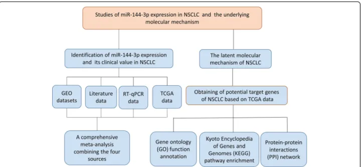
This study investigated the correlations between miR-144-3p expression and clinical characteristics through data collected from Gene Expression Omnibus (GEO) mi-croarrays, the relevant literature, The Cancer Genome Atlas (TCGA), and real-time quantitative real-time PCR (RT-qPCR) analyses to determine the clinical role of
miR-144-3p in NSCLC. The latent mechanism of action in NSCLC was subsequently examined by using 12 predictive programs to forecast the genes targeted by miR-144-3p. In addition, bioinformatics analyses, which included Gene Ontology (GO), Kyoto Encyclopedia of Genes and Ge-nomes (KEGG), and protein–protein interaction (PPI) net-work analyses, were performed.
Materials and methods Data collection
A microarray search of miR-144-3p in NSCLC was conducted in the GEO database (http://www.ncbi.nlm.ni h.gov/geo/) with the following keywords: (MicroRNA OR “Micro RNA” OR “non-coding RNA” OR ncRNA OR “small RNA” OR miRNA) AND (Lung OR pulmonary) AND (cancer OR tumor OR neoplasm OR malignancy OR carcinoma OR adenocarcinoma OR AC OR SCC OR NSCLC). The entry type was restricted to“series,”and the organism was filtered by“Homo sapiens.”The criteria for inclusion were as follows: (1) patients diagnosed with NSCLC and its subtypes were investigated; (2) cancerous and noncancerous samples were involved; (3) the healthy and malignant groups included at least three samples in the form of tissue, blood, or plasma; (4) and the expression profiling data for miR-144-3p were available. Related stud-ies were retrieved from the PubMed, Google Scholar, China National Knowledge Infrastructure (CNKI), Chongqing VIP electronic (VIP) and Chinese Wanfang databases. Figure1shows the workflow for the study.
MicroRNA-144-3p expression data from the Cancer genome atlas
TCGA (https://cancergenome.nih.gov/) was used for obtaining detailed information about the expression value of miR-144-3p in NSCLC and noncancerous sam-ples. The differences in the miR-144-3p expression in the NSCLC samples and the normal controls were cal-culated with IBM SPSS Statistics V22.0 software.
Quantitative real-time PCR
Statistical analysis and comprehensive meta-analysis After miR-144-3p was log2-transformed, the expression
profiling information was used to calculate the number, mean (M) and standard deviation (SD) for each control and experimental group with IBM SPSS Statistics V22.0 software. In addition, Stata 12.0 software was used for performing a comprehensive meta-analysis of data ag-gregated from multiple sources (microarray, literature, miRNA sequencing, and RT-qPCR). The analysis of the miR-144-3p expression in the NSCLC and tumor-free specimens was displayed on forest plots that illustrate the standardized mean difference (SMD) and the 95% confidential interval (CI). The chi-squared test of Q and the I2 statistic were calculated to assess heterogeneity across the studies and to determine the appropriateness of applying either a random effects model or fixed ef-fects model to the pooling process. To measure publica-tion bias, Egger’s and Begg’s tests and a funnel plot, for which significance wasp< 0.05, were performed.
Latent targets of microRNA-144-3p in non-small cell lung cancer
MiRWALK2.0, an online archive of data on miRNA-target interactions [20], was mined to forecast the genes targeted by miR-144-3p. In total, 12 servers with miRWalk, miRMap, MicroT4, miRNAMap, TargetScan PICTAR2, miRBridge, PITA, miRanda, RNAhybrid, miRDB, RNA22 were used. Only those genes projected by more than six of the servers were recognized as target genes. The high-expressed genes in LUAD and LUSC were
acquired through Gene Expression Profiling Interactive Analysis (GEPIA). The overlapping genes among the up-regulated genes in LUAD and LUSC and the predicted target genes, were viewed as promising targets of miR-144-3p in NSCLC. A review of the literature on the specific target genes of miR-144-3p in NSCLC was conducted. The target genes determined by previous studies and the pre-dicted target genes, namely promising target genes, were used in the functional analysis.
Functional analysis for promising target genes
The GO vocabularies, which include biological processes (BPs), cellular components (CCs), and molecular functions (MFs), were enriched by Metascape (http://metascape.org/ gp/). The functional annotation of the underlying target genes was then elucidated by KEGG pathway analysis with Metascape tool. In addition, a PPI network was con-structed to reveal the hub genes of the potential target genes on STRING, a web portal for undermining the inte-grated function of multiple genes [21].
Expression of hub genes from the Cancer genome atlas and the genotype-tissue expression database
To further confirm the function of hub genes in NSCLC and their relationship to miR-144-3p, a search of TCGA and the Genotype-Tissue Expression (GTEx) database was performed to determine the expression pattern of the hub genes in NSCLC. Box plots of the hub genes in the NSCLC and non-cancer samples were developed through GEPIA.
Results
Confirmation of the expression and clinical value of microRNA-144-3p in non-small cell lung cancer, based on gene expression omnibus datasets
MicroRNA-144-3p expression in non-small cell lung cancer obtained through gene expression omnibus microarrays
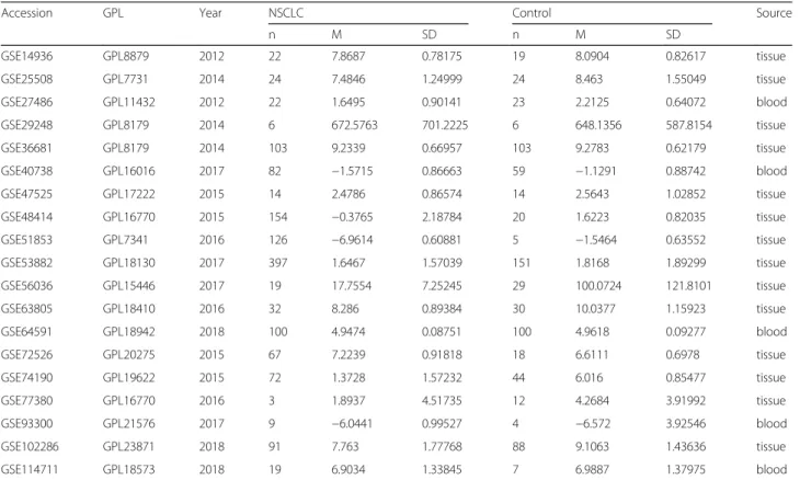
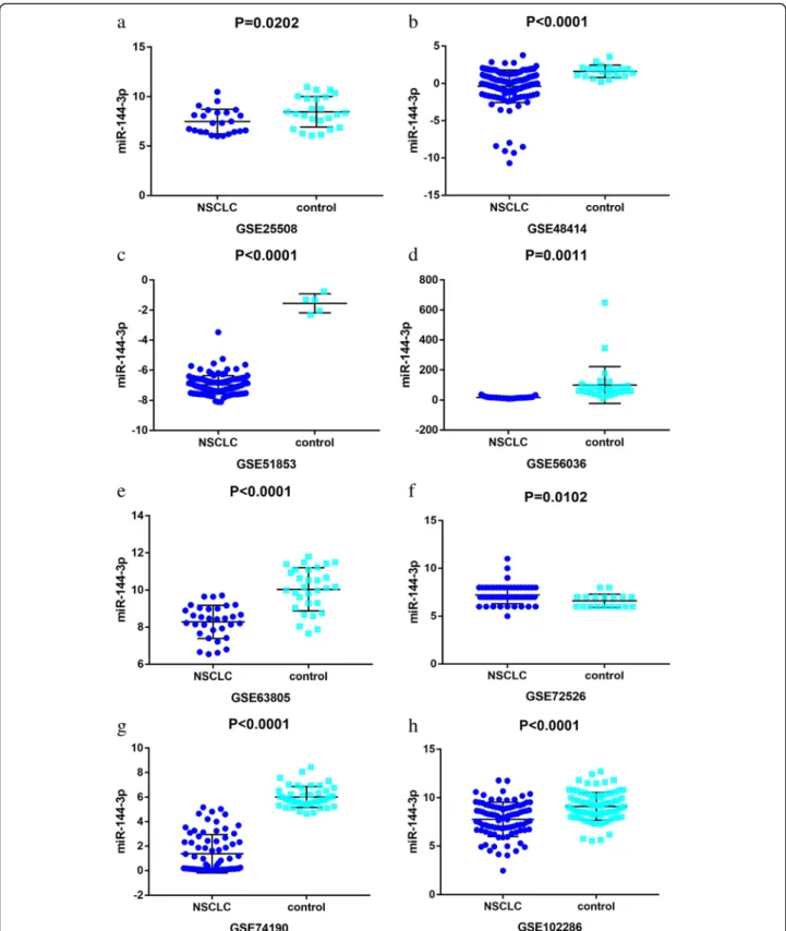
A total of 19 microarrays from the GEO database met the entry criteria. The features of the included GEO datasets are depicted in Table 1. Of the microarrays, 14 were obtained from tissue, and 5 were derived from blood (GSE27486, GSE40738, GSE64951, GSE93300, and GSE114711). In addition, the expression data from the NSCLC and control groups were collected on the basis of the GEO database. With respect to the data from the tissue samples, the NSCLC groups had a significantly lower level of miR-144-3p expression than the control groups in GSE25508, GSE48414, GSE51853, GSE56036, GSE63805, GSE72526, GSE74190, and GSE102286 (p= 0.0202,p< 0.0001,p< 0.0001,p= 0.0011, p< 0.0001, p= 0.0102, p< 0.0001, and p< 0.0001, respectively (Fig.2)). In contrast, no notable distinction in miR-144-3p expression was detected between the NSCLC and the control groups in the other microarrays (GSE14936, GSE29248, GSE36681, GSE47525, GSE53882, and GSE77380). Regarding the data from the blood samples, miR-144-3p expression in NSCLC was found
to decrease significantly in GSE27486 and GSE40738 (p= 0.0196,p= 0.0036, respectively (Fig.3)).
Results of meta-analysis of gene expression omnibus datasets
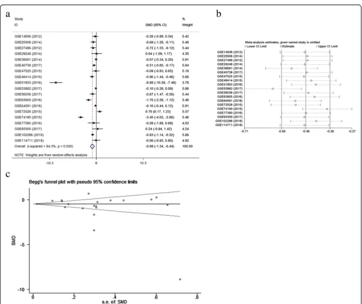
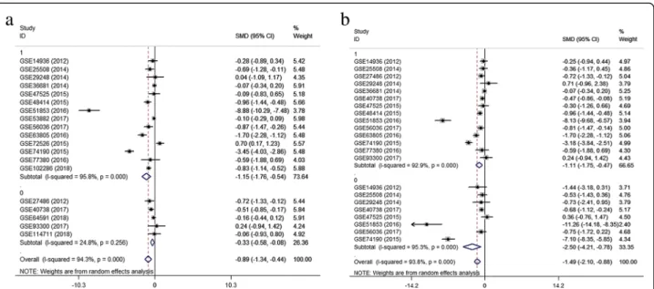
A meta-analysis was conducted on the basis of the 19 in-cluded microarrays from the GEO database. The results are demonstrated in Fig. 4a. Given the apparent hetero-geneity (p< 0.05, I2= 94.3%), a random effects model was applied, and remarkable down-regulation (SMD =−0.89; 95% CI−1.34,−0.44;p= 0.000) of miR-144-3p was found in the NSCLC groups. A sensitivity analysis was later con-ducted to explore whether a particular microarray played a vital role in significant heterogeneity (Fig.4b). After an individual study was removed each time of meta-analysis, the combined effect was compared to the previous one. No study was found to have played a crucial role in any of the enrolled studies.
A funnel plot was generated to estimate publication bias (Fig.4c). To further clarify the heterogeneity source, a subgroup analysis was performed. It was based on multiple characteristics: sample source (tissue vs. blood), and cancer type (adenocarcinoma vs. squamous cell carcinoma). As is illustrated in Fig.5, significant hetero-geneity was observed in the tissue subgroup (I2= 95.8%, p= 0.000). Significant heterogeneity was also found in adenocarcinoma (I2= 92.9%, p= 0.000) and squamous
Table 1Features of the enrolled Gene Expression Omnibus datasets
Accession GPL Year NSCLC Control Source
n M SD n M SD
GSE14936 GPL8879 2012 22 7.8687 0.78175 19 8.0904 0.82617 tissue
GSE25508 GPL7731 2014 24 7.4846 1.24999 24 8.463 1.55049 tissue
GSE27486 GPL11432 2012 22 1.6495 0.90141 23 2.2125 0.64072 blood
GSE29248 GPL8179 2014 6 672.5763 701.2225 6 648.1356 587.8154 tissue
GSE36681 GPL8179 2014 103 9.2339 0.66957 103 9.2783 0.62179 tissue
GSE40738 GPL16016 2017 82 −1.5715 0.86663 59 −1.1291 0.88742 blood
GSE47525 GPL17222 2015 14 2.4786 0.86574 14 2.5643 1.02852 tissue
GSE48414 GPL16770 2015 154 −0.3765 2.18784 20 1.6223 0.82035 tissue
GSE51853 GPL7341 2016 126 −6.9614 0.60881 5 −1.5464 0.63552 tissue
GSE53882 GPL18130 2017 397 1.6467 1.57039 151 1.8168 1.89299 tissue
GSE56036 GPL15446 2017 19 17.7554 7.25245 29 100.0724 121.8101 tissue
GSE63805 GPL18410 2016 32 8.286 0.89384 30 10.0377 1.15923 tissue
GSE64591 GPL18942 2018 100 4.9474 0.08751 100 4.9618 0.09277 blood
GSE72526 GPL20275 2015 67 7.2239 0.91818 18 6.6111 0.6978 tissue
GSE74190 GPL19622 2015 72 1.3728 1.57232 44 6.016 0.85477 tissue
GSE77380 GPL16770 2016 3 1.8937 4.51735 12 4.2684 3.91992 tissue
GSE93300 GPL21576 2017 9 −6.0441 0.99527 4 −6.572 3.92546 blood
GSE102286 GPL23871 2018 91 7.763 1.77768 88 9.1063 1.43636 tissue
GSE114711 GPL18573 2018 19 6.9034 1.33845 7 6.9887 1.37975 blood
Fig. 2Down-regulation of microRNA-144-3p in in the other microarrays tissues, based on Gene Expression Omnibus datasets. Notes:aGSE25508.
Fig. 3Low expression of miR-144-3p in non-small cell lung cancer chips derived from blood sample. Notes:aGSE27486.bGSE40738
cell carcinoma (I2= 95.3%, p= 0.000). These results sug-gest that sample source and cancer type might be sources of heterogeneity.
Literature
A review of the literature relevant to miR-144-3p was conducted through searches in the PubMed, Google Scholar, CNKI, VIP, and Wanfang databases. Neither the M nor the SD of miR-144-3p in the NSCLC and normal groups was provided in the literature; therefore, no available data could be obtained from the existing studies.
Confirmation of the expression and clinical effects of microRNA-144-3p in non-small cell lung cancer, based on the Cancer genome atlas data
MicroRNA-144-3p expression and prognostic value in non-small cell lung cancer tissues
TCGA contained 376 samples for LUSC patients and 488 samples for LUAD patients. Regarding LUSC, miR-144-3p expression was remarkably downregulated in comparison with the normal controls (2.8193 ± 1.40600 vs. 5.5678 ± 1.27693, p< 0.001 (Fig. 6a and Table 2)). In terms of LUAD, the expression level of miR-144-3p was obviously less than that in the healthy tissue (2.8959 ± 1.35967 vs. 5.2775 ± 1.64708,p< 0.001 (Fig. 6b and Table 3)). The data from LUSC and LUAD were combined for further examination of the miR-144-3p expression in NSCLC. As is illustrated in Fig. 6c and Table 4, miR-144-3p was significantly reduced in the
NSCLC tissue compared to the non-cancerous lung tissue (2.8632 ± 1.37928 vs. 5.4243 ± 1.4702, p< 0.0001). A Kaplan–Meier curve was later used to identify the effects of the expression of miR-144-3p on survival time. As is shown in Fig. 7, the pvalues for the three Kaplan–Meier curves were all greater than 0.05, thus indicating no signifi-cant difference in survival time between the group with low levels of miR-144-3p and the one with high levels.
Relationships between microRNA-144-3p and clinical pathology of non-small cell lung cancer, based on the Cancer genome atlas data
As can be seen in Tables2 and3, the clinical character-istics of 332 LUSC patients and 445 LUAD patients were downloaded from TCGA. Regarding LUSC, a significant difference in miR-144-3p was found for stage (p= 0.040) and primary tumor (T) (p= 0.035). LUSC patients in Stages III–IV (2.4469 ± 1.41079) had a lower expression of miR-144-3p than those in Stages I–II (2.8878 ± 1.39556). The miR-144-3p expression of LUSC in T3– T4 (2.5088 ± 1.36178) was more significantly decreased than T1–T2 (2.9023 ± 1.40854). In terms of LUAD, a sig-nificant difference of miR-144-3p expression was ob-served for stage (p= 0.031). Patients in Stages III–IV (3.1868 ± 1.45395) had higher expression values of miR-144-3p. The data on LUAD and LUSC, based on TCGA, were pooled for further validation. As is illus-trated in Table4, the significance in the statistics for the T stage was based on the lower miR-144-3p expression in patients in T3–T4 (p< 0.05).
Quantitative real-time PCR analysis
The microRNA-144-3p expression and its significance in non-small cell lung cancer prognoses
Using RT-qPCR, the clinical expression value of miR-144-3p in 125 matched tissues was evaluated. As is il-lustrated in Fig.8a and Table5, the NSCLC samples exhib-ited a significantly lower expression level of miR-144-3p than the non-cancerous samples (2.808 ± 1.303 vs. 4.813 ± 2.618, p< 0.001). Next, miR-144-3p expression was then analyzed for LUAD and for LUSC. As is shown in Fig.8b and c, unlike what was found in the adjacent non-cancer-ous tissue, apparently lowly expressed miR-144-3p was ob-served in both LUSC and LUAD (p= 0.0004,p< 0.0001). A Kaplan–Meier curve was generated to assess whether miR-144-3p is appropriate for the prognosis prediction of LUAD. As is depicted in Fig.9, the LUAD patients who ex-hibited lower expression values of miR-144-3p might have worse outcomes (p= 0.397).
Correlations between microRNA-144-3p expression and clinical characteristic for non-small cell lung cancer patients
The miR-144-3p expression in NSCLC cases was signifi-cantly different in lymph node metastasis and vascular inva-sion (Table5). The lower value of miR-144-3p was found in patients with lymph node metastasis but not in those
without it (Table5). Patients with vascular invasion main-tained a miR-144-3p value of 2.1400 ± 1.2263, and the ex-pression level of those without vascular invasion was 3.0682 ± 1.24395 (Table 5). To further validate the correl-ation of miR-144-3p and clinicopathological characteristics, the NSCLC cases were divided into LUSC and LUAD groups. For the LUSC group (Table6), no statistical differ-ences were seen for smoking, vascular invasion, or lymph node metastasis. However, for LUAD, the statistical analyses indicated significant differences for smoking, vascular inva-sion, and lymph node metastasis. In comparison to patients without vascular invasion, those with vascular invasion had lower miR-144-3p expression values. The miR-144-3p expression in patients with a smoking habit was signifi-cantly down-regulated over that of patients without the habit (p= 0.027, Table7). Besides, the miR-144-3p expres-sion in NSCLC patients who were considered to have lymph node metastasis was markedly reduced.
Meta-analysis of combination of gene expression omnibus, the Cancer genome atlas, and quantitative real-time PCR data
samples were extracted for integrated meta-analyses with a random effects model because of high hetero-geneity (I2= 95.4%, p= 0.000). The differences in indi-viduals, NSCLC subtype, and sample source were considered the sources of heterogeneity. As is shown in Fig. 10a, the combined SMD of miR-144-3p was−0.95 with 95% CI of (−1.37, −0.52), indicating that less miR-144-3p was expressed in the NSCLC tissue than in the normal tissue. The sensitivity analysis (Fig.10b) in-dicated significant differences among the studies; how-ever, no specific study had a significant effect on high heterogeneity. The evaluation of publication bias was performed with Begg’s and Egger’s tests and a funnel plot (Fig.10c). In general, the funnel plot was symmet-rical, and the p values obtained from the Begg’s and Egger’s tests were 0.833 and 0.335, respectively. In sum, the results indicate that publication bias for the studies was controlled passably.
Bioinformatic analyses Promising target genes collection
From miRWALK2.0, 1635 genes targeted by miR-144-3p in NSCLC predicted by more than six algo-rithms were obtained. A total of 1109 overexpressed genes in LUAD were collected on the basis of GEPIA. In addition, 1922 overexpressed genes in LUSC were acquired from GEPIA. After intersection, 34 predicted target genes were selected. Four specific targets of miR-144-3p were verified in previous studies related to
LC (Table8). The TIGAR is also known as the C12orf5 gene. Accordingly, a total of 37 potential target genes were collected.
Gene ontology and Kyoto encyclopedia of genes and genomes analyses
For further interpretation of the function of the prom-ising genes targeted by miR-144-3p in NSCLC, KEGG, and GO annotations were performed in Metascape. For the GO analysis, three categories were used: BP, CC, and MF. For the BP, renal system development (GO: 0072001) and the nucleobase-containing small mol-ecule metabolic process (GO: 0055086) were the top two pathways (Fig. 11a). For the CC, the potential tar-get differentially expressed genes (DEGs) were predom-inantly enriched in the centriole (GO: 0005814), Golgi membrane (GO: 0000139), and mitochondrial envelope (GO: 0005740) (Fig. 11c). For MF, the three signifi-cantly involved items were RNA polymerase II proximal promoter sequence-specific DNA binding (GO: 0000978); cofactor binding (GO: 0048037); and trans-ferase activity, transferring glycosyl groups (GO: 0016757) (Fig. 11b). Regarding KEGG, the top two enriched pathways were the protein digestion and ab-sorption (hsa04974) and the thyroid hormone signaling pathways (hsa04919) (Fig.12). PPI revealed five genes— C12orf5, CEP55, E2F8, STIL, and TOP2A—as hub genes with the threshold value of 6 (Fig.13).
Table 2Association between microRNA-144-3p expression and clinicopathological parameters in lung squamous cell carcinoma, based on The Cancer Genome Atlas data
Clinicopathological feature n mean ± SD p-value
Tissue Adjacent non-cancerous tissue 44 5.5678 ± 1.27693 < 0.001*
LUSC 332 2.8193 ± 1.40600
Age (years) < 60 59 2.7458 ± 1.32704 0.739
≥60 267 2.8106 ± 1.42109
Gender Female 84 2.8195 ± 1.49697 0.999
Male 248 2.8192 ± 1.37698
Tumor location Central lung 106 2.7551 ± 1.27303 0.848
Peripheral lung 73 2.7968 ± 1.51724
Stage Stage I-II 276 2.8878 ± 1.39556 0.04*
Stage III-IV 53 2.4469 ± 1.41079
T T1-T2 262 2.9023 ± 1.40854 0.035*
T3-T4 70 2.5088 ± 1.36178
N No 213 2.8637 ± 1.43682 0.302
Yes 113 2.6987 ± 1.33482
M No 252 2.7345 ± 1.38222 0.895
Yes 3 2.9144 ± 2.09106
SDStandard deviation,LUSCLung squamous cell carcinoma *
Expression of hub genes from the Cancer genome atlas and the genotype-tissue expression datasets
Of the five hub genes, not including C12orf5, that occupied the central region of the PPI network, four genes (CEP55, E2F8, STIL, and TOP2A) were significantly up-regulated in the NSCLC group compared to the control group (Fig.14).
Discussion
Although previous studies have documented the expres-sion of miR-144-3p in NSCLC, correlations of the clinical features with NSCLC and miR-144-3p have been seldom reported. This study used a larger number of sam-ples for a systematic investigation of the relationships. A
Table 4Association between microRNA-144-3p expression and clinicopathological parameters in non-small cell lung cancer, based on The Cancer Genome Atlas data
Clinicopathological feature n mean ± SD p-value
Tissue Adjacent non-cancerous tissue 87 5.4243 ± 1.4702 < 0.001*
LUSC+LUAD 777 2.8632 ± 1.37928
Age (years) < 60 179 2.919 ± 1.37944 0.330
≥60 573 2.8038 ± 1.37438
Gender Female 323 2.8799 ± 1.37826 0.776
Male 454 2.8513 ± 1.38139
Tumor location Central lung 160 2.6915 ± 1.24372 0.604
Peripheral lung 186 2.7666 ± 1.41787
Stage Stage I-II 625 2.8494 ± 1.3589 0.630
Stage III-IV 144 2.9145 ± 1.47731
T T1-T2 647 2.9128 ± 1.37596 0.008*
T3-T4 127 2.5658 ± 1.33528
N No 504 2.8585 ± 1.39145 0.803
Yes 258 2.8324 ± 1.35039
M No 535 2.7722 ± 1.34822 0.124
Yes 22 3.277 ± 1.45564
SDStandard deviation,LUADLung adenocarcinoma,LUSCLung squamous cell carcinoma *
p< 0.05 was considered statistically significant
Table 3Association between microRNA-144-3p expression and clinicopathological parameters in lung adenocarcinoma, based on The Cancer Genome Atlas data
Clinicopathological n mean ± SD pvalue
Tissue Adjacent non-cancerous tissue 43 5.2775 ± 1.64708 < 0.001*
LUAD 445 2.8959 ± 1.35967
Age (years) < 60 120 3.0042 ± 1.40207 0.168
≥60 306 2.7979 ± 1.33460
Gender Female 239 2.9011 ± 1.33672 0.931
Male 206 2.8898 ± 1.38905
Tumor location Central lung 54 2.5665 ± 1.1857 0.382
Peripheral lung 113 2.747 ± 1.3564
Stage Stage I-II 349 2.8191 ± 1.33042 0.031*
Stage III-IV 91 3.1868 ± 1.45395
T T1-T2 385 2.9199 ± 1.35515 0.133
T3-T4 57 2.6359 ± 1.3106
N No 291 2.8546 ± 1.35977 0.553
Yes 145 2.9367 ± 1.35784
M No 283 2.8058 ± 1.31875 0.126
Yes 19 3.3343 ± 1.4003
SDStandard deviation,LUADLung adenocarcinoma *
highlight of this study was the use of the computational biology method to explore the latent mechanism of miR-144-3p in NSCLC.
A decreased expression of miR-144-3p was found in the NSCLC cases, as presented by the GEO, TCGA, and RT-qPCR. A comprehensive meta-analysis was the focus of the current study. Data were obtained from multiple
sources: namely, microarrays, previous studies, RT-qPCR, and TCGA. The meta-analysis results revealed that the decline of miR-144-3p in NSCLC was consistent across studies [19,22]. It was therefore concluded that there was a considerable decrease of miR-144-3p expression in NSCLC. Based on the data from TCGA, the miR-144-3p expression level in LUSC and LUAD was related to stage,
Fig. 8Patterns of 144-3p expression in clinical samples, based on quantitative real-time PCR data. Note: Lowly expressed microRNA-144-3p was observed ina23 lung squamous cell carcinoma tissues,b101 lung adenocarcinoma tissues, andc125 non-small cell lung cancer tissues
and the predominant difference for T was found in LUAD only.
The results revealed that miR-144-3p could be in-volved in the occurrence and development of NSCLC, and low miR-144-3p could indicate the promotion of
NSCLC. Regarding RT-qPCR, miR-144-3p expression was related to lymph node metastasis and vascular inva-sion. Moreover, the patients with lymph node metastasis and vascular invasion had a tendency to down-regulate miR-144-3p expression. Regarding LUAD, the amount of miR-144-3p differed greatly depending on the smoking status of the patient, the existence of lymph node metas-tasis, and the presence of vascular invasion. However, no statistical differences were found for LUSC. In sum, miR-144-3p might serve as a marker to monitor the pro-gression of NSCLC.
No differences were observed in the survival times for the low-miR-144-3p group and the high-miR-144-3p group, according to TCGA data. In contrast, the PCR results revealed a trend that suggests that LUAD patients with lower miR-144-3p levels might have worse outcomes although not to a significant extent. A study by Wu et al. [23] revealed that miR-144-3p was one of the independent prognostic risk factors for LUAD patients, with those with low miR-144-3p having poor prognoses. Further investiga-tions are required for corroboration.
At the present time, the specific molecular mechanism of NSCLC is not widely understood. Therefore, bioinfor-matics analyses were performed to discover the inherent mode of NSCLC activity at the molecular level. Based Fig. 9A Kaplan–Meier curve for microRNA-144-3p in clinical lung
adenocarcinoma samples. Note: Thepvalue of the Kaplan–Meier curve for the clinical lung adenocarcinoma patients was 0.397, highlighting a trend of possibly longer overall survival rates in the high microRNA-144-3p group. (green curve: low expression, blue curve: high expression)
Table 5Associations between microRNA-144-3p expression and clinicopathological features in non-small cell lung cancer based on quantitative real-time PCR data
Clinicopathological feature n mean ± SD pvalue
Tissue Adjacent non-cancerous tissue 125 4.813 ± 2.618 < 0.001*
NSCLC 125 2.808 ± 1.303
Age (years) < 60 57 2.7879 ± 1.33904 0.874
≥60 68 2.8254 ± 1.28196
Gender Female 50 2.9786 ± 1.40038 0.247
Male 75 2.6948 ± 1.23055
Tumor size (cm) ≤3 60 2.9242 ± 1.44526 0.346
> 3 65 2.7014 ± 1.15768
Smoke No 38 3.2489 ± 1.48252 0.166
Yes 30 2.7767 ± 1.29623
Lymph node metastasis No 56 3.0875 ± 1.38065 0.033*
Yes 69 2.5817 ± 1.19935
Vascular invasion No 90 3.0682 ± 1.24395 < 0.001*
Yes 35 2.1400 ± 1.22630
TNM I-II 54 2.9615 ± 1.27286 0.251
III-IV 71 2.6918 ± 1.32268
Histological type ADC 101 2.7917 ± 1.30837 0.561
SCC 23 2.9643 ± 1.26398
LCLC 1 0.9000
SDStandard deviation,NSCLCNon-small cell lung cancer,ADCAdenocarcinoma,SCCSquamous cell carcinoma,LCLCLarge-cell lung carcinoma *
on miRWALK2.0 and TCGA data, the candidate targets of miR-144-3p were projected. In an attempt to explore the roles of these genes further, KEGG and GO annota-tion analyses were performed. According to GO enrich-ment, the candidate targets of miR-144-3p might have
an important effect on the progression of NSCLC by modulating several cellular biology processes, such as renal system development. Moreover, these genes could also have an important effect, such as cofactor binding, on MF. The results of the KEGG analysis also showed
Table 7Associations between microRNA-144-3p expression and clinicopathological features in lung adenocarcinoma, based on quantitative real-time PCR data
Clinicopathological feature n mean ± SD p-value
Tissue Adjacent non-cancerous tissue 101 4.693 ± 2.607 < 0.001*
LUAD 101 2.792 ± 1.308
Age (years) < 60 41 2.8032 ± 1.31708 0.942
≥60 60 2.7838 ± 1.31347
Gender Female 45 2.9789 ± 1.46340 0.211
Male 56 2.6413 ± 1.16082
Tumor size (cm) ≤3 53 2.9115 ± 1.41270 0.332
> 3 48 2.6594 ± 1.18327
Smoke No 26 3.5146 ± 1.56261 0.027*
Yes 18 2.5722 ± 1.16539
Lymph node metastasis No 45 3.0982 ± 1.3707 0.037*
Yes 56 2.5454 ± 1.21274
Vascular invasion No 70 3.1109 ± 1.30906 < 0.001*
Yes 31 2.0710 ± 0.99515
TNM I-II 44 2.9577 ± 1.35229 0.269
III-IV 57 2.6635 ± 1.27056
SDStandard deviation,LUADLung adenocarcinoma *
p< 0.05 was considered statistically significant
Table 6Associations between microRNA-144-3p expression and clinicopathological features in lung squamous cell carcinoma, based on quantitative real-time PCR data
Clinicopathological feature n mean ± SD pvalue
Tissue Adjacent non-cancerous tissue 23 5.405 ± 2.684 0.0004*
LUSC 23 2.964 ± 1.264
Age (years) < 60 15 2.872 ± 1.39748 0.611
≥60 8 3.1375 ± 1.03086
Gender Female 5 2.976 ± 0.6827 0.974
Male 18 2.9611 ± 1.39922
Tumor size (cm) ≤3 7 3.02 ± 1.79763 0.915
> 3 16 2.94 ± 1.02398
Smoke No 12 2.6733 ± 1.1468 0.263
Yes 11 3.2818 ± 1.36222
Lymph node metastasis No 11 3.0436 ± 1.48816 0.784
Yes 12 2.8917 ± 1.08163
Vascular invasion No 20 2.919 ± 0.99752 0.667
Yes 3 3.2667 ± 2.82194
TNM I–II 10 2.978 ± 0.89643 0.963
III–IV 13 2.9538 ± 1.52513
SDStandard deviation,LUSCLung squamous cell carcinoma *
the roles of the candidate targets of miR-144-3p in NSCLC. The top two enriched pathways were the pro-tein digestion and absorption and the thyroid hormone signaling pathways. This suggests that the promising targets of miR-144-3p could be involved in the
aforementioned pathways to influence the occurrence and progression of NSCLC.
The protein digestion and absorption pathway is a key pathway in several human cancers. A study by Shi et al. showed that this pathway might contribute to the pul-monary metastasis of osteosarcoma patients [24]. It might be involved in the up-regulation of differentially expressed genes in breast cancer [25], and it has already been associated with the down-regulated differentially expressed genes in pancreatic neuroendocrine tumors [26]. The protein digestion and absorption pathway is connected mainly to differentially expressed genes, and this affects the occurrence and development of enchon-dromas [27]. Additionally, B-cell malignancies are relevant Table 8Identified target genes derived from the literature
validated target gene PMID
BLACAT1 PMID: 28885863
GLUT1 PMID: 27313692
TIGAR PMID: 25660220
ZEB1 PMID: 26191328
to the protein digestion and absorption pathway [28]. However, few studies have been conducted on the protein digestion and absorption pathway in NSCLC. Therefore, more studies are required to further validate the specific molecular mechanism in NSCLC.
According to the PPI network data, five genes (TIGAR, CEP55, E2F8, STIL, and TOP2A) have been recognized as hub genes in NSCLC, thus proving their roles as ideal candidates for miR-144-3p targets. The current study found that four of the 5 hub genes— CEP55, E2F8, STIL, and TOP2A—were more greatly upregulated in the NSCLC cases than in the normal cases. In addition, the decreased miR-144-3p expression
in NSCLC was validated in this study. Therefore, the overexpression of the four hub genes in NSCLC was in-direct proof that these genes might be the targets of miR-144-3p. By targeting the TIGAR, miR-144-3p sup-presses proliferation, mediates programmed cell death, and increases autophagy in LC cells [18]. Studies have found that CEP55 might have an important effect on the proliferation of LC [29, 30]. The role of the E2F8 gene has been studied in several cancers. Sun et al. [31] provided evidence that E2F8 was targeted by miR-144-3p in papillary thyroid cancer; however, a connec-tion between E2F8 and miR-144-3P in NSCLC has not been reported so far.
Fig. 11Distribution of gene ontology terms for the genes targeted by microRNA-144-3p in non-small cell lung cancer.aBiological process.b
The current study has limitations. Because a large-scale clinical sample was not used, more investigations with large clinical samples are required for further confirm-ation of the function of miR-144-3p in NSCLC prognoses. In addition, experiments were not conducted for the detection of the expression of hub genes. The expression of these genes needs to be verified by additional
well-designed studies. Although bioinformatics analyses were performed, the specific molecular mechanisms were not identified.
In conclusion, the current study validated that miR-144-3p was lowly expressed in NSCLC. More importantly, miR-144-3p might function as a latent tumor biomarker in the prognosis prediction for
Fig. 13The protein–protein interaction networks of the promising target genes of microRNA-144-3p in non-small cell lung cancer. Notes: Edges represent protein–protein associations, and colored nodes represent query proteins and the first shell of interactors
NSCLC. The results of bioinformatics analyses may present a new method for investigating the pathogen-esis of NSCLC.
Abbreviations
BP:Biological process; CC: Cellular component; CI: Confidential interval; GEO: Gene Expression Omnibus; GO: Gene Ontology; KEGG: Kyoto Encyclopedia of Genes and Genomes; LC: Lung cancer; LCC: Lung large cell carcinoma; LUAD: Lung adenocarcinoma; LUSC: Lung squamous cell carcinoma; M: Mean; MF: Molecular function; NSCLC: Non-small cell lung cancer; PPI: Protein–protein interaction network; qRT-PCR: Quantitative real-time PCR; SD: Standard deviation; SMD: Standardized mean difference; TCGA: The Cancer Genome Atlas
Acknowledgments
None.
Funding
The current study was supported by the future academic star project of Guangxi Medical University (2017).
Availability of data and materials
One hundred twenty-five (125) paired non-small cell lung cancer specimens were obtained from the First Affiliated Hospital of Guangxi Medical University (Guangxi, China).
Authors’contributions
TQG and GC conceived and designed the study. YJC, WQX, and HMH drafted the manuscript. YJC, HMH, KS and SPH participated in the data collection, analysis and interpretation, and statistical analysis. YJC, YNG and WQX revised the manuscript. TQG, GC, KLW and ZYL were responsible for quality control. All of the authors have read and approved the manuscript.
Ethics approval and consent to participate
This study was approved by the Ethics Committee of the First Affiliated Hospital of Guangxi Medical University.
Consent for publication
Not applicable.
Competing interests
The authors declare that they have no competing interests.
Publisher’s Note
Springer Nature remains neutral with regard to jurisdictional claims in published maps and institutional affiliations.
Author details
1Department of Medical Oncology, Second Affiliated Hospital of Guangxi
Medical University, Daxuedong Road, Nanning, Guangxi Zhuang Autonomous Region 530021, People’s Republic of China.2Department of Pathology, Second Affiliated Hospital of Guangxi Medical University, Daxuedong Road, Nanning, Guangxi Zhuang Autonomous Region 530021, People’s Republic of China.3Department of Pathology, First Affiliated
Hospital of Guangxi Medical University, Shuangrong Road, Nanning, Guangxi Zhuang Autonomous Region 530021, People’s Republic of China.
Received: 17 September 2018 Accepted: 31 January 2019
References
1. Siegel RL, Miller KD, Jemal A. Cancer statistics, 2018. CA Cancer J Clin. 2018; 68:7–30.
2. Inamura K. Lung cancer: understanding its molecular pathology and the 2015 who classification. Front Oncol. 2017;7:193.
3. Hirsch FR, Scagliotti GV, Mulshine JL, Kwon R, Curran WJ Jr, Wu YL, et al. Lung cancer: current therapies and new targeted treatments. Lancet. 2017; 389:299–311.
4. Yun YH, Kim YA, Sim JA, Shin AS, Chang YJ, Lee J, et al. Prognostic value of quality of life score in disease-free survivors of surgically-treated lung cancer. BMC Cancer. 2016;16:505.
5. Tang Y, Qiao G, Xu E, Xuan Y, Liao M, Yin G. Biomarkers for early diagnosis, prognosis, prediction, and recurrence monitoring of non-small cell lung cancer. Onco Targets Ther. 2017;10:4527–34.
6. Rios J, Gosain R, Goulart BH, Huang B, Oechsli MN, McDowell JK, et al. Treatment and outcomes of non-small-cell lung cancer patients with high comorbidity. Cancer Manag Res. 2018;10:167–75.
7. Beermann J, Piccoli MT, Viereck J, Thum T. Non-coding RNAs in development and disease: background, mechanisms, and therapeutic approaches. Physiol Revs. 2016;96:1297.
8. Bracken CP, Scott HS, Goodall GJ. A network-biology perspective of microRNA function and dysfunction in cancer. Nat Rev Genet. 2016;17:719. 9. Zhou Q, Huang SX, Zhang F, Li SJ, Liu C, Xi YY, et al. MicroRNAs: a novel
potential biomarker for diagnosis and therapy in patients with non-small cell lung cancer. Cell Prolif. 2017.https://doi.org/10.1111/cpr.12394. 10. Kang L, Tao X, He BS, Pan YQ, Sun HL, Peng HX, et al. MicroRNA expression
profiles predict progression and clinical outcome in lung adenocarcinoma. Onco Targets Ther. 2016;9:5679–92.
11. Zhang SY, Lu ZM, Lin YF, Chen LS, Luo XN, Song XH, et al. miR-144-3p, a tumor suppressive microRNA targeting ETS-1 in laryngeal squamous cell carcinoma. Oncotarget. 2016;7:11637.
12. Li B, Zhang S, Hao S, Li C. MicroRNA-144-3p suppresses gastric cancer progression by inhibiting epithelial-to-mesenchymal transition through targeting PBX3. Biochem Biophys Res Commun. 2017;484:241–7. 13. Liang H, Ye Z, Yin S, Mo W, Wang H, Zhao J, et al. A comprehensive insight
into the clinicopathologic significance of miR-144-3p in hepatocellular carcinoma. Onco Targets Ther. 2017;10:3405–19.
14. Li J, Sun P, Yue Z, Zhang D, You K, Wang J. miR-144-3p induces cell cycle arrest and apoptosis in pancreatic cancer cells by targeting proline-rich protein 11 expression via the mitogen-activated protein kinase signaling pathway. DNA Cell Biol. 2017;36:619.
15. Xiao W, Lou N, Ruan H, Bao L, Xiong Z, Yuan C, et al. Mir-144-3p promotes cell proliferation, metastasis, sunitinib resistance in clear cell renal cell carcinoma by downregulating ARID1A. Cell Physiol Biochem. 2017;43:2420–33.
16. Zhang LY, Hofun LV, Wong AM, Kwong DL, Zhu YH, Dong SS, et al. MicroRNA-144 promotes cell proliferation, migration and invasion in nasopharyngeal carcinoma through repression of PTEN. Carcinogenesis. 2013;34:454.
17. Ye JJ, Cao J. MicroRNAs in colorectal cancer as markers and targets: recent advances. World J Gastroenterol. 2014;20:4288.
18. Chen S, Li P, Li J, Wang Y, Du Y, Chen X, et al. MiR-144 inhibits proliferation and induces apoptosis and autophagy in lung cancer cells by targeting TIGAR. Cell Physiol Biochem. 2015;35:997–1007.
19. Liu M, Gao J, Huang Q, Jin Y, Wei Z. Downregulating microRNA-144 mediates a metabolic shift in lung cancer cells by regulating GLUT1 expression. Oncol Lett. 2016;11:3772.
20. Dweep H, Gretz N. miRWalk2.0: a comprehensive atlas of microRNA-target interactions. Nat Methods. 2015;12:697.
21. Szklarczyk D, Franceschini A, Wyder S, Forslund K, Heller D, Huerta-Cepas J, et al. STRING v10: protein–protein interaction networks, integrated over the tree of life. Nucleic Acids Res. 2015;43:D447–52.
22. Zhang G, An H, Fang X. MicroRNA-144 regulates proliferation, invasion, and apoptosis of cells in malignant solitary pulmonary nodule via zinc finger E-box-binding homeobox 1. Int J Clin Exp Pathol. 2015;8:5960–7.
23. Wu C, Xu B, Zhou Y, Ji M, Zhang D, Jiang J, Wu C. Correlation between serum IL-1βand miR-144-3p as well as their prognostic values in LUAD and LUSC patients. Oncotarget. 2016;7:85876–87.
24. Shi Z, Zhou H, Pan B, Lu L, Wei Z, Shi L, et al. Exploring the key genes and pathways of osteosarcoma with pulmonary metastasis using a gene expression microarray. Mol Med Rep. 2017;16:7423–31.
25. Dong LF, Xu SY, Long JP, Wan F, Chen YD. RNA-sequence analysis reveals differentially expressed genes (DEGs) in patients exhibiting different risks of tumor metastasis. Med Sci Monit. 2017;23:2842–9.
26. Wang DD, Liu ZW, Han MM, Zhu ZM, Tu YL, Dou CQ, et al. Microarray based analysis of gene expression patterns in pancreatic neuroendocrine tumors. Eur Rev Med Pharmacol Sci. 2015;19:3367–74.
27. Shi Z, Zhou H, Pan B, Lu L, Kang Y, Liu L, et al. Exploring the key genes and pathways in enchondromas using a gene expression microarray. Oncotarget. 2017;8:43967–77.
28. Hsu LI, Briggs F, Shao X, Metayer C, Wiemels JL, Chokkalingam AP, et al. Pathway analysis of genome-wide association study in childhood leukemia among Hispanics. Cancer Epidemiol Biomark Prev. 2016;25:815–22. 29. Ma XP, Zhang W, Wu BQ, Qin J. Correlations between mRNA levels of
Centrosomal protein 55 (CEP55) and clinical features of patients with lung cancer. Med Sci Monit. 2018;24:3093–7.
30. Liu L, Mei Q, Zhao J, Dai Y, Fu Q. Suppression of CEP55 reduces cell viability and inducesapoptosis in human lung cancer. Oncol Rep. 2016;36:1939–45. 31. Sun J, Shi R, Zhao S, Li X, Lu S, Bu H, et al. E2F8, a direct target of miR-144,