The University of San Francisco
USF Scholarship: a digital repository @ Gleeson Library |
Geschke Center
Master's Theses Theses, Dissertations, Capstones and Projects
Winter 12-22-2014
Epigenetic Regulation of Nuclear Hormone
Receptor DAX-1
Michael B. Heskett
University of San Francisco, [email protected]
Follow this and additional works at:https://repository.usfca.edu/thes
Part of theBiochemistry, Biophysics, and Structural Biology Commons,Biology Commons,Cell and Developmental Biology Commons, and theGenetics and Genomics Commons
This Thesis is brought to you for free and open access by the Theses, Dissertations, Capstones and Projects at USF Scholarship: a digital repository @ Gleeson Library | Geschke Center. It has been accepted for inclusion in Master's Theses by an authorized administrator of USF Scholarship: a digital repository @ Gleeson Library | Geschke Center. For more information, please [email protected].
Recommended Citation
Heskett, Michael B., "Epigenetic Regulation of Nuclear Hormone Receptor DAX-1" (2014).Master's Theses. 116.
2
TABLE OF CONTENTS
LIST OF ABBREVIATIONS 3
LIST OF FIGURES 6
ABSTRACT
8
CHAPTER 1: INTRODUCTION 9
CHAPTER 2: DETERMINATION OF DAX EXPRESSION 22
CHAPTER 3: DETERMINATION OF DAX-1 METHYLATION STATUS 35
CHAPTER 4: DAX-1 INTERACTION WITH EPIGENETIC FACTORS 60
CHAPTER 5: SUMMARY
77
3
LIST OF ABBREVIATIONS
5-mC - 5-methylCytosine
AHC - Adrenal Hypoplasia Congenita AR - Androgen Receptor
ATCC - American Type Culture Collection bp - base pairs
cDNA - complimentary Deoxyribonucleic Acid CG - Cytosine and Guanine
ChIP - Chromatin Immunoprecipitation CpG - Cytosine phosphate Guanine
CXXC - Cysteine - any amino acid - any amino acid - Cysteine
DAX-1/NR0B1 - Dosage sensative sex reversal, Adrenal hypoplasia congentia, on chromosome X, gene 1/ Nuclear Receptor subfamily 0 group B member 1
DNA - Deoxyribonucleic Acid DNMT - DNA Methyltransferase DSS - Dosage Sensitive Sex Reversal DBD - DNA Binding Domain
ER(α/β) - Estrogen Receptor (alpha/beta)
ER+ - Estrogen Receptor Positive ES - Embryonic Stem
4
FAS - Fatty Acid Synthase
GAPDH - Glyceraldehyde 3-Phosphate Dehydrogenase H3 - Histone H3
HMT - Histone Methyl Transferase HPA - Hypothalamic/Pituitary/Adrenal HRE - Hormone Response Element IgG - Immunoglobulin G
LBD - Ligand Binding Domain LRH-1 - Liver Receptor Homolog -1 LXR - Liver X Receptor
LxxLL - Leucine - any amino acid - any amino acid - Leucine - Leucine MBD1 - Methyl CpG binding Domain protein 1
MeCP2 - Methyl CpG binding Protein 2 N-CoR - Nuclear Receptor Co-Repressor 1
NCBI - National Center for Biotechnology Information NR - Nuclear Receptor or Nuclear Hormone Receptor PCR - Polymerase Chain Reaction
qPCR - quantitative Polymerase Chain Reaction RNA - Ribonucleic Acid
5
SF-1 - Steroidogenic Factor - 1 SHP - Small Heterodimeric Partner
SMRT - Silencing Mediator for Retinoid or Thyroid hormone receptors / Nuclear Receptor Co-Repressor 2.
6
LIST OF FIGURES
Figure 1-1. The accepted model of nuclear receptor action. 10
Figure 1-2. The structure of a typical nuclear receptor. 12
Figure 1-3. The retioid X receptor complex. 14
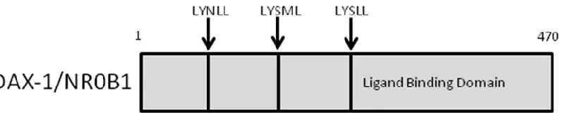
Figure 1-4. The structure of the unusual nuclear hormone receptor DAX-1. 16
Figure 1-5. Alingment of human and mouse DAX-1 amino acid sequences. 17
Figure 1-6. A simplified model of epigenetic regulation by DNA methylation. 20
Figure 2-1. Relative DAX-1 expression in many tissue types. 23
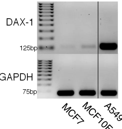
Figure 2-2. DAX-1 PCR expression analysis in MCF7, MCF10F, and A549. 28
Figure 2-3. DAX-1 quantitative PCR expression analysis: MCF7 and MCF10F. 29
Figure 2-4. DAX-1 quantitative PCR expression analysis: MCF7 and A549. 30
Figure 2-5. DAX-1 quantitative PCR expression analysis: MCF10F and A549. 31
Figure 2-6. DAX-1 expression data from the Cancer Cell Line Encyclopedia. 33
Figure 3-1. Methylation of Cytosine by DNA Methyltransferase. 36
Figure 3-2. Bisulfite Sequencing scheme. 38
Figure 3-3. CpG site analysis of the entire DAX-1 gene region. 46
7
Figure 3-5. Comparison of mouse and human DAX-1 CpG islands. 48
Figure 3-6. Methylation sensitive restriction digest of DAX-1 genomic DNA. 49
Figure 3-7. Locations of three bisulfite sequencing fragments. 51
Figure 3-8. Bisulfite sequencing fragment 1. 52
Figure 3-9. Bisulfite sequencing fragment 2. 53
Figure 3-10. Bisulfite sequencing fragment 3. 54
Figure 3-11. DAX-1 expression in mouse stem and somatic cell lines. 57
Figure 3-12. Methylation analysis of DAX-1 in mouse stem and somatic cell lines. 58
Figure 4-1. Epigenetic modifications and the organization of the eukaryotic chromosome. 61
Figure 4-2. Three proposed mechanisms for modulation of gene expression by DNA methylation. 63
Figure 4-3. Cytosine Methylation Cycle. 66
Figure 4-4. Chromatin immunoprecipitation targeting MeCP2 on DAX-1. 71
Figure 4-5. Proposed model of DAX-1 epigenetic regulation mechanism by MeCP2. 72
Figure 4-6. Chromatin immunoprecipitation targeting MeCP2 and MBD1 on DAX-1. 73
8
ABSTRACT
DAX-1 (NR0B1) is an orphan nuclear receptor that plays a key role in the development and maintenance of steroidogenic tissue in mammals. Dax-1 is also expressed in mouse embryonic stem (ES) cells and is required to maintain pluripotency. Duplication of the X-chromosome in the region containing the NR0B1 gene results in sex reversal, and mutations in NR0B1 cause adrenal hypoplasia congenita. DAX-1 has been observed to act as a corepressor of other nuclear receptors including androgen receptor (AR), estrogen receptor (ER), and steroidogenic factor 1 (SF-1). In addition to pluripotent ES cells, DAX-1 is primarily expressed in select tissues of the body such as testes, ovaries, adrenal cortex and breast. In some cases, changes in DAX-1 expression may serve as an indicator of aberrant growth. For example, DAX-1 expression is greatly reduced in ER+ breast cancer patient samples and several ER+ cell lines; however the mechanism leading to this change in DAX-1 expression is unknown. Here, we propose that expression of DAX-1 may be governed by epigenetic mechanisms.
9
CHAPTER 1: INTRODUCTION
Nuclear Receptors
Nuclear Receptors (NRs) are a large group of transcription factors that are characterized by their ability to bind steroid hormones and other lipophilic non-steroid hormones and regulate gene expression by directly binding to the promoter and regulatory regions of specific target genes1 (Fig. 1-1). Nuclear receptors play a major role in differential gene expression and are found in diverse animal species from invertebrates to primates2. Evidence suggests that nuclear receptors may have first appeared around the same time animals appeared in the fossil record,
approximately 635 million years ago2. There exists a loose correlation between the number and complexity of tissue types and the number of NR genes an organism has, ranging from only a few NR genes in sponges to 48 distinct NR genes in humans. In humans, these 48 receptors of varied structure and function comprise the nuclear receptor superfamily. Nuclear receptor ligands include steroid hormones such as estrogen and testosterone, Vitamin A compounds known as retinoids, tyrosine-based hormones known as thyroid hormones, and Vitamin D3 2.
10
Figure 1-1. A typical nuclear receptor binds a hormone ligand, dissociates heat shock proteins, homodimerizes, then translocates to the nucleus where it directly binds to a hormone response elementon the DNA in association with coactivators and RNA polymerase to affect the levels of gene expression. Created by Boghog2. Nuclear_receptor_action. Accessed 9/10/2014 at
11
Nuclear receptors were first discovered to have modular substructures by Wrange et al in 19784.
Since then, the structure of nuclear hormone receptors has been elucidated. A typical nuclear receptor contains an N-terminal domain of varied function, a zinc finger DNA binding domain (DBD) that is important in mediating direct contact to the DNA, a flexible hinge region, a ligand binding domain (LBD) containing a ligand binding pocket, and a C-terminal domain5 (Fig. 1-2). Nuclear receptors are responsible for a wide variety of physiological functions including cell growth and differentiation, fatty acid synthesis, and cancer initiation and progression6,7,8. During mammalian development, estrogen receptor (ER) is expressed in reproductive tissue and upon binding to estrogens stimulates cell proliferation by activating transcription of cyclins6. In stem cells, orphan nuclear receptors such as LRH-1, SF-1, and DAX-1 play an important role for the maintenance of pluripotency7. In lipid metabolism, the orphan receptor liver X receptor (LXR) regulates key genes in cholesterol and fatty acid pathways including fatty acid synthase (FAS), and sterol regulatory element binding protein 1c 8. Nuclear receptors are especially intriguing to biologists because they combine the function of receptors, which are typically membrane-bound, and transcription factors into a modular protein that can affect gene expression without the need for a long cascade of signal transduction.
Nuclear receptor mechanism of action
12
13
presence in the nucleus at all times and form heterodimers with other nuclear receptors, most commonly partnered with retinoid X receptor (RXR)10(Fig. 1-3). Type III receptors are similar to type I receptors in mode of action except they bind to HREs with direct repeats instead of inverted repeats1. Type IV nuclear receptors distinguish themselves by only binding to a single half-site in the target gene HRE1. In most cases, nuclear receptors do not act alone and instead form a complex including coactivator and corepressor proteins11.
Nuclear receptor coregulators
Biochemical and proteomic analyses has shown that nuclear receptors do not act alone. Nuclear receptor coregulators are proteins that form protein-protein interactions to make large complexes with NRs on and off the DNA11. A survey of the literature finds over 450 reported NHR
coregulator species12. Coregulators typically belong to one of two subgroups; coactivators or corepressors. Coactivators are generally defined as molecules that are found bound with ligand-activated NRs and lead to an increase in target gene expression12. Corepressors are typically found bound to unliganded NRs and decrease target gene expression. Collectively, nuclear receptor coregulators, or SRCs (steroid hormone coregulators), share a conserved basic helix-loop-helix that allows interaction with other coregulators and transcription factors and contains a nuclear localization signal13. In the N-terminal region is a leucine rich (LxxLL) domain that facilitates binding to NRs. SRCs play a role in regulating many aspects of gene regulation
including transcription, cofactor recruitment, and post translational modifications13. SRC activity can be regulated by phosphorylation of specific residues of the SRC proteins themselves13. Nuclear receptor coactivators were originally found in complex with nuclear hormone receptors bound to nuclear DNA and are required for optimal activation of a ligand-bound nuclear
14
15
molecules, NR coactivators may be hundreds in number, and contribute to cancer biology and regulation of metabolism12,13. Nuclear receptor corepressors are also important for regulating healthy levels of hormone responsive genes . The two founding members of the corepressor class SMRT and N-CoR were discovered in relation with thyroid hormone resistance14, a rare syndrome where some or all of the body's tissues are resistant to the normal effects of thyroid hormone. Point mutations in SMRT or N-CoR cause ligand-activated thyroid hormone receptor to be unable to release these corepressors, resulting in a continuously inactive receptor and a host of medical complications such as goiter and tachycardia14. Expression levels of NHR
coactivators also appear to be a critical factor in NR function. Notable pathology related to NR coactivator dysfunction includes several types of cancers caused by overexpression of hormone-regulated genes due to coactivator overexpression14.
DAX-1
First identified in association with X-linked adrenal hypoplasia15 (AHC) and dosage sensitive sex reversal (DSS)16, DAX-1 is classified as an orphan nuclear receptor, as no ligand has been
16
17
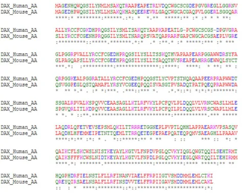
Figure 1-5. The DAX-1 amino acid sequence shares 66% identity between mouse and human proteins.
An * (asterisk) indicates an identical amino acid sequence. A : (colon) indicates conservation between
18
expression pattern with steroidogenic factor-1 (SF-1)5. DAX-1 has a slightly shorter splice variant, DAX-1A, which is spliced replacing exon 2 with exon 2A5. DAX-1A is expressed at a lower level and its significance remains unknown5.
DAX-1 function is well accepted as a negative coregulator of other nuclear receptors including SF-1, a key transcriptional regulator of steroidogenic and reproductive tissues18. Missense mutations in the DAX-1 gene of AHC patients affect DAX-1’s ability to translocate to the nucleus and its ability to bind corepressors to form an active corepressor complex18. In DSS, increased expression of DAX-1 is thought to antagonize SRY (TDF, testis determining factor) leading to development of female reproductive tissues and structures19. In 2000, Zhang et al.
observed DAX-1 binding to ligand-activated estrogen receptor alpha (ERα) and estrogen
receptor beta (ERβ) via the leucine rich motif, repressing ER activity by preventing its ability to bind DNA20. In 2008, Sablin et al. solved the crystal structure of the DAX-1 LBD
homodimerized and bound to LRH-1 via a repressor helix motif, PCFXXLP21. Some evidence shows DAX-1 may act at the gene level as part of a larger repressor complex. In 2010, Lanzino
et al. found that after stimulation with dihydrotestosterone (DHT), an androgen hormone, DAX-1 and HDAC-DAX-1, as part of an AR-mediated repressor complex were able to reduce the levels of cyclin D1 in MCF7 breast cancer cells22. In summary, DAX-1 repressive function in normal individuals is achieved through activity of both the LXXLL motif and LBD, and is necessary for normal development and maintenance of steroidogenic tissues.
19
(Tzagarakis-Foster, unpublished). Overexpression of small heterodimeric partner (SHP/ NR0B2), an orphan nuclear receptor closely related to DAX-1, in hepatocellular carcinoma inhibited tumor growth and increased sensitivity to apoptosis24.
Epigenetic regulation of Nuclear Hormone Receptors
In mammals and other higher eukaryotes, genes can be regulated by epigenetic mechanisms including DNA methylation at cytosine bases and histone post-translational modifications (further discussed in chapter 3,4). Epigenetic changes are heritable from cell to cell and from organism to organism. Epigenetic regulation of nuclear receptors may partially explain their precisely regulated schedule of expression and absence during development34. Aberrant changes in DNA methylation at NR promoters may disrupt the balance of the NR gene-regulation
network, leading to dramatic physiological effects25. Increased methylation of promoter DNA is thought to repress gene expression by several mechanisms including steric blockage of
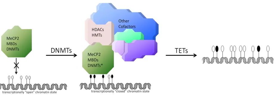
transcriptional activators, and recruitment of methyl-DNA binding proteins that can remodel chromatin to a tightly bound, inactive state26 (Fig. 1-6).
Clinical Relevance of Nuclear Receptors
20
Figure 1-6. Simplified schematic of cytosine methylation cycle in higher eukaryotes. Filled circles represent methylated CpG sites and empty circles represent unmethylated CpG sites.
Left, unmethylated DNA in the 5’ regulatory region of a gene allows an open chromatin state and
active transcription. Middle, CpG sites are methylated by DNA methyltransferases (DNMTs) and recognized by methylated DNA binding proteins such as MeCP2. Right, CpG are oxidized by ten-eleven translocation (TET) family proteins and return to unmodified status.
21
22
CHAPTER 2: DETERMINATION OF DAX-1 EXPRESSION
INTRODUCTION
DAX-1 expression
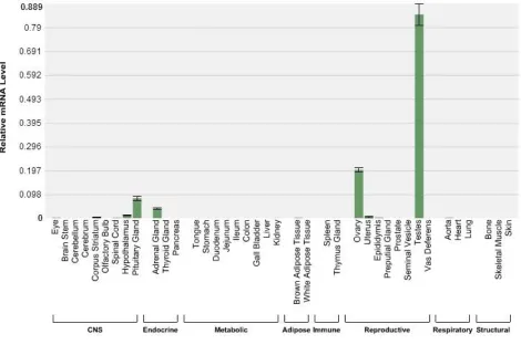
DAX-1 has a restricted expression pattern mainly present in the
Hypothalamic/Pituitary/Gonadal axis (HPA-axis), as well as other tissues involved in steroid hormone production and reproduction such as Leydig and Sertoli cells in the testis and granulosa cells in the ovary18(Fig. 2-1). DAX-1 expression co-localizes with steroidogenic factor-1 (SF-1), a transcriptional activator involved in sex determination and adrenal development. DAX-1 acts as an anti-steroidogenic factor by repressing the actions of SF-1 and liver receptor homolog-1 (LRH-1) and decreasing the levels of steroidogenic pathway genes such as aromatase29. Duplication of the region of the X chromosome containing DAX-1, and thus higher DAX-1 expression, leads to a syndrome in which an XY individual develops as female16. In contrast, loss of DAX-1 protein function due to inactivating mutations causes improper adrenal
development and adrenal hormone insufficiency15.
DAX-1 expression in stem cells
23
24
1 into differentiated cells is not sufficient to induce pluripotency, Dax-1 has been observed bound to promoters of essential pluripotency factors such as Oct3/4, suggesting Dax-1 has a major role in repression of differentiation-regulated genes31.
DAX-1 expression in cancer
DAX-1 expression is also altered in cancer cells. The levels of DAX-1 in adrenocortical neoplasms are inversely related to the steroidogenic activity, suggesting that DAX-1 inhibits steroidogenesis32. Another observation of DAX-1 by immunoreactivity was directly correlated to androgen receptor (AR) status in breast cancer33. This finding suggests that DAX-1 has the ability to bind and sequester AR in the cytoplasm, changing the activity of an important nuclear receptor in breast cancer diagnosis and treatment. Transfection of DAX-1 into cells of the well classified MCF7 breast cancer cell line reduces proliferation in vitro and tumor size in a mouse xenograft model (Tzagarakis-Foster, unpublished). Additionally, patients whose breast tumors express DAX-1 have a more favorable prognosis34. Loss of ER regulation in breast cancer cells by DAX-1 may lead to upregulation of ER target genes such as cyclin D, which controls the G1/S transition of the cell cycle and subsequent replication of the cell22.
DAX-1 may also contribute to lung cancer tumorigenicity by maintaining cancer stem cells35. DAX-1 is highly expressed in the human lung adenocarcinoma cell line A54936, making it a routinely utilized cell line for DAX-1 research.
Investigation into DAX-1 expression
25
has DAX-1 expression been directly measured between MCF7/MCF10F and A549 cell lines in a focused single gene study. DAX-1 expression data will lay the groundwork for further studies into the epigenetic regulation of DAX-1 in these cell lines.
MATERIALS AND METHODS
Cell culture
MCF7, MCF10F, and A549 cells were obtained from the American Type Culture Collection (ATCC). MCF7 cells are known to express estrogen receptor and are responsive to estrogen, so were grown in media lacking phenol red because of its potential endocrine disruptive actions. Base DMEM/F-12 media was supplemented with 10% FBS and other growth factors as
described by ATCC. Cells were grown in T25 and T75 cell culture flasks with vented caps, and passaged 2-4 times per week, as described by ATCC recommended culture and subculture protocols. Cells in T25 flasks were grown with 3 ml media, and cells in T75 flasks grown with 12-15 ml media. Average cell number counted from a near confluent T75 flask was between 3-6 million cells, depending on cell line and growth characteristics. Cell lineages were discarded after 20 passages.
RNA isolation
MCF7, MCF10F, and A549 cells were grown to 50-70% confluency after 24hrs in 6-well plates. Cells were lysed directly on the plate with buffer RLT from the QIAGEN RNeasy RNA isolation kit (Valencia, CA). RNA was isolated with QIAGEN spin column technology following the standard protocol including optional RNAse-free DNA digestion to remove genomic DNA contaminants. RNA was immediately used as template for cDNA synthesis, or stored at -80
°C
26
cDNA synthesis
cDNA synthesis was performed with standard protocol using the Applied Biosystems high capacity cDNA reverse transcription kit (Thermo Fisher Scientific, Waltham, MA). cDNA was synthesized using random primers and a three step incubation: 25 °C for 10 min, 37 °C for 120 min, 85 °C for 5 min. cDNA yield produced from one well of 50-70% confluent cells in a 6-well plate was on average, >1000ng/µl.
Endpoint-PCR and visualization
Endpoint-PCR was performed with Promega GoTaq2x green master mix. Endpoint-PCR was performed with a thermal cycler using the following conditions: 95°C denaturation step for 3 min (1 time), 95°C for 30s -> 58°C for 30s -> 72°C for 10s (32 times), 72°C for 4 min (1 time). Primers used to assay DAX-1 expression were: forward: 5’ –
ATCAGTACCAAGGAGTACGCC – 3’, reverse: 5’ – GTTCCCCACTGGAGTCCCTG – 3’, at an annealing temperature of 58°C. Primers used to assay GAPDH expression were: foward: 5' - TTGCCATCAATGACCCCTTCA - 3, reverse: 5' - CGCCCCACTTGATTTTGGA - 3', at an annealing temperature of 58°C. Endpoint-PCR products were separated by electrophoresis through a 2% agarose gel with ethidium bromide added, and visualized by ultraviolet radiation.
Quantitative reverse transcriptase PCR
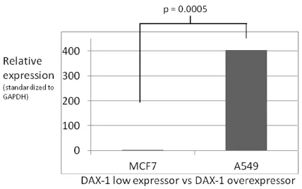
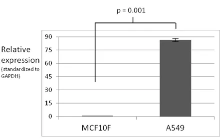
qPCR was performed with the BioRad (Hercules, CA) CFX-96 system using iQ SYBR-Green supermix. High amounts of cDNA per sample (1-2μg) were used to ensure reproducible amplification of DAX-1, a low abundance transcript in MCF10F and MCF7 cell lines. Primers were designed to span the Exon1-Exon2 junction, with a 125 bp amplicon. Fold difference in expression was calculated using the delta Ct method:
27
For relative gene expression comparisons, GAPDH was used as a standard reference gene. GAPDH expression was uniform across all samples, and melting curves were checked to ensure the absence of primer-dimer. PCR conditions and primers used were the same as used in
endpoint-PCR (see above).
P-values were calculated using a paired, one-tailed student's t-test to compare the relative
expression across the three cell lines. Error bars were created using the standard deviation of the relative expression across the three cell lines. Results are representative of three replicates of qPCR gene expression analyses using input cDNA synthesized from mRNA collected from separate cell populations.
RESULTS
28
29
30
31
32
DISCUSSION
These results demonstrate a reliable system to study the epigenetic regulation of DAX-1, the goal of research conducted in later chapters of this report. The MCF7/MCF10F comparison
represents a medically relevant disease model where a low DAX-1 expressor in a cancer environment (MCF7) can be compared to a physiological DAX-1 expressor in a normal
environment (MCF10F). DAX-1 as a nuclear receptor repressor is especially important in breast tissue due to its ability to repress the activity of activated estrogen receptor (ER), a biomarker of the majority of breast cancers. Although beyond the direct scope of this research, the loss of ER repression due to diminished levels of DAX-1 is a valuable hypothesis that may be further studied. These studies demonstrate the A549 cell line expresses DAX-1 at a level much higher than MCF7 or MCF10F. I was also able to validate that this finding agrees with relative
expression levels found in the Cancer Cell Line Encyclopedia (Fig 2-6). A549 represents a valuable research tool where the effects of DAX-1 function, and the repercussions of loss of DAX-1 by genetic manipulations, may be easily studied. Here, the inclusion of the A549 cell line represents a predicted control for methylation studies, in that we expect no epigenetic repression of DAX-1 in this cell line. DAX-1 is expressed at a level much higher than MCF7, and if the DAX-1 gene is regulated by methylation, one would expect the methylation levels of DAX-1 DNA to be very different in these cell lines. Following the cancer methylation
33
34
CONCLUSION
35
CHAPTER 3: DETERMINATION OF DAX-1 METHYLATION STATUS
INTRODUCTION
Epigenetics and DNA methylation
Epigenetics can be defined as a heritable change in cellular phenotype that is caused by
mechanisms independent of a change in the DNA sequence. Although the term epigenetics may encompass many cellular processes, it is usually used to refer to DNA methylation, a well classified epigenetic modification that can have drastic changes on the cellular phenotype. DNA methylation, first observed in humans in 194837, is now considered by many to be the fifth base of the human genome. DNA methylation occurs in humans on the 5’ carbon of the cytosine ring (Fig 3-1) primarily when cytosine is paired with guanosine, as a part of a ‘CpG’ dinucleotide38. Lower eukaryotes and bacteria use different epigenetic schemes including modifications of several bases26. 5-methylcytosine (5mC) is not uniformly spread throughout the genome26. The CpG dinucleotide is present in a much lower frequency than predicted, except for in CG rich regions denoted ‘CpG islands’, often present in the 5’ regulatory regions of genes38
. 5-mC presence in CpG islands is often associated with transcriptional repression39,40, and high levels of CpG methylation is responsible for transcriptional silencing of tumor suppressor genes in many types of cancer41. Non CpG methylation, discovered more recently, is found in some human tissues42. Well documented in the nervous system, non-CpG methylation may suggest a broader role of gene regulation by DNA methylation occuring outside of CpG islands42. In the world of epigenetics, there exist ‘readers, ‘writers’, and ‘erasers’ 43. Writers such as the DNA
36
37
Readers such as MeCP2 are able to selectively bind methylated DNA and recruit other factors that affect cell phenotype through chromatin remodeling44. Erasers such as the Ten-eleven translocation (TET) family of enzymes oxidize 5mC,which is the first step in removal of the methyl group45.
Scope of Epigenetic Modifications
It has recently become apparent that the “epigenome” or the set of all epigenetic modifications to the genome plays a large role in cellular and organismal phenotype. Studies have shown
epigenetic links to common diseases such as obesity46 and cancer47. Epigenetic modifications are sensitive to the environment and behavior of an individual48. Behaviors such as smoking tobacco49 affect modifications to DNA and have the potential to be passed on to gametes. The methylation patterns of pluripotent cells are unique, including robust methylation of specific developmental regulator genes50. Different types of pluripotent stem cells are defined by their epigenetic hallmarks50, including non-CG methylation and 5-hydroxymethylcytosine, a ‘sixth’ base pair whose function is not yet clear40,45,51. A recent study with importance to regenerative medicine analyzed methylation patterns between several variations of induced pluripotent stem cells (iPS) to determine which method of generation produce an iPS product most similar to embryonicstem cells52. The methylation patterns of stem cells, and potentially many types of cells, can be used to judge the differences between populations of the same types of cells.
Methylation analysis
38
Figure 3-2. Bisulfite sequencing scheme. Filled black ovals represent methyl groups. Traditional sequencing of untreated DNA gives no information about methylation status (left). After
39
Methylated cytosines remain unmodified and are observed as expected in a sequencing reaction. Recently, knowledge about the importance of epigenetics have come from epigenome wide association studies (EWAS), where novel high throughput methylation assays can be used to map associations to disease and physical traits53.
A major next-generation methylation analysis tool uses methylated DNA hybridization to assay many CpG sites in the human genome; however, the tool only assays a mere 2 CpG sites within DAX-1, providing little meaningful data about the methylation status of the DAX-1 gene and promoter. Much research is needed into the mechanism and functions of DNA methylation and other epigenetic modifications.
DAX-1 methylation in human cells
To study methylation levels of DAX-1, the well classified and commonly used cancer cell lines MCF7 and A549 were used. MCF7 represents a breast cancer environment where DAX-1 expression is low, and A549 represents a lung cancer environment where DAX-1 expression is very high (see: Chapter 2, DAX-1 expression). The MCF10F normal breast cell line was used to represent a normal level of DAX-1expression in adult cells. DAX-1 genomic DNA was
analyzed for regions that satisfy the definition of a CpG island. To examine the methylation levels of DAX-1, a simple qualitative assay was first employed with a methylation sensitive restriction digest. To further study the methylation levels of DAX-1 at regions of interest within the CpG island, bisulfite sequencing was performed with three cell lines, mentioned above, known to express DAX-1 differentially. The aim of work in this chapter is to analyze
40
differentially methylated regions within the CpG island that may be where mechanistic pathways of transcriptional regulation are focused.
MATERIALS AND METHODS
Prediction of DAX-1 CpG Island
DAX-1 genomic DNA sequences (ID:190) from Homo sapiens and Mus musculus were obtained from the National Center for Biotechnology Information’s (NCBI) GenBank database
Calculation and visualization of a CG rich region in the promoter region of the DAX-1 genomic DNA sequences in human and mouse was accomplished with MethPrimer54 software, using default prediction parameters. CpG islands were defined as a DNA segment with a minimum length of 100bp, >50% CG content, and an observed-to-expected CpG ratio greater than 60%. Observed-to-expected CpG ratio is calculated where:
observed = (number of CpGs/length of sequence),
expected = (((number of C + number of G) / length of sequence) / 2)2.
Sequence alignment
DAX-1 human and mouse coding sequence DNA were obtained from the NCBI Nucleotide database and aligned with the Clustal Omega multiple sequence alignment program using default parameters. Genomic sequence was not aligned between mouse and human due to low homology between non-coding sequences, including the DAX-1 intron and region upstream of
41
Methylation Sensitive Restriction Digest (MSRD)
MCF7, MCF10F, and A549 cells were grown to 70-90% confluency (see: cell culture methods, Chapter 2) and genomic DNA was extracted with the Sigma-Aldrich (St. Louis, MO)
mammalian genomic DNA miniprep kit. Genomic DNA from each cell line was digested with HpaII for 12-16 hours at 37°C. Digested DNA was used as a template for endpoint-PCR and methylation trends were qualitatively analyzed by gel electrophoresis using 2% agarose gels with ethidium bromide. Photographs of agarose gel DNA visualizations were selected as
representatives of the average of three independent experiments. Primer sequences are below: Forward Reverse
MSRD Fragment 1 CCCTCCCAATAAAGGGAAC A
GCTGGAAATGGAAGA ACAGC
MSRD Fragment 2 CCGTGGCACTCCTGTACC GGTAGCGCCTCTTTAC CCC
MSRD Fragment 3 CTGTTCGTGCGGCTCTGAT ACCAGAGGAGGTGTC CCAC
GAPDH TTGCCATCAATGACCCCTTC A
CGCCCCACTTGATTTT GGA
Endpoint-PCR
42
time), 95°C for 30s -> 58°C for 30s -> 72°C for 15s (30 times), 72°C for 4 min (1 time). Endpoint-PCR products were separated by electrophoresis through a 2% agarose gel with ethidium bromide added and visualized by ultraviolet radiation.
Bisulfite Sequencing Primer Selection
Bisulfite specific primers were picked using guidelines discussed in Li L.C. et al.54: 1. to avoid biased amplification the primer sequence must not contain a CG dinucleotide, 2. the primer sequence must contain a minimum amount of non-CpG C's to decrease the chance of amplifying a non-bisulfite converted DNA fragment, 3. the primer sequence should be at least 25
nucleotides long. Only a small number of regions in the DAX-1 CpG island satisfied these requirements, and several potential bisulfite sequencing primers were tested with an annealing temperature gradient PCR amplification program before three validated primer sets (bisulfite fragments 1-3: see below) were chosen for further use. Annealing temperature gradient PCR involved standard endpoint-PCR conditions described above, however a gradient of 48-60°C was applied across 8 separate duplicate samples during the annealing phase. After gel electrophoresis and visualization, primers that produced a single, bright band were selected and others discarded.
Bisulfite Conversion and Cloning Bisulfite Amplicons
43
extraction kit according to the manufacturer’s protocol. 3μL of the purified PCR product was ligated into the Promega (Madison, WI) pGEM T-easy vector system with the supplied T4 DNA ligase overnight at 4°C, following the manufacturer’s protocol. The ligation product was used to transform Promega JM109 competent E. coli cells. Transformed E. coli were grown on LB-Amp plates for 12-16 hours at 37°. Potentially transformed colonies were selected based on size and isolation and were subject to colony PCR with bisulfite specific primers to confirm presence of the insert of interest. Colonies containing the insert of interest, as confirmed by colony PCR, were picked and grown overnight under ampicillin selection. Cultures were grown at 37°C for 12-16 hours in a shaker incubator and plasmid DNA was isolated from cells with the Zymo Research Corporation (Irvine, CA) Zyppy plasmid miniprep kit. Concentration and purity of plasmid DNA was measured by a GE Healthcare Nanovue spectrophotometer. Samples with a concentration >60μg/μL and 260/280nm absorption ratio near 1.8 were used as a template for direct sequencing by Molecular Cloning Laboratories (South San Francisco, CA).
Forward Reverse
Bisulfite Fragment 1 GTTGTTGTTTTTTTATTTTT AGTTTTTAAAGA
AACCCAATTCTACCCAATAA CTACCTTTTAAA
Bisulfite Fragment 2 GTAGTTATTGGGTAGAATT GGGTTA
AACAACATCCTCTACAACAT ACTAAC
Bisulfite Fragment 3 AGTGGTAGGGTAGTATTTT TTATAATATGT
44
Colony PCR
To confirm the ligation of an insert of interest into the pGEM vector system, transformed E. coli
cells were picked off of selective media (LBamp) plates and were directly used for PCR amplification. All PCR were conducted using Promega GoTaq2x green master mix. A longer initial denaturing step (3:30 at 95°C) was used to ensure lysis of cells and denaturing of DNA. After PCR, reactions were gel electrophoresed and the presence of a strong band of the
appropriate size was confirmed before colonies were used for overnight cultures and plasmid DNA isolation
Bisulfite Converted DNA Sequence analysis
45
RESULTS
CpG Island Identification
The 5' region of DAX-1 upstream of the transcriptional start site and into the first exon were found to contain a CG rich region containing many CpG sites, with total CG content between ~60-80% (Fig. 3-3). Downstream of the first 1500bp of the DAX-1 gene, the CG content drops sharply to the ~20-50% range, and only a few CpG sites are found for the next ~3500bp through a large intron to the end of exon 2 and the genic region.
To determine if this CG content and CpG pattern was conserved across species or just a phenomenon in humans, the DAX-1 coding sequence was first aligned with coding sequence from Mus musculus (Fig. 3-4). The DAX-1 coding region in human and mouse have 73% sequence similarity. Next, the CpG islands of the coding regions were identified and compared (Fig. 3-5). Both mouse and human DAX-1 coding region sequences contain CpG rich regions of similar position and CpG density. However, the mouse CpG islands are slightly smaller and less CpG rich than those in human. The mouse NR0B1 coding region contains 76 CpG sites
distributed between four separate CpG islands. The human NR0B1 coding region contains 105 CpG sites distributed between four separate CpG islands, each larger and containing more CpG sites than their corresponding similarly positioned CpG island in the mouse coding region.
Methylation Sensitive Restriction Digest
46
47
Figure 3- 4. Alignment of DAX-1 coding sequences. Homo sapiens: top, Mus musculus: bottom.
48
49
Figure 3-6. Methylation sensitive restriction digest of genomic DNA showing digested (D) and undigested (U) samples. The presence of a band in the digested sample indicates high
50
MCF10F templates were much more protected from digestion and produced PCR products similar in intensity to the undigested samples, suggesting that this DNA is methylated. MCF10F template in the fragment 3 region shows a slightly less intense band in the digested sample suggesting the restriction sites may have been partially unmethylated.
Bisulfite Sequencing
An ~700bp region of the DAX-1 CpG island was divided into 3 sections referred to as fragment 1-3, and are labeled in order of most 5' to most 3'. The fragments represent large sections of the DAX-1 CpG island where bisulfite specific primers could be properly designed and validated for use (Fig. 3-7). For bisulfite sequencing analysis, P-values shown compare methylation of each cell line against MCF10F. Differential methylation between the MCF7, MCF10F, and A549 cell line was observed in fragment 1 (Fig. 3-8). MCF7 shows high methylation typically between 80-100%, whereas MCF10F methylation is lower at most CpG sites. Fragment 2 of the CpG island shows the greatest differential methylation between MCF7 and MCF10F, and A549 is entirely unmethylated (Fig. 3-9). Fragment 3 is the longest and most 3' region observed within the DAX-1 CpG island, and shows differential methylation of MCF7 and MCFDAX-10F between CpG sites DAX- 1-19 (Fig. 3-10). CpG sites 20-38 of fragment 3 are not differentially methylated in MCF7 and MCF10F cells (p > .05); however, most CpG sites of fragment 3 are entirely unmethylated in A549 cells.
DISCUSSION
The methylome, or methylation status of the genome, is important in regulation of gene
51
52
MCF7 p = 3.8 x 10⁻⁵
A549 p = 2.68 x 10⁻¹²
53
MCF7 p = 1.59 x 10⁻¹⁰
A549 p = 8.24 x 10⁻¹¹
Fig 3-9. Bisulfite fragment 2. MCF7, MCF10F, and A549 DNA are differentially methylated at the DAX-1 CpG island. MCF7 shows highest methylation percentages across most CpG, and A549 is entirely unmethylated. Fragment 2 of the DAX-1 CpG island shows the greatest
differential methylation between MCF7, A549, and MCF10F cell lines. P-values were calculated using a student's t-test to determine the significance of differential methylation between
54
MCF7 p = 1.82 x 1';';0-7*
A549 p = 4.5 x 10⁻⁷*
Figure 3-10. Bisulfite fragment 3. MCF7, MCF10F, and A549 DNA are differentially
55
needed, including epigenetic analysis of breast and lung cancer patients. In the MCF7/MCF10F breast cancer model, we propose a mechanism to explain how loss of DAX-1 may contribute to the tumorigenic phenotype of these cells. Estrogen receptor (ER) is a biomarker of the majority of breast cancer cases and accounts for the hormone sensitive, unregulated growth of breast cancers6. DAX-1 is known to bind to and repress activity of ligand activated ER20, and perhaps loss of DAX-1 by excessive methylation allows ER activity to ascend to a dangerously high level. This hypothesis could be tested in future studies by selectively demethylating DAX-1, and noting effects on MCF7 cell proliferation and tumor growth in a mouse xenograft model.
In A549 cells, one hypothesis is that increased DAX-1 levels may be responsible for maintaining pluripotency of a small side population of cancer stem cells. Additionally, knockdown of DAX-1 in A549 reduces cell proliferation and invasion through matrigel35, suggesting that high levels of DAX-1 in A549 may result in repression of tumor supressor genes that are typically active under normal levels of DAX-1.
The question remains: why is DAX-1 highly methylated in one cancer environment and unmethylated in another? Expression of epigenetically controlled genes is a function of the activity of the epigenetic readers, writers, and erasers that are expressed in a particular
56
DAX-1 expression and methylation in mouse cells
The inverse relationship between DAX-1 expression and methylation found here has also been observed in mice when the CpG methylation status of DAX-1 in several Mus musculus cell lines was recently mapped (unpublished data, George Tzertzinis, New England Biolabs). Of four cell lines assayed, DAX-1 expression was found in E14 and D3 cells (Fig. 3-11). E14 and D3 cells are mouse embryonic stem cells, cell types where DAX-1 is highly expressed30. Two somatic cell lines, NIH 3T3 and I10 did not show detectable DAX-1 expression. Within the mouse DAX-1 CpG island, a groupof CpG sites labeled fragment 2 was identified to have the strongest differential methylation between the cell lines assayed (Fig. 3-12). Although the mouse CpG island contains fewer CpG sites, the trend of an inverse relationship between DAX-1 expression and methylation is conserved between many human and mouse cell lines.
Clinical relevance of DAX-1 epigenetics
Epigenetic aberrations in disease provide a new avenue of medical treatment. DNA methylation modifiers are an interesting route to treat disease caused by abnormal methylation patterns, and one methylation inhibitor, 5-azacytidine, is already on the market used to treat myelodysplastic syndrome. However, 5-azacytidine is a global methylase inhibitor and follows the paradigm of non-targeting chemotherapy with unwanted side effects, demethylating the entire genome, with the potential to strongly affect gene expression. It may soon be possible to edit the epigenome, altering the methylation state of single genes to a level considered physiologically normal for a specific tissue in the body.
CONCLUSION
57
58
Figure 3-12. DAX-1 methylation status in 4 mouse cell lines, as observed by bisulfite PCR and direct sequencing. Colored ovals represent CpG sites. Methylation status was determined by analyzing sequence chromatogram CpG peaks for presence of C only (methylated), C and T peak (mixed), or T only peak (unmethylated). Genomic region ‘Fragment 2’ shows mixed or
59
exon 1 of DAX-1, contains CG methylation sites that are differentially methylated between a breast cancer and normal breast tissue cell line model system, and are completely unmethylated in the A549 lung cancer cell line. The methylation pattern observed shows an inverse
60
CHAPTER 4: DAX-1 INTERACTION WITH EPIGENETIC FACTORS
INTRODUCTION
Epigenetic modifications regulate gene expression
A simplified model of eukaryotic chromatin describes DNA wrapped around several histone proteins akin to beads on a string43 . Upon closer look, both the DNA and histone proteins are chemically modified in many ways, known as epigenetic modifications43 (Fig. 4-1). These many different modifications have been identified as being associated with either transcriptional activation or repression of a gene at a given time. Epigenetic modifications are reversible, and can change over the lifetime of an individual organism, tissue, or cell. The most common epigenetic modifications are DNA methylation on cytosine residues. To affect gene expression, an epigenetic mark may passively change the physical architecture of the chromatin26, and/or attract epigenetic 'readers' that actively recruit chromatin remodeling complexes and
transcriptional cofactors. Acetylation of histone proteins is generally thought to prevent DNA from wrapping tightly around the core histones, leading to an accessible chromatin
environment56. Methylation of histone proteins have been shown to have different effects on transcription depending on which residue of a specific histone protein is modified43,56. Modifications to DNA and chromatin are created, recognized, and reversed by DNA binding factors deemed the 'readers', 'writers', and 'erasers' of epigenetics.
MeCP2 ('Reader')
61
62
methylated CpGsite58 and acts as a link between DNA methylation and transcriptional repression by recruiting chromatin remodeling factors such as histone deacetylases44 (Fig. 4-2).
MeCP2 is known to be associated with Rett syndrome, an X-linked dominant
neurodevelopmental disease that primarily affects girls59,60. Mutations in MeCP2 are responsible for approximately 80% of Rett syndrome cases, but the exact mechanism by which a
non-functional MeCP2 leads to mental retardation is unknown60.
MeCP2 is also implicated in cancer. MeCP2 has been observed binding to hypermethylated tumor suppressor gene promoters in many cancer types44. MeCP2 is also required for prostate cancer cell growth61 where silencing of tumor suppressor genes by DNA methylation through MeCP2 is a well-recognized process62.
MBD1 ('Reader')
Methyl CpG binding domain protein 1 (MBD1), a 55kDa protein, utilizes an MBD domain and multiple cysteine rich domains (CXXC)63,64 to bind methylated DNA. MBD1 can recruit SETDB1, a histone methyl transferase (HMT) that creates a repressive chromatin environment through H3K9 methylation65,66.
MBD1 is also found occupying the promoters of several epigenetically silenced tumor suppressor genes in cancer44. MBD1 is overexpressed in prostate cancer67, and knockdown of MBD1 in prostate cancer derived cells dramatically reduces cell proliferation in vitro68. MBD1 can recruit histone deacetylases to remodel chromatin67, but more research is needed to elucidate a canonical mechanism of gene silencing by MBD1.
63
64
First isolated in humans in 199269, DNA Methyltransferases (DNMTs) are responsible for methylating the 5' carbon of cytosine when followed by a guanine (CpG)70. DNMTs are able to methylate DNA both de novo, and in the hemi-methylated state which can occur after DNA replication. DNMT1 preferentially methylates hemi-methylated substrate DNA, whereas DNMT3a and DNMT3b can methylate unmethylated and methylated DNA71,72. DNMT1, termed the ‘maintenance’ methyltransferase, has shown a 10-40x preference for hemimethylated DNA substrates. DNA methylation, catalyzed by the DNA methyltransferases, is the most common modification of eukaryotic DNA, and it is responsible for affecting gene regulation at many stages of development, and during adulthood26,73. DNMT1 is required for embryonic development in mice, demonstrated by the arrested development of a DNMT1 deficient strain74. Other members of the DNMT family include DNMT3a and 3b, proteins responsible for genome wide de novo methylation72, a process also crucial to early mouse development and
gametogenesis. DNMTs methylate and transcriptionally silence genes important for genomic stability, including imprinted genes, transposable elements, and genes on the inactivated X -chromosome75. DNMTs are also associated with many cancers26,75,76. Inactivation of DNA-damage response genes by aberrant promoter hypermethylation by DNMTs is closely linked to colorectal, breast, lung cancers, and glioma76.
Ten-Eleven Translocation (TET) enzymes ('Erasers')
Although there are many possible passive mechanisms by which methylated DNA may become demethylated, a family of enzymes identified as the Ten-eleven translocation methylcytosine dioxygenases (TETs)77,78 have been shown to be responsible for active demethylation of
65
(5-CaC)79 (Fig. 4-3). 5-hmC is a key step in DNA demethylation, and may be passively depleted through DNA replication or through DNA-repair by thymine DNA glycosylase base-excision repair79.
Chromatin Modifications
Epigenetics also refers to the current state of histone post-translational modifications. DNA-wrapped histone proteins can be differentially acetylated or methylated at different loci, or at one locus in between individual cells or tissue types43,80. By adding or relieving steric hindrance, modification of nucleosomal histone proteins modulates 'tightness' of the wrap of DNA around the histone core complex, and the transcriptional activity of a specific genomic location80. The current model suggests acetylation of histone tails prevents DNA from wrapping tightly around the histone core protein, creating a transcriptionally active region, traditionally known as euchromatin43,80,81. Methylation of histone proteins may be associated with repression or activation depending on which residue within the histone protein becomes modified43.
Promoters of many tumor suppressor genes are occupied by MBD proteins in cancer cells
66
67
DAX-1 is differentially methylated in human cell lines
In Chapter 3, the differential methylation of the CG rich region of the DAX-1 gene was
described. DAX-1 genomic DNA was analyzed for the methylation status in MCF7, MCF10F, and A549 cell lines. Relative DAX-1 gene expression was found to be low in MCF7 breast cancer cells, with a 4.6 fold increase in MCF10F breast normal cells, and an over 400 fold increase in A549 lung cancer cells. Methylation status was inversely proportional to gene expression, with A549 methylation low or non-detectable, MCF10F containing mixed
methylated/unmethylated DNA, and MCF7 almost entirely methylated. This phenomenon was also observed in four Mus musculus cell lines, including two embryonic stem cell lines, D3 and E14 (high DAX-1 expression), and two somatic cell lines that do not detectably express DAX-1 (unpublished, George Tzertzinis, New England Biolabs).
An analysis of the occupancy of two MBD proteins on the DAX-1 CpG Island
To begin to investigate the mechanism bridging the hypermethylation of DAX-1 observed in MCF7 cells to reduced gene expression of DAX-1, chromatin immunoprecipitation (ChIP) assays were utilized. In order to determine the factors that play a key role in regulating the methylation status of the DAX-1 gene, we sought to examine occupancy of two well classified epigenetic readers, MeCP2 and MBD1. In addition, the presence of an epigenetic mark of active transcription, AcH3 was examined.
MATERIALS AND METHODS
Cell culture
68
media lacking phenol red because of its potential endocrine disruptive actions. Base DMEM/F-12 media was supplemented with 10% FBS and other growth factors as described by ATCC. Cells were grown in T25 and T75 cell culture flasks with vented caps, and passaged 2-4 times per week, as described by ATCC recommended culture and subculture protocol. Cells in T25 flasks were grown with 3 ml media, and cells in T75 flasks grown with 12-15 ml media. Average cell number counted from a near confluent T75 flask was between 3-6 million cells, depending on cell line and growth characteristics. Cell lineages were discarded after 20 passages.
Sonication Optimization
Sonication conditions were optimized to produce DNA fragments of length 200-800bp in length. 200µL of a 10ng/ul solution of human gDNA was used to optimize power level and sonication time with the Misonix S-4000 water bath sonicator in 1.5mL microtubes. Starting at 50% power and 2 minutes sonication time, power and time were incremented up 5% and 30 seconds,
respectively, until the desired fragment length was achieved. Fragment length was analyzed by visualization on an ethidium bromide-stained 2% agarose gel and conditions that produced the greatest proportion of bands in the 200-800bp range were selected.
Chromatin Immunoprecipitation (ChIP)
69
input material for each immunoprecipitation consisted of a pool of chromatin isolated from ~5 x 10⁵ cells.
ChIP Antibodies List
anti-RNAPII, monoclonal Epigentek A-2032-050 anti-AcH3, polyclonal Epigentek A-4021-025 anti-MeCP2, polyclonal AbCam ab2828 anti-IgG, polyclonal Epigentek P-2002 anti-MBD1, polyclonal AbCam ab2846
Note: anti-RNAPII antibody recognizes both phosphorylated and non-phosphorylated RNAPII. PCR Analysis
Qualitative ChIP-PCR of DAX-1 was performed with DAX-1 primers designed to amplify a 463bp genomic sequence including 330bp upstream of the transcriptional start site, as well as 133bp of exon 1. Forward primer sequence used for ChIP: 5’ AGATGCGAGGGTTCAATGGA 3’, and the reverse primer sequence used was: 5’ CCCAGCACTGATCCACCA 3’.
70
were as follows: 95°C denaturation step for 3 min (1 time), 95°C for 30s -> 60°C for 30s -> 72°C for 15s (35 times), 72°C for 4 min (1 time).
RESULTS
Using the ChIP assay followed by PCR visualization, we visualized the occupancy of DNA-binding proteins on DAX-1. Three cell lines were analyzed for promoter occupancy by ChIP analysis: MCF7 breast cancer cells, MCF10F non-transformed breast cells, and A549 lung carcinoma cells.MeCP2 is associated with the DAX-1 promoter region in the MCF7 and MCF10F cells, but not in the A549 cells (Figure 4-4, lane 4). Each of the cell lines examined demonstrate the presence of AcH3 on the DAX-1 promoter.
In order to examine other factors that may also associate with the DAX-1 promoter, the presence of MBD1 was assayed. MBD1 was strongly abundant on the DAX-1 promoter in MCF7 cells and weakly detected or undetectable in MCF10F and A549 cells, respectively(Fig. 4-5, lane 5). All three cell lines examined showed the presence of RNAPII on the DAX-1 5' region (Fig. 4-5, lane 3).
DISCUSSION
71
Fig 4-4. Endpoint-PCR of immunoprecipitated chromatin (lanes 2-4). Input (lane 1) is a positive PCR control using a standardized portion of the total isolated chromatin. MeCP2 bound to the DAX-1 promoter is immunoprecipitated from MCF7 and MCF10F, but not A549 chromatin. All three cell lines show evidence of an epigenetic mark associated with active transcription,
72
Fig 4-5. Endpoint PCR of immunoprecipitated chromatin (lanes 2-5). Input (lane 1) is a positive PCR control using a standardized portion of the total isolated chromatin. MeCP2 bound to the DAX-1 promoter is immunoprecipitated from MCF7 and MCF10F chromatin, but not from A549 (lane 4(. MBD1 bound to the DAX-1 promoter is immunoprecipitated strongly from MCF7 chromatin (lane 5), weakly from MCF10F chromatin, and not at all in A549. RNAPII is immunoprecipitated from chromatin from all three cell lines (lane 3). IgG is included to
73
Fig 4-6. Proposed model of DAX-1 epigenetic regulation mechanism by MeCP2, based on ChIP-PCR data. a) In MCF7, MeCP2 may be bound to DAX-1 in most cells in the population, leading to significantly reduced expression. b) In MCF10F, MeCP2 may be bound to DAX-1 in few cells in the population, leading to an observed normal physiological level of DAX-1 expression. c) MeCP2 is not likely bound to the DAX-1 locus in A549 cells and the gene is openly
74
methylated. MeCP2 only requires one methylated cytosine to bind to DNA, of which there are many in both MCF7 and MCF10F cell lines at the DAX-1 locus, but nearly zero in A549 cells. Thus, presence of a moderate amount of methylation of DAX-1 in MCF10F may allow MeCP2 to play a role in regulating normal expression levels, but may be aberrantly affecting DAX-1 regulation in MCF7 due to higher methylation and stronger repression. The data suggests that at physiologically normal levels for the breast tissue cell line, DAX-1 is epigenetically
regulated;however, ChIP-PCR is not a sensitive enough assay to make a strong conclusion about the differential roles of MeCP2 in the two differentially methylated environments. It is possible that a slight difference in the levels of MeCP2 occupancy of the DAX-1 CpG island can lead to major differences in gene expression. A potential follow up experiment would be to knock down MeCP2 in both MCF7 and MCF10F cell lines and comparatively observe the effect on DAX-1 expression.
The results presented here also show MBD1 is bound to the DAX-1 promoter in MCF7 cells, and less so in MCF10F relative to the input controls for each cell line. This result is validated by the presence of MBD1 protein in MCF7 cells84. Compared to MeCP2, which only requires a single methylated CpG site to bind, MBD1 requires multiple methylated CpG sites in order to bind to DNA63.64. It is possible that MBD1 recognition sequences occur less often in the less-methylated DAX-1 gene in MCF10F, leading to lesser or no repression activity of MBD1 on the DAX-1 promoter in MCF10F (Fig. 4-7).
The results shown here lay a foundation for further research into the mechanism of epigenetic regulation of DAX-1. Future experiments could include a large scale screen of promoter
75
76
of one or several factors that appear on DAX-1 in MCF7 but absent in MCF10F. It may be especially useful to probe for the presence of epigenetic 'erasers' on the DAX-1 promoter in the unmethylated environment of the A549 cell line, compared to MCF7 and MCF10F cells. Additionally, more quantitative methods to assay promoter occupation could be utilized. With further optimization, it may be possible to quantitatively assess the enrichment of epigenetic factors on the DAX-1 promoter using a stringent quantitative real-time PCR protocol or by high throughput methods such as ChIP-seq. High throughput analysis of immunoprecipitated
chromatin, although not specifically targeted at DAX-1, may give quantitative insight due to the normalization of sample by ligation of adapter oligonucleotides in the library preparation phase of deep sequencing, and use of non-gene-specific primers. ChIP-seq would be a valuable tool for future experiments involving quantification of epigenetic factors on the DAX-1 promoter between cell lines. Because ChIP-seq uses whole-genome sequencing, it would also allow a broader investigation of DAX-1 regulation pathways such as observing the epigenetic status of DAX-1 target genes.
In summary, the occupancy of the DAX-1 promoter by two well classified epigenetic readers was observed, showing a striking difference between MCF7 and A549 cells, and a potentially meaningful difference between MCF7 and MCF10F cells. Further investigation into the
77
CHAPTER 5: SUMMARY
78
To validate these findings outside of a cell line model system, future research into the epigenetic regulation of DAX-1 should include methylation analysis of breast and lung cancer tissue from human patients. Additionally, a large scale screen of the occupancy of epigenetic writers and erasers on the DAX-1 CpG island should be performed. With this knowledge, knockouts of epigenetic factors in cancer cell lines could be created, and the expression and methylation of DAX-1 could be analyzed and compared to a potential change in tumorigenicity of the cells. The end goal of this research is to determine if aberrant epigenetic regulation of DAX-1 in cancer or other disease environments may contribute to disease phenotypes, giving the scientific
community knowledge that could be used to pursue new avenues of treatment.
Because DAX-1 has no known ligand, and a diminished binding pocket, attempting to design a synthetic modulator of DAX-1 would be impractical. Instead, future researchers and
bioengineers could use promising new technologies such as gene therapy or gene editing to regulate the levels of DAX-1 in abnormal environments with the hope of restoring normal phentoypes. In addition, the future may hold technology allowing the epigenetic status of small genomic regions in vivo to be programmable, allowing scientists to fine tune gene expression pathways by selectively adding or removing methylation.
In summary, the results presented here represent a base of information about the epigenetic regulation of DAX-1 that can be used to justify and guide further research into the epigenetics of nuclear hormone receptors, and could provide a new avenue of development for cancer
medicine.
79
REFERENCES
1. Mangelsdorf, D.J. et al. The Nuclear Receptor Superfamily: the second decade. Cell 83, 835-839 (1995).
2. Sladek, F.M. What are nuclear receptor ligands? Mol Cell Endocrinol 334, 3-13 (2011). 3. Giguere, V. Orphan nuclear receptors: from gene to function. Endocrine Reviews 20,
689-725 (1999).
4. Wrange, O., Gustafsson J. A separation of the hormone and DNA-binding sites of the hepatic glucocorticoid receptor by means of proteolysis. J Biol Chem 253, 856-865 (1978). 5. Ehrlund, A., Treuter, E. Ligand-independent actions of the orphan receptors/corepressors
DAX-1 and SHP in metabolism, reproduction and disease. JSBMB 130, 169-179 (2012). 6. Shoker, B.S. Estrogen receptor-positive proliferating cells in the normal and precancerous
breast. Am J Pathol 155, 1811-1815 (1999).
7. Mullen E.M., Gu, P., Cooney, A.J. Nuclear receptors in regulation of mouse ES cell pluripotency and differentiation. PPAR Res 61563, doi: 10.1155/2007/61563 (2007).
8. Baranowski M. Biological role of liver X receptors. J Physiol Pharmacol59, suppl 7:31-55 (2008).
9. Linja M.J. et al. Expression of androgen receptor coregulators in prostate cancer. Clin Cancer Res 10, 1032-1040 (2004).
10. Klinge C.M. et al. Binding of type II nuclear receptors and estrogen receptor to full and half site estrogen response elements in vitro. Nucleic Acids Res 25, 1903-1912 (1997).
11. Lonard, D. M. and O'Malley, B.W. Nuclear Receptor coregulators: modulators of pathology and therapeutic targets. Nat Rev Endocrinol 8, 598-604 (2012).
12. Dasgupta S., Lonard D.M., O'Malley B.W. Nuclear Receptor coactivators: master regulators of human health and disease. Annu Rev Med65, 279-292 (2014).
13. York, B. and O'Malley B.W. Steroid receptor Coactivator (SRC) family: Masters of systems biology. J Biol Chem 285, 38743-38750 (2010).
14. Lonard, D.M, and O'Malley B.W. Nuclear receptor coregulators: judges juries, and executioners of cellular regulation. Mol Cell 27, 691-700 (2007).
80
16. Bardoni, B. et al. A dosage sensitive locus at chromosome Xp21 is involved in male to female sex reversal. Nat Genet7, 497-501 (1994).
17. Niakan K.K., and McCabe E.R.B DAX1 origin, function, and novel role. Mol Gen and Metabol 86, 70-83 (2005).
18. Lalli, E. Role of orphan nuclear receptor DAX-1/NR0B1 in development, physiology, and disease. Adv in Biol 2014, http://dx.doi.org/10.1155/2014/582749 (2014).
19. Goodfellow, P.N., and Camerino G. DAX-1 an 'antitestis' gene. Cell Mol Life Sci 55, 857-863 (1999).
20. Zhang H. et al. DAX-1 functions as an LXXLL-containing corepressor for activated estrogen receptors. J Biol Chem 275, 39855-39859 (2000).
21. Sablin, E. et al. The structure of corepressor Dax-1 bound to its target nuclear receptor LRH-1. Proc Natl Acad Sci 105, 18390-18395 (2008).
22. Lanzino M. et al. Inhibition of cyclin D1 expression by androgen receptor in breast cancer cells--identification of a novel androgen response element. Nuc Acids Res38, 5351-5365 (2010).
23. Kinsey, M., Smith, R., Lessnick, S.L. NR0B1 is required for the oncogenic phenotype mediated by EWS/FLI in Ewing's sarcoma. Mol Cancer Res 4, 851-859 (2006).
24. He N. et al. Epigenetic inhibition of nuclear receptor small heterodimer partner is associated with and regulates hepatocelluar carcinoma growth. Gastroent 134, 793-802 (2008).
25. Kato, S., Yokoyama, A., Fujiki, R. Nuclear receptor coregulators merge transcriptional coregulation with epigenetic regulation. Trends in Biochem Sci 36, 272-281 (2011). 26. Singal, R., Ginder G.D. DNA Methylation. Blood 93, 4059-4070 (1999).
27. Gronemeyer H., Gustafsson J., Laudet V. Principles for modulation of the nuclear receptor superfamily. Nat Rev Drug Disc 3, 950-964 (2004).
28. Sladek, F.M. Nuclear receptors as drug targets: new developments in coregulators, orphan receptors and major therapeutic areas. Expert Opinion Therapeutic Targets 7, 1-6 (2003). 29. Lanzino, M. et al. DAX-1 as an androgen-target gene, inhibits aromatase expression: a novel
mechanism blocking estrogen-dependent breast cancer cell proliferation. Cell Death Dis4, e724 (2013).
81
31. Khalfallah O. et al. Dax-1 knockdown in mouse embryonic stem cells induces loss of pluripotency and multilineage differentiation. Stem cells 27, 1529-1537 (2009).
32. Reincke M. et al. DAX-1 expression in human adrenocortical neoplasms: implications for steroidogenesis. J Clin Endocrinol Metab 83, 2597-2600 (1998).
33. Conde, I. et al. DAX-1 expression in human breast cancer: comparison with estrogen
receptors ER-α, ER-β and androgen receptor status. Breast Cancer Res 6, R140-R148 (2004). 34. Zhang, H. et al. The prognostic value of the orphan nuclear receptor DAX-1 (NR0B1) in
node-negative breast cancer. Anticancer Res 31, 443-449 (2011).
35. Oda, T. et al. Tumorigenic role of orphan nuclear receptor NR0B1 in lung adenocarcinoma.
Am J Pathol 175, 1235-1245 (2009).
36. Seo, D.C. et al. Gene expression profiling of cancer stem cell in human lung adenocarcinoma A549 cells. Mol Cancer 6, 75 (2007).
37. Hotchkiss, R.D. The quantitative separation of purines, pyrimidines and nucleosides by paper chromatography. J Biol Chem 175, 315-332 (1948).
38.Jabbari, K., Bernardi, G. Cytosine methylation and CpG, TgP (CpA) and TpA frequencies.
Gene 333, 143-149 (2004).
39. Colot, V., Rossignol, J.L. Eukaryotic DNA methylation as an evolutionary device. Bioessays
21, 402-411 (1999).
40. Pfeifer, G. et al. 5-Hydroxymethylcytosine and its potential roles in development and cancer. Epigenetics & Chromatin 6, 10 (2013).
41. Esteller M. Cancer epigenomics: DNA methylomes and histone-modification maps. Nat Rev Genet 8, 286-298 (2007).
42. Guo, J.U. et al. Distribution, recognition, and regulation of non-CpG methylation in the adult mammalian brain. Nat Neurosci 17, 215-222 (2014).
43. Jakovcevski, M., Akbarian, S. Epigenetic mechanisms in neurological disease. Nature Medicine 18, 1194-1204 (2012).
44. Lopez-Serra, L., Esteller, M. Proteins that bind methylated DNA and human cancer: reading the wrong words. Br J Cancer 98, 1881-1885 (2008).