and its Importance in Critically Ill Patients
ISBN: 978-94-6182-534-6
Title: Peripheral perfusion in relation to systemic hemodynamics and its importance in critically ill patients.
Cover: The heart is a muscular organ which pumps blood through the blood vessels of the circulatory system. Made possible by PULSION Medical Systems (PiCCO).
Cover design: Martine Pannekoek.
Lay-out and printing: Off Page (www.offpage.nl)
Copyright © Michel E. van Genderen, Rotterdam 2015, The Netherlands. All rights reserved. No parts of this thesis may be published or transmitted in any form or by any means, electronic or mechanical, including photocopying, without written permission of the copyright owner.
The printing of this manuscript was financially supported by the Dutch Heart Foundation and J.E. Jurriaanse Stichting.
and its Importance in Critically Ill Patients
Perifere circulatie bij ernstig zieke patiënten en de relatie met de systemische circulatie
Proefschrift
ter verkrijging van de graad van doctor aan de Erasmus Universiteit Rotterdam
op gezag van de rector magnificus prof.dr. H.A.P Pols
en volgens besluit van het College voor Promoties. De openbare verdediging zal plaatsvinden op
13 maart 2015 om 13.30 uur Michel Egide van Genderen
Overige leden: Prof.dr. R. Zietse
Prof.dr. A.B.J. Groeneveld
Prof.dr. J.G. van der Hoeven
the extra ounce of power it takes to win when the match is even.” Cassius Marcellus Clay -Muhammad
Ali-“No one is born hating another person because of the color of his skin, or his background, or his religion.”
Nelson Rolihlahla Mandela
Voor mijn grootouders, mijn moeder, vader en broertje
PARt A Introduction 11
Chapter 1 General introduction and outline of the thesis 13
Chapter 2a Monitoring peripheral perfusion in critically ill patients 23 at the bedside
Curr Opin Crit Care 2012; 18:273-279
Chapter 2b Perifere circulatie bij ernstig zieke patiënten non-invasieve 35 methoden voor beoordeling van de perifere perfusie
Ned Tijdschr Geneeskd. 2013;157:A5338
PARt B Validation of peripheral perfusion parameters 47
Chapter 3 Peripheral perfusion index as an early predictor for 49 central hypovolemia
Anesth Analg 2013; 116:351-356
Chapter 4 Peripheral vasoconstriction influences thenar oxygen 63 saturation as measured by near-infrared spectroscopy
Intensive Care Med 2012; 38:606-611
PARt C Monitoring the peripheral perfusion in critically 73 ill patients
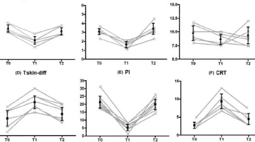
Chapter 5 Persistent peripheral and microvascular perfusion 75 alterations after out-of- hospital cardiac arrest are
associated with poor survival
Crit Care Med 2012; 40:2287-2294
Chapter 6 Clinical assessment of peripheral perfusion to predict 93 postoperative complications after major abdominal
surgery early: a prospective observational study in adults
Crit Care. 2014;18(3):R114
Chapter 7 Evolution of capillary refill time and the peripheral 115 perfusion index after septic shock resuscitation
Chapter 8 Postoperative sublingual microcirculatory derangement 129 following esophagectomy is prevented with dobutamine
Clin Hemorheol Microcirc 2011; 48:275-283
Chapter 9 Microvascular perfusion as a target for fluid resuscitation 141 in experimental circulatory shock
Crit Care Med. 2014;42:e96-e105
Chapter 10 Early peripheral perfusion-guided fluid therapy in 159 patients with septic shock
Am J Respir Crit Care Med 2015; in press
PARt E General discussion, summary and conclusions 175
Chapter 11 General discussion and future research perspectives 177
Chapter 12 Summary and conclusions 187
Chapter 13 Samenvatting en conclusie 191
PARt F Appendices 195
A Authors and affiliations 197
B List of publications and oral presentations 203
C Acknowledgements- Dankwoord 209
D PhD Portfolio 215
Introduction
1
General introduction
and outline of the thesis
1
“To him medicine owes the art of clinical inspection and observation.” Hippocrates (460 BC – 370 BC).1
The practice of physical examination, diagnosis and prognosis is introduced by the Egyptians and the Babylonians. The Diagnostic Handbook written by the ummânū (chief scholar) Esagil-kin-apli is known as one of the earliest and most extensive Babylonian medical texts (1069-1046 BC). In this handbook it was suggested that the patient’s disease could be determined by use of clinical inspection and physical examination.2 Later it was Hippocrates (460-370 BC) “father of Western medicine” who in, a structured fashion, described many diseases and medical conditions. He is well known as a major contributor of descriptions of the symptomatology and physical findings of many different diseases.1,3 Despite this ancient knowledge, how many physicians today would have the courage to write, unaided, a text on the physical examination, diagnosis, and treatment of different diseases at the bedside? It was the Dutch physician Isidore Snapper (1889-1973), who first shared his knowledge regarding the use of Bedside Medicine.4 Before that, Jordan Furneaux, a British surgeon, gave one of the first elaborated descriptions of alterations in peripheral perfusion during ‘shock’ conditions (1827).
Although I feel fortunate to live in the 21th century, with incredible innovations in medical technology and progress in patient-centered care, the emphasis and dependence on technology in modern medicine has generated an unquestioned faith in this technology, which in turn may undermine the clinician’s confidence in their own clinical skills. This may result in a decline of traditional bedside patient assessment at the expense of an increased use of technical diagnostic procedures. The danger of such unwavering confidence in technology (even in the absence of a solid evidence base for its benefits) may eventually reduce the status of traditional clinical bedside assessment to that of a “medical folklore”.5
Circulatory shock
Nowadays ‘shock’ is best defined as a life-threatening, generalized form of acute circulatory failure associated with inadequate oxygen utilization by the cells. It is a state in which the circulation is unable to deliver sufficient oxygen to meet the demands of the tissues, resulting in cellular dysfunction. The result is cellular dysoxia, i.e. the loss of the physiological independence between oxygen delivery and oxygen consumption, associated with increased lactate levels.6 Circulatory shock frequently consists of inadequate tissue blood flow and if untreated, proceeds from organ dysfunction to organ failure and eventually death. It is therefore one of the most common reasons to admit patients to the Intensive Care Unit.7
According to Weil and Shubin,8 founders of the understanding and management of circulatory (dys)function, shock can be classified into four main categories: 1. Hypovolemic (decreased circulating volume), 2. Cardiogenic (cardiac pump failure), 3. Obstructive (cardiovascular circuit obstruction), and 4. Distributive (vascular dysfunction) causes (Figure 1)9.
Of these four types, the first three are characterized by decreased cardiac output (i.e. low flow state) and as such can be regarded typical ‘shock’ conditions. The last
type, distributive shock (of which septic shock is the most common example), is typically associated with high cardiac output (hyperdynamic flow) and as such provides a huge challenge in terms of understanding the underlying biologic features and identifying the ‘ideal’ endpoints for hemodynamic therapy. It is the result of several mediators, such as pathogen factors (i.e. microbial load and virulence) leading to a host–pathogen interaction and an exaggerated inflammation resulting in loss of autoregulation and a severe disbalance between vasodilation and vasoconstriction. Sepsis, severe sepsis, and septic shock are all used to describe the increasing severity of a systemic inflammatory response to infection accompanied by systemic symptoms (i.e. fever, leucocytosis, hyperventilation, tachycardia).10 These alterations finally cause inadequate regional oxygen delivery, tissue injury and/or refractory hypotension and acute organ dysfunction, accompanied by a normal or even of a supranormal cardiac output.11,12
Hemodynamic monitoring is essential for diagnosis and management of shock. In time, many techniques have been developed: invasive or noninvasive, continuous or intermittent, and looking at different physiologic variables, directly or indirectly (through signal processing).13
Hemodynamic monitoring
“Hemodynamic monitoring plays an important role in the management of today’s acutely ill patient.” 13
Hemodynamic monitoring emerged as a distinct scientific discipline late in the 19th century when Scipione Riva-Rocci developed an easy-to-use version of the sphygmomanometer to measure arterial blood pressure. Almost 70 years later in 1964, dr. Ronald Bradley introduced miniature diagnostic catheters for intravascular use.14 This inspired Jeremy Swan and William Ganz in 1968 to develop an intravascular flow-directed catheter, which could be percutaneously inserted and positioned in the pulmonary artery for the measurement of cardiac output with thermodilution.15 With their landmark study detailing the human use of this catheter, the era of modern Figure 1. Schematic representation of the four types of shock. The classification of the different shock types, reproduced from Vincent et al.9, with permission of the NEJM.
Distributive shock Hypovolemic shock Cardiogenic shock Obstructive shock
Obstruction Pericardial tamponade Loss of plasma or blood volume Vasodilatation Ventricular failure
1
hemodynamic monitoring begun. 16 Pulmonary artery catheterization highlighted the importance of (semi)continuous monitoring of hemodynamic parameters at the bedside. Concurrently, the introduction of monitors attached to digital computers to monitor hemodynamic measurements was a defining moment in the development of critical care medicine.17 The combination of this latter technology and pulmonary artery catheters provided intensivists with a powerful platform to (semi)continuously monitor heart function and cardiac output. Despite the widespread use of this invasive catheter in critically ill patients in the 1980’s and 1990’s, its use has declined in the past years and frequent complications were highligted.17-20 Nowadays, there is a trend toward less invasive methods of hemodynamic monitoring. Current hemodynamic monitoring tools need to be “reliable, continuous, noninvasive, operator-independent, cost-effective, and should have a fast response time (beat-to-beat).”21 Therefore alternative techniques have been developed, for instance: the transpulmonary thermodilution with integrated pulse contour analysis (PiCCO TM) monitoring system (PULSION Medical System, Munich, Germany).22 This device is less invasive than pulmonary artery catheterization because it allows the assessment of cardiopulmonary hemodynamics without contact between the catheter and different cardiopulmonary structures.
Despite these advanced state-of-the-art hemodynamic monitoring techniques, simply targeting parameters related to the systemic circulation does not resuscitate various organ system tissues and might lead to increased use of medical interventions and hospital mortality.23 Surprisingly, the quest towards advanced hemodynamic monitoring techniques (central venous, arterial and pulmonary artery catheters) left a great study in 1969 unattended. In this study it was demonstrated that circulatory shock patients admitted to the Intensive Care Unit with a toe temperature ≤ 27°C had more chance to die during their intensive care unit stay.24 The importance of peripheral perfusion assessment at the bedside was highlighted with this study already in the 20th century.
The 21
stcentury
Welcoming a new era of hemodynamic monitoring: Expanding from the macro to the microcirculation*.25
It is known that all shock states, including distributive shock, are characterized by inadequate tissue perfusion and consequent tissue ischemia. In this latter type of shock, a combination of hypovolemia, reduced ventricular function, and pronounced vasodilation can lead to inadequate tissue perfusion. Because each of these determinants can cause hypotension, monitoring of cardiac function and volume status is essential for selecting the proper therapy. However, regional tissue hypoperfusion may persist, despite the normalization of systemic hemodynamics. During circulatory shock, blood flow is diverted from less important tissues to vital organs (heart, brain, and kidneys) to maintain vital organ perfusion at the cost of peripheral circulation, resulting in poor outcome in various pathophysiologic conditions. 26,27 Sympathetic activity, which is induced by the baroreceptor reflex as a response to systemic hypotension, leads to increased vasomotor tone. As sympathetic neuroactivity predominates in the skin and
muscle, the sympathetic neurohumoural response-induced vasoconstriction manifests primarily as decreased peripheral perfusion.28 Therefore, the rationale of peripheral perfusion monitoring is based on the concept that peripheral tissues are the first to reflect hypoperfusion during shock, and the last to reperfuse during resuscitation.29.
Several studies demonstrated that inadequate systemic circulation in both hypodynamic and hyperdynamic shock states can be accompanied by impaired tissue and microcirculatory perfusion. The importance of monitoring the peripheral microcirculatory tissue perfusion in different types of shock was further expanded upon by the work of Creteur, DeBacker, and their coworkers,30,31 who used visualization of microvessels and sublingual capnometry. These abnormalities can be determined noninvasively at the bedside using several clinical assessments. Since we now have entered an era where modern medical practitioners seek improved diagnostic techniques, that are easy to apply at the bedside, have low-risk for complications, and are non-invasive; noninvasive clinical assessment of peripheral perfusion gained interest.32,33 To date, many issues, to the quest that already started in 1969, remain to be elucidated. For instance, the clinical significance of the various noninvasive peripheral perfusion methods in different patient populations as well as its role as resuscitation endpoint.
Aim of the thesis
The aim of this thesis was to investigate the value of noninvasive assessment of the peripheral perfusion at the bedside for the recognition and treatment of critically ill patients.
Outline of the thesis
First, we start this thesis with a recapitulation of the currently available methods that can be used at the bedside for the noninvasive monitoring of the peripheral circulation in critically ill patients. We also discuss the potential role of peripheral perfusion monitoring for outcome prediction and resuscitation strategies, Chapter 2A and Chapter 2B (Dutch version) respectively.
In the next part of this thesis we discuss how peripheral perfusion parameters act under different circulatory conditions. We first evaluated the effect of central hypovolemia on the pulse oximeter-derived peripheral perfusion index (PPI) in awake healthy volunteers (Chapter 3). Second (Chapter 4), as blood flow in different vascular beds is regulated by local vasomotor tone, we explored the effect of peripheral cooling on the different peripheral perfusion parameters in healthy volunteers.
The third part of this thesis focuses on the clinical value of peripheral circulation assessment at the bedside in different patient populations. In Chapter 5 we evaluated the relative contribution of systemic blood flow and peripheral vasomotor tone (i.e., vasoconstriction) to sublingual and peripheral perfusion parameters before, during, and after therapeutic hypothermia in patients following out-of-hospital cardiac arrest, and the relation to outcome. We further explored the value of peripheral perfusion assessment for the prediction of complications following major abdominal surgery (Chapter 6). Finally in this part, we studied the association between the evolution of
1
different peripheral perfusion parameters with outcome in patients admitted to the intensive care unit, after undergoing initial septic shock resuscitation (Chapter 7).
In the fourth part of this thesis we aimed to answer the quintessential question: can we incorporate the different peripheral perfusion parameters into a resuscitation protocol that could benefit the patient. We therefore studied, in a prospective randomized controlled fashion, whether alterations in peripheral perfusion could be prevented in postoperative esophagectomy patients admitted to the Intensive Care Unit (Chapter 8). Next, we investigated in an experimental setting, whether changes in the perfusion of different regional vascular beds are dependent on the type of circulatory shock (obstructive vs. septic) and whether the different microvascular and peripheral perfusion parameters can be used to assess the adequacy of hemodynamic resuscitation during these different types of shock (Chapter 9). Finally, we conducted a single-center prospective randomized controlled pilot-study in septic shock patients admitted to the Intensive Care Unit. In this study our primary goal was to compare early goal directed fluid resuscitation based on clinical assessment of peripheral perfusion with standard fluid therapy, to investigate whether peripheral perfusion guided resuscitation is feasible and would lead to less fluid administration (Chapter 10).
In Chapter 11, 12, and 13 we respectively discuss the main results of each chapter, provide recommendations for future work, and summarize the most important findings of this thesis.
References
1. Garrison, Fielding H: An introduction to the history of medicine. 4th Edition. Philadelphia & London: W. B. Saunders, 1966., 1966 2. Herman F.J.Horstmanshoff, Marten
Stol CRVT: Magic And Rationality In Ancient Near Eastern And Graeco-roman Medicine. Brill Publishers, 2004
3. Louden I: Western Medicine: An Illustrated History. Oxford university press, 1997 4. van Gijn J, Gijselhart JP: [Isidore Snapper
(1889-1973) and Bedside medicine]. Ned
Tijdschr Geneeskd 2011; 155:A2647
5. Knight P: Physics and medicine--two tips
for a long and happy marriage. Lancet
2012; 379:1463-1464
6. Cecconi M, De Backer D, Antonelli M, et al: Consensus on circulatory shock and hemodynamic monitoring. Task force of the European Society of Intensive Care
Medicine. Intensive Care Med 2014;
40:1795-1815
7. Rivers E, Nguyen B, Havstad S, et al: Early goal-directed therapy in the treatment of
severe sepsis and septic shock. N Engl J
Med 2001; 345:1368-1377
8. Weil MH, Shubin H: Proposed reclassification of shock states with special
reference to distributive defects. Adv Exp
Med Biol 1971; 23:13-23
9. Vincent JL, De Backer D: Circulatory
shock. N Engl J Med 2014; 370:583
10. Dellinger RP, Levy MM, Rhodes A, et al: Surviving sepsis campaign: international guidelines for management of severe
sepsis and septic shock, 2012. Intensive
Care Med 2013; 39:165-228
11. Angus DC, van der Poll T: Severe sepsis
and septic shock. N Engl J Med 2013;
369:2063
12. Boldt J, Ince C: The impact of fluid therapy on microcirculation and tissue oxygenation
in hypovolemic patients: a review. Intensive
Care Med 2010; 36:1299-1308
13. Vincent JL, Rhodes A, Perel A, et al: Clinical review: Update on hemodynamic
monitoring--a consensus of 16. Crit Care
2011; 15:229
14. Braley RD: Diagnosting right/heart catheterisation with miniature catheters in
15. Palmieri TL: The inventors of the Swan-Ganz catheter: H. J. C. Swan and William
Ganz. Curr Surg 2003; 60:351-352
16. Swan HJ, Ganz W, Forrester J, et al: Catheterization of the heart in man with use of a flow-directed balloon-tipped
catheter. N Engl J Med 1970; 283:447-451
17. Weil MH, Shubin H, Rand W: Experience with a digital computer for study and improved management of the critically ill.
JAMA 1966; 198:1011-1016
18. Hadian M, Pinsky MR: Evidence-based review of the use of the pulmonary artery catheter: impact data and complications.
Crit Care 2006; 10 Suppl 3:S8
19. Polanco PM, Pinsky MR: Practical issues of hemodynamic monitoring at the bedside.
Surg Clin North Am 2006; 86:1431-1456
20. Sandham JD, Hull RD, Brant RF, et al: A randomized, controlled trial of the use of pulmonary-artery catheters in high-risk
surgical patients. N Engl J Med 2003;
348:5-14
21. de Waal EE, Wappler F, Buhre WF:
Cardiac output monitoring. Curr Opin
Anaesthesiol 2009; 22:71-77
22. Oren-Grinberg A: The PiCCO Monitor. Int
Anesthesiol Clin 2010; 48:57-85
23. Kelm DJ, Perrin JT, Cartin-Ceba R, et al: Fluid Overload in Patients with Severe Sepsis and Septic Shock Treated with Early-Goal Directed Therapy is Associated with Increased Acute Need for Fluid-Related Medical Interventions and Hospital Death.
Shock 2014;
24. Joly HR, Weil MH: Temperature of the great toe as an indication of the severity of
shock. Circulation 1969; 39:131-138
25. Weil MH, Tang W: Welcoming a new era of hemodynamic monitoring: expanding from the macro to the microcirculation.
Crit Care Med 2007; 35:1204-1205
26. Guyton AC: The relationship of cardiac output and arterial pressure control.
Circulation 1981; 64:1079-1088
27. Poeze M, Solberg BC, Greve JW, et al: Monitoring global volume-related hemodynamic or regional variables after initial resuscitation: What is a better predictor
of outcome in critically ill septic patients? Crit
Care Med 2005; 33:2494-2500
28. Lima A, Bakker J: Noninvasive monitoring
of peripheral perfusion. Intensive Care
Med 2005; 31:1316-1326
29. Poeze M, Solberg BC, Greve JW, et al: Monitoring global volume-related hemodynamic or regional variables after initial resuscitation: What is a better predictor
of outcome in critically ill septic patients? Crit
Care Med 2005; 33:2494-2500
30. Creteur J, De Backer D, Sakr Y, et al: Sublingual capnometry tracks microcirculatory changes
in septic patients. Intensive Care Med 2006;
32:516-523
31. De Backer D, Ospina-Tascon G, Salgado D, et al: Monitoring the microcirculation in the critically ill patient: current methods
and future approaches. Intensive Care
Med 2010; 36:1813-1825
32. Morris P, Perkins A: Diagnostic imaging.
Lancet 2012; 379:1525-1533
33. Pugsley J, Lerner AB: Cardiac output monitoring: is there a gold standard and how
do the newer technologies compare? Semin
Monitoring peripheral
perfusion in critically
ill patients at the bedside
Curr Opin Crit Care 2012; 18:273-279
M. E. van Genderen, A. Lima, J. van Bommel
Dept. of Intensive Care, Erasmus MC, Rotterdam, The Netherlands
Abstract
Purpose of review
The holy grail of circulatory monitoring is the use of an accurate, continuous and noninvasive method that can easily assess tissue perfusion under clinical conditions. As peripheral tissues are sensitive to alterations in perfusion, the noninvasive monitoring of peripheral circulation could be used as an early marker of systemic haemodynamic derangement. We therefore aim to discuss the currently available methods that can be used at the bedside as well as the role of peripheral perfusion monitoring in critically ill patients.
Recent findings
The deterioration of peripheral circulation has frequently been observed in critically ill patients with the use of subjective assessment and several optical techniques. In various patient categories, more severe and persistent alterations have been associated with worse outcomes, and these associations were independent of systemic haemodynamic parameters. Interventions aimed at systemic parameters have an unpredictable effect on peripheral circulation parameters, especially during hyperdynamic conditions. Thus, it appears that changes in peripheral perfusion reflect changes in regional vasomotor tone rather than systemic blood flow.
Summary
Subjective assessments and optical techniques provide important information regarding peripheral circulation. Moreover, these techniques are relatively easy to implement and interpret at the bedside and can be applied during acute conditions. Further research is warranted to investigate the effects of therapeutic interventions on peripheral perfusion parameters and patient outcome.
2
a
Introduction
The conventional classification for the causes of haemodynamic instability discriminates between a lack of circulating volume, insufficient pump function, obstruction of blood flow and loss of blood flow regulation.1 Although corresponding treatment modalities have been established, selecting the correct method can be very difficult when a proper diagnosis cannot be made. Especially in patients with severe sepsis, a combination of hypovolaemia, reduced ventricular function, and pronounced vasodilation can lead to inadequate tissue perfusion. Because each of these determinants can cause hypotension, monitoring of cardiac function and volume status is essential for selecting the proper therapy. However, regional tissue hypoperfusion may persist, despite the normalization of systemic haemodynamics.
During circulatory shock, blood flow is diverted from less important tissues to vital organs (heart, brain, and kidneys) to maintain vital organ perfusion at the cost of peripheral circulation, resulting in poor outcome in various pathophysiologic conditions. Sympathetic activity, which is induced by the baroreceptor reflex as a response to systemic hypotension, leads to increased vasomotor tone. As sympathetic neuroactivity predominates in the skin and muscle, the sympathetic neurohumoural response-induced vasoconstriction manifests primarily as decreased peripheral perfusion.2 Therefore, the rationale of peripheral perfusion monitoring is based on the concept that peripheral tissues are the first to reflect hypoperfusion during shock, and the last to reperfuse during resuscitation.3
It is well known that inadequate systemic circulation in both hypodynamic and hyperdynamic shock states can be accompanied by impaired peripheral circulation.4 These abnormalities can be determined noninvasively using simple clinical assessments,5 skin temperature measurements,5 and optical monitoring devices.6,7 These ‘peripheral’ techniques each involve the use of ‘abnormal’ values, which are generally associated with worse outcome in critically ill patients, do not need an intravascular catheter, and can be used directly at the bedside without entry into the body through orifices or skin or mucosal tissue punctures (Table 1). In the following section, we will discuss the currently available and commonly used techniques for assessing peripheral circulation in clinical conditions.
Clinical assessment of peripheral perfusion
The cutaneous vascular bed plays an important role in the thermoregulation of the body, and this process can result in skin perfusion alterations that have direct effects on skin temperature and colour, that is, a cold, clammy, white, and mottled skin.
Capillary refill time
Capillary refill time (CRT) is defined as the time required for a distal capillary bed (i.e., the nailbed) to regain its colour after pressure has been applied to cause blanching. This concept was first introduced by Champion et al.8 in 1981 as a component of the international trauma severity score for the rapid and structured cardiopulmonary
table 1.
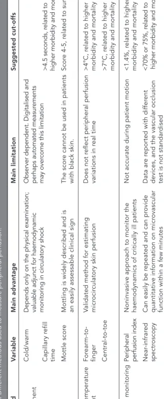
Dif
fer
ent methods used to measur
e peripheral perfusion. Method V ariable Main advantage Main limitation Suggested cut-of fs Clinical assessment Cold/war m
Depends only on the physical examination; valuable adjunct for haemodynamic monitoring in cir
culatory shock
Observer dependent. Digitalised and perhaps automated measur
ements
may over
come this limitation
Capillary r efill time >4.5 seconds, r elated to
higher morbidity and mortality
Mottle scor
e
Mottling is widely described and is
an easily assessable clinical sign
The scor
e cannot be used in patients
with black skin.
Scor e 4-5, r elated to survival Body temperatur e gradient For ear m-to-finger
Validated method for estimating micr
ocir
culatory skin perfusion
Does not r
eflect peripheral perfusion
variations in r
eal time
>4°C, r
elated to higher
morbidity and mortality
Central-to-toe
>7°C, r
elated to higher
morbidity and mortality
Optical monitoring
Peripheral perfusion index
Noninvasive appr
oach to monitor the
haemodynamics of critically ill patients
Not accurate during patient motion
<1.4%, r
elated to higher
morbidity and mortality
Near -infrar ed spectr oscopy Can easily be r
epeated and can pr
ovide
quantitative infor
mation on micr
ovascular
function within a few minutes
Data ar
e r
eported with dif
fer
ent
devices, and the vascular occlusion test is not standar
dised
<70% or 75%, r
elated to
2
a
assessment of critically ill patients. Because CRT assessment is an easily applicable method in many circumstances, it is most useful for assessing peripheral perfusion and predicting unfavourable outcomes in both critically ill adult and paediatric patients. For instance, in paediatric patients, Bohnhorst et al.9 showed that among several clinical signs present at the very first instance of suspected infection, a prolonged CRT demonstrated the most sensitive correlation with proven infection. In a recent review of clinical features that are used to confirm or exclude the possibility of serious infection in paediatric patients presented to ambulatory care, a prolonged CRT was shown to be one of the strongest indications of serious infection.10 In these patients, there may be alterations in the balance of vasoconstrictor and vasodilator substances and in the cross-talk between endothelial cells, which could result in impaired microvascular tissue blood flow regulation that is related to significant dehydration, serious infection, and severe organ dysfunction.11
In a healthy adult population, Schriger and Baraff12 reported that the upper limit of a normal CRT is 4.5 s. After applying this cutoff in critically ill patients, we demonstrated that a delayed return of normal colour (>4.5 s) can be regarded as decreased peripheral perfusion. Moreover, we found that following initial haemodynamic optimization during the first 24 h of ICU admission, the CRT could be used to discriminate patients with more severe organ dysfunction. In addition, a prolonged CRT (>4.5 s) was related to tissue hypoperfusion and a greater likelihood of worsening organ failure in the following days, compared with patients with a normal CRT.5,13 Although interobserver variability remains a matter of debate, an upper normality limit of more than 4.5 s appears to be highly reproducible for critically ill patients admitted to the ICU.14
Skin temperature
Skin temperature is best estimated using the dorsal surface of the hands or fingers of the medical examiner, as these areas are most sensitive to temperature perception. Patients are considered to have cool extremities if all examined extremities are considered cool by the examiner or if only the lower extremities are cool despite warm upper extremities in the absence of peripheral vascular occlusive disease.15 It has been demonstrated that subjectively determined variations in skin temperature correspond to objective measures of peripheral skin perfusion.13,16 Similarly, fingertip temperature estimations correlated well with objective assessments of fingertip blood flow.13,17 Accordingly, patients with a subjectively determined ‘abnormal’ peripheral perfusion following initial haemodynamic resuscitation have been associated with higher lactate levels and more severe organ dysfunction.5
Mottling
Mottling of the skin is easily recognized and is often encountered in critically ill patients. It is defined as a bluish skin discoloration that typically manifests near the elbows or knees and has a distinct patchy pattern. Mottling is the result of heterogenic small vessel vasoconstriction and is thought to reflect abnormal skin perfusion. To analyse mottling objectively, Ait-Oufella et al.18 recently developed a clinical scoring system
(from 0 to 5) based on the area of mottling from the knees to the periphery (Fig. 1). This group reported that a higher mottling score within 6h after initial resuscitation was a strong predictive factor of 14-day mortality during septic shock, and this was independent of systemic haemodynamics. This scoring system is very easy to learn, has good interobserver agreement, and can be used at the bedside.
From these studies, it is clear that the clinical assessment (Fig. 1) of peripheral perfusion is a valuable adjunct for the haemodynamic monitoring of critically ill patients and should be incorporated into future multimodal monitoring strategies to adequately monitor optimal circulatory shock resuscitation.
Body temperature gradient
Although skin temperature has been shown to be an easily accessible parameter for circulatory shock severity,19 later research demonstrated that body temperature gradients can better reflect changes in cutaneous blood flow than the absolute skin temperature itself in critically ill patients.20,21 Body temperature gradients are determined by the temperature difference between two measurement points, such as peripheral-to-ambient, central-to-toe, and forearm-to-fingertip (Tskin-diff). Increased vasoconstriction during circulatory shock leads to decreased skin temperature and a diminished ability of the core
Peripheral Perfusion Index
Capillary refill time
Pressure is applied to nailbed until it turns white Blood returned to tissue Score 2 Score 5 Mottle Score Near-infrared spectroscopy
Body temperature gradient
Delta TEMPERATURE
Tforearm - Tfingertip =
Tskindiff
Figure 1. Peripheral perfusion methods used in clinical practice. Different methods are used to measure peripheral perfusion, and these provide quantitative real-time information regarding peripheral circulation at the bedside.
2
a
to regulate its temperature before hypothermia occurs. Consequently, core temperature is maintained at the cost of the periphery to maintain vital organ perfusion, resulting in an increased central-to-peripheral temperature difference, when vasoconstriction decreases fingertip blood flow. This concept permits the establishment of central-to-toe temperature difference as an indicator of peripheral perfusion in critically ill patients, and a normal temperature gradient of 3–7°C occurs once the patient’s haemodynamics have been optimized.22 Because the effect of the operating room environment on skin and body temperature changes during surgery, especially with the use of cardiopulmonary bypass, Tskin-diff may be a more reliable measurement for critically ill patients.23 The use of Tskin-diff is based on assumption that the reference temperature is a skin site exposed to the same ambient temperature producing little change in the gradient (Fig. 1). Experimental studies have suggested Tskin-diff thresholds of 0°C for initiating vasoconstriction and 4°C for severe vasoconstriction.23,24 In critically ill adult patients, Tskin-diff measurements conducted simultaneously with clinical observation have helped to address the reliability of subjective peripheral perfusion assessment, and are able to indicate abnormal peripheral perfusion in the postresuscitation period.5
Optical monitoring
Optical methods apply light with different wavelengths directly to the tissue to assess various tissue states. There are several research techniques (described elsewhere25) that apply optical methods to visualize the microcirculation, assess oxygen availability, measure Pco2, and assess microvascular function in different tissues. Commonly used optical methods in the clinical setting that are able to monitor peripheral perfusion at the bedside include finger photoplethysmography and near-infrared spectroscopy (NIRS). Although these techniques are particularly promising, as objective numerical information can be obtained within a couple of minutes, the interpretations should be considered in combination with the clinical examination and additional peripheral perfusion measurements.13
Peripheral perfusion index
The peripheral perfusion index (PPI) is derived from the photoelectric plethysmographic signal of the pulse oximeter. Pulse oximetry, a standard of care in the ICU, allows for the measurement of arterial haemoglobin oxygen saturation and pulse rate monitoring. This noninvasive tool uses two wavelengths of light (red and infrared) that are transmitted through the distal phalanx of the index finger, resulting in the display of a pulsatile photoplethysmographic waveform. Analogous to an arterial pulse contour analysis, several variables can be derived from the plethysmographic waveform, such as the PPI (Fig. 1). The PPI is the ratio of the pulsatile part to the nonpulsatile part of the curve, expressed as a percentage. Because the size of the pulsatile portion increases with vasodilation and decreases with vasoconstriction, changes in the PPI reflect changes in peripheral vasomotor tone. This was first demonstrated in a model of axillary plexus-induced vasodilatation, and the analgesic effect of this nerve block could be predicted within minutes using the increase in PPI as a measure of
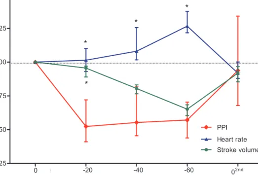
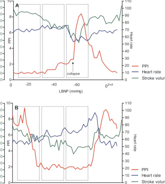
concomitant peripheral vasodilatation in patients undergoing hand surgery.26 Similarly, the PPI was shown to be rapidly reduced following sympathetic response-induced vasoconstriction after the introduction of a nociceptive skin stimulus27 or an intravenous injection of epinephrine28 or norepinephrine.29 Furthermore, in a lower body negative pressure model, the PPI also rapidly decreased following sympathetic activation in healthy volunteers who underwent stepwise decreases in venous return.30
In a large population of healthy volunteers, the median PPI value was 1.4%.7 In critically ill patients, the same value was found to represent a very sensitive cutoff point for determining abnormal peripheral perfusion, as defined by a prolonged CRT and an increased skin temperature difference.5,7,31 Therefore, this easily obtainable and noninvasive method can be used for monitoring peripheral perfusion in critically ill patients.
Near-infrared spectroscopy
NIRS is a noninvasive technique that enables the determination of tissue oxygenation based on the spectrophotometric quantitation of oxyhaemoglobin and deoxyhaemoglobin within a tissue. Although this technique can be applied to any tissue, it is primarily used to monitor peripheral oxygenation of muscle tissue in critically ill patients. The variables that are analysed using NIRS can either be directly calculated or derived from physiological interventions, such as an arterial and venous vascular occlusion test (VOT). As a result, information regarding muscle oxygen saturation (StO2) and the absolute tissue haemoglobin index, an indicator of blood volume in the microvasculature tissue region, can be obtained.2 In addition, changes in StO
2 levels during a VOT have been used to represent microvascular reactivity.6,32-34 The utility of NIRS for managing critically ill patients remains a matter of debate. Increasing publications using NIRS have described profound alterations in microvascular function in patients suffering from different pathophysiological conditions, such as sepsis and traumatic shock.32-35 In a study by Shapiro et al.36, the dynamic NIRS variables collected during a VOT were strongly associated with the severity of organ dysfunction and mortality in patients with septic shock. In this study, the StO2 recovery slope was most sensitive for the prediction of mortality. This is of special interest because there is a lack of agreement on standardization for the appropriate method for performing a
VOT37,38. When measured at the thenar eminence, NIRS-derived measurements are
influenced by the peripheral circulation condition (Fig. 1).13 Nevertheless, when used in conjunction with other peripheral perfusion methods, repeated StO2 monitoring has the potential to assess the effect of therapeutic intervention on the peripheral microvascular circulation in various shock states. Similarly, Colin et al.39 investigated the dynamic response of StO2 at different sites during the first 6h of severe sepsis resuscitation, and argued that StO2 values measured at the masseter muscle may better relate to patient outcome and may be a more powerful indicator for monitoring the effect of resuscitation, compared with measurements taken at the thenar eminence.
Although NIRS can potentially be very useful for tissue oxygenation and perfusion assessments, additional studies are still being conducted to clarify its role in the clinical management of ICU patients.
2
a
Potential therapeutic interventions to resuscitate
peripheral perfusion
Although the relationship between systemic and peripheral circulation is not always well defined, the assessment of peripheral perfusion during peripheral cooling-induced vasoconstriction in healthy volunteers has shown that profound changes in the peripheral circulation can occur independently of systemic haemodynamic parameters, such as blood pressure and cardiac output.31 Similar observations have been made during therapeutic hypothermia following cardiac arrest.40 In patients with severe sepsis, this discrepancy between systemic and peripheral circulation can become even larger; regional vasoconstriction appears to be independent of systemic blood flow in these patients.41 Considering that peripheral vasoconstriction during septic shock is an effect of increased sympathetic activity, the origin of the latter is unclear; it is likely that baroreceptor reflex activity, systemic inflammatory response, and the loss of parasympathetic activity each may play a role.42,43
Although the mechanism involved in sepsis resuscitation is not yet fully understood, it is clear that the persistence of impaired peripheral perfusion is associated with worse patient outcomes.5,6 It can be hypothesized that interventions specifically aimed at the peripheral vascular bed could resuscitate these alterations. For instance, based on the central-to-toe temperature, Boerma et al.44 showed that the administration of nitroglycerine to septic patients following early resuscitation significantly improved peripheral perfusion, independently of systemic haemodynamics. Although there was a trend to increasing mortality, this was accompanied by reduced morbidity in the nitroglycerine-treated patient group compared with the placebo group; as a result, vasodilatory agents may be promising treatment modalities.
Although fluid resuscitation is the first line therapy for sepsis-induced hypoperfusion, few studies have evaluated its effect on peripheral perfusion. Futier et al.45 recently showed that the administration of a fluid challenge had positive effects on peripheral tissue oxygenation in patients undergoing major abdominal surgery.
Whether these interventions are capable of resuscitating different peripheral perfusion parameters is, however, still unknown. Current studies to determine the effects of these interventions on peripheral circulation in critically ill patients are ongoing.
Conclusion
The rationale for monitoring peripheral perfusion is based on the concept that the peripheral circulation is the first to reflect a disturbance of the circulation that may lead to shock. Monitoring peripheral circulation not only provides a different point of reference for patient circulation but it also does not require invasive techniques and can be used directly at the bedside. Moreover, it is a simple approach that can be rapidly applied throughout the hospital, including the emergency department, operating room, wards, and ICU.
1. Weil MH, Shubin H: Proposed reclassification of shock states with special reference to
distributive defects. Adv Exp Med Biol 1971;
23:13-23
2. Lima A, Bakker J: Noninvasive monitoring
of peripheral perfusion. Intensive Care
Med 2005; 31:1316-1326
3. Poeze M, Solberg BC, Greve JW, et al: Monitoring global volume-related hemodynamic or regional variables after initial resuscitation: What is a better predictor
of outcome in critically ill septic patients? Crit
Care Med 2005; 33:2494-2500
4. Bonanno FG: Clinical pathology of the
shock syndromes. J Emerg Trauma Shock
2011; 4:233-243
5. Lima A, Jansen TC, van Bommel J, et al: The prognostic value of the subjective assessment of peripheral perfusion in
critically ill patients. Crit Care Med 2009;
37:934-938
6. Lima A, van Bommel J, Jansen TC, et al: Low tissue oxygen saturation at the end of early goal-directed therapy is associated with worse outcome in critically ill patients.
Crit Care 2009; 13 Suppl 5:S13
7. Lima AP, Beelen P, Bakker J: Use of a peripheral perfusion index derived from the pulse oximetry signal as a noninvasive
indicator of perfusion. Crit Care Med
2002; 30:1210-1213
8. Champion HR, Sacco WJ, Carnazzo AJ,
et al: Trauma score. Crit Care Med 1981;
9:672-676
9. Bohnhorst B, Lange M, Bartels DB, et al: Procalcitonin and valuable clinical symptoms in the early detection of neonatal late-onset bacterial infection.
Acta Paediatr 2012; 101:19-25
10. Van den Bruel A, Haj-Hassan T, Thompson M, et al: Diagnostic value of clinical features at presentation to identify serious infection in children in developed countries: a systematic
review. Lancet 2010; 375:834-845
11. Pickard A, Karlen W, Ansermino JM: Capillary refill time: is it still a useful clinical
sign? Anesth Analg 2011; 113:120-123
12. Schriger DL, Baraff L: Defining normal capillary refill: variation with age, sex,
and temperature. Ann Emerg Med 1988;
17:932-935
13. Lima A, van Bommel J, Sikorska K, et al: The relation of near-infrared spectroscopy
with changes in peripheral circulation in
critically ill patients. Crit Care Med 2011;
39:1649-1654
14. Lima A, van Genderen M, Boerstra T, et al. Is bedside clinical examination of capillary refill time reproducible in critically ill patients between different observers? A inter-rater variability study. Intensive Care Med. 37[Supplement 1]:215
15. De Backer D, Dubois MJ: Assessment of the microcirculatory flow in patients in the
intensive care unit. Curr Opin Crit Care
2001; 7:200-203
16. Pearson J, Low DA, Stohr E, et al: Hemodynamic responses to heat stress in the resting and exercising human leg: insight into the effect of temperature on skeletal
muscle blood flow. Am J Physiol Regul Integr
Comp Physiol 2011; 300:R663-R673
17. Carrillo AE, Cheung SS, Flouris AD: A novel model to predict cutaneous finger blood flow via finger and rectal temperatures.
Microcirculation 2011; 18:670-676
18. Ait-Oufella H, Lemoinne S, Boelle PY, et al: Mottling score predicts survival in septic
shock. Intensive Care Med 2011; 37:801-807
19. Joly HR, Weil MH: Temperature of the great toe as an indication of the severity of
shock. Circulation 1969; 39:131-138
20. Akata T, Kanna T, Yoshino J, et al: Reliability of fingertip skin-surface temperature and its related thermal measures as indices of peripheral perfusion in the clinical
setting of the operating theatre. Anaesth
Intensive Care 2004; 32:519-529
21. Rubinstein EH, Sessler DI: Skin-surface temperature gradients correlate with fingertip blood flow in humans.
Anesthesiology 1990; 73:541-545
22. Curley FJ, Smyrnios NA: Routine
monitoring of critically ill patients. J
Intensive Care Med 1990;153-174
23. House JR, Tipton MJ: Using skin temperature gradients or skin heat flux measurements to determine thresholds of
vasoconstriction and vasodilatation. Eur J
Appl Physiol 2002; 88:141-145
24. Sessler DI: Skin-temperature gradients are a validated measure of fingertip perfusion.
Eur J Appl Physiol 2003; 89:401-402
25. Sakr Y: Techniques to assess tissue oxygenation in the clinical setting.
Transfus Apher Sci 2010; 43:79-94
2
a
26. Galvin EM, Niehof S, Verbrugge SJ, et al: Peripheral flow index is a reliable and early indicator of regional block success.
Anesth Analg 2006; 103:239-43, table
27. Takeyama M, Matsunaga A, Kakihana Y, et al: Impact of skin incision on the pleth
variability index. J Clin Monit Comput
2011; 25:215-221
28. Mowafi HA, Ismail SA, Shafi MA, et al: The efficacy of perfusion index as an indicator for intravascular injection of epinephrine-containing epidural test dose in propofol
- anesthetized adults. Anesth Analg 2009;
108:549-553
29. Biais M, Cottenceau V, Petit L, et al: Impact of norepinephrine on the relationship between pleth variability index and pulse pressure variations in ICU adult patients.
Crit Care 2011; 15:R168
30. van Genderen ME, Bartels SA, Lima A, et al: Peripheral perfusion index as an early predictor for central hypovolemia in
awake healthy volunteers. Anesth Analg
2013; 116:351-356
31. Lima A, van Genderen M, Klijn E, et al: Peripheral vasoconstriction influences thenar oxygen saturation as measured by
near-infrared spectroscopy. Intensive Care
Med 2012; 38:606-611
32. Doerschug KC, Delsing AS, Schmidt GA, et al: Impairments in microvascular reactivity are related to organ failure in
human sepsis. Am J Physiol Heart Circ
Physiol 2007; 293:H1065-H1071
33. Gomez H, Torres A, Polanco P, et al: Use of non-invasive NIRS during a vascular occlusion test to assess dynamic tissue
O(2) saturation response. Intensive Care
Med 2008; 34:1600-1607
34. Skarda DE, Mulier KE, Myers DE, et al: Dynamic near-infrared spectroscopy measurements in patients with severe
sepsis. Shock 2007; 27:348-353
35. De Blasi RA, Palmisani S, Alampi D, et al: Microvascular dysfunction and skeletal muscle oxygenation assessed by phase-modulation near-infrared spectroscopy in
patients with septic shock. Intensive Care
Med 2005; 31:1661-1668
36. Shapiro NI, Arnold R, Sherwin R, et al: The association of near-infrared spectroscopy-derived tissue oxygenation measurements
with sepsis syndromes, organ dysfunction and mortality in emergency department
patients with sepsis. Crit Care 2011; 15:R223
37. Damoisel C, Payen D: Vascular occlusion
tests: do we need another definition? Crit
Care Med 2011; 39:2587-2588
38. Mayeur C, Campard S, Richard C, et al: Comparison of four different vascular occlusion tests for assessing reactive hyperemia using near-infrared spectroscopy.
Crit Care Med 2011; 39:695-701
39. Colin G, Nardi O, Polito A, et al: Masseter tissue oxygen saturation predicts normal central venous oxygen saturation during early goal-directed therapy and predicts mortality in patients with severe sepsis.
Crit Care Med 2011;
40. van Genderen ME, Lima A, Akkerhuis M, et al: Persistent peripheral and microcirculatory perfusion alterations after out-of-hospital cardiac arrest are
associated with poor survival*. Crit Care
Med 2012; 40:2287-2294
41. Boerma EC, Kuiper MA, Kingma WP, et al: Disparity between skin perfusion and sublingual microcirculatory alterations in severe sepsis and septic shock: a
prospective observational study. Intensive
Care Med 2008; 34:1294-1298
42. Convertino VA, Ludwig DA, Cooke WH: Stroke volume and sympathetic responses to lower-body negative pressure reveal new insight into circulatory shock in
humans. Auton Neurosci 2004;
111:127-134
43. Cooke WH, Convertino VA: Association between vasovagal hypotension and low sympathetic neural activity during
presyncope. Clin Auton Res 2002;
12:483-486
44. Boerma EC, Koopmans M, Konijn A, et al: Effects of nitroglycerin on sublingual microcirculatory blood flow in patients with severe sepsis/septic shock after a strict resuscitation protocol: a double-blind randomized placebo controlled trial.
Crit Care Med 2010; 38:93-100
45. Futier E, Christophe S, Robin E, et al: Use of near-infrared spectroscopy during a vascular occlusion test to assess the microcirculatory response during fluid
2
b
Perifere circulatie bij ernstig
zieke patiënten non-invasieve
methoden voor beoordeling
van de perifere perfusie
[Peripheral circulation in critically
ill patients: non-invasive methods
for the assessment of the
peripheral perfusion]
Ned Tijdschr Geneeskd. 2013;157:A5338
M. E. van Genderen, A. Lima, J. Bakker, J. van BommelAbstract
Peripheral tissues, such as skin and muscles, are sensitive to alterations in perfusion. During circulatory shock, these tissues are the first to receive less blood and the last to recover after treatment. By monitoring peripheral circulation, disturbance of the systemic circulation can be detected at an early stage. Peripheral perfusion is often disturbed in critically ill patients. Peripheral perfusion may remain disturbed, even if conventional hemodynamic parameters such as blood pressure and heart frequency normalize after treatment. Persistent abnormal peripheral perfusion is related to a poorer clinical course. With current non-invasive methods, peripheral circulation in critically ill patients can easily be assessed at the bedside. Interventions that improve peripheral circulation may speed up recovery in critically ill patients.
Samenvatting
• Perifere weefsels, zoals huid en spieren, zijn gevoelig voor veranderingen in de perfusie. Tijdens circulatoire shock worden deze weefsels als eerste minder doorbloed en na behandeling herstellen ze als laatste.
• Door het monitoren van de perifere circulatie kan daarom al in een vroeg stadium een verstoring van de systemische circulatie worden opgespoord.
• Bij ernstig zieke patiënten is de perifere perfusie vaak verstoord.
• De perifere perfusie kan verstoord blijven, ook als de waarden van conventionele hemodynamische parameters, als bloeddruk en hartfrequentie, na behandeling normaliseren.
• Een blijvend afwijkende perifere perfusie is gerelateerd aan een slechter klinisch beloop.
• Met de huidige non-invasieve methoden kan de perifere circulatie bij ernstig zieke patiënten eenvoudig worden beoordeeld aan het bed.
• Mogelijk kunnen interventies die de perifere circulatie verbeteren het klinisch herstel bespoedigen bij ernstig zieke patiënten.
2
b
Introduction
Al sinds 1969 is bekend dat de temperatuur van de grote teen een maat is voor de ernst van circulatoire shock.1 De onderzoekers lieten zien dat patiënten met een teentemperatuur ≤ 27°C bij opname op de IC een slechtere hartfunctie en een groter risico op overlijden hadden. Anno 2013 zijn er andere methoden om de doorbloeding van de perifere circulatie te bepalen aan het bed van de patiënt. Een slechtere perifere doorbloeding, zoals bepaald met deze nieuwere methoden, is geassocieerd met een verhoogd risico op orgaanfalen en op overlijden.2-5 Mogelijk wordt het verbeteren van de perifere doorbloeding een belangrijk doel bij de behandeling van ernstig zieke patiënten.6
In dit artikel presenteren we de huidige non-invasieve methoden om de doorbloeding van de perifere circulatie te bepalen en de implicaties hiervan voor de kliniek.
Waarom de perifere perfusie meten
Tijdens de initiële periode van circulatoire shock vindt een herverdeling van het hartminuutvolume plaats, van de perifere weefsels (huid en spier) naar de vitale organen (hersenen en hart). Dit is het gevolg van verschillende compensatiemechanismen en heeft als doel de doorbloeding van de vitale organen te garanderen. Door een daling van de arteriële bloeddruk worden de vasomotorische centra in het ruggenmerg gestimuleerd via de arteriële baroreceptoren en de hersenstam. Dit leidt achtereenvolgens tot een sterke sympathische reflex, stimulatie van perifere chemoreceptoren, vasoconstrictie in arteriolen en venen in de niet-vitale organen en een verminderde doorbloeding van de huid en het spierweefsel. Deze mechanismen treden al in een vroeg stadium in werking, nog voordat veranderingen optreden in de waarden van conventionele hemodynamische parameters zoals bloeddruk en hartfrequentie. De perfusie van deze perifere weefsels is daarom een gevoelige indicator voor de vroege opsporing van een verstoring van de systemische circulatie, zoals tijdens acute circulatoire shock.
Als de juiste behandeling wordt gestart en de circulatie zich herstelt, zal de activiteit van de compensatiemechanismen bij de meeste patiënten verminderen. De vasoconstrictie in het perifere vaatbed neemt af en de perfusie van de huid en het spierweefsel herstelt zich weer; dit is een teken van een succesvolle behandeling. Bij sommige patiënten blijven de compensatiemechanismen – en dus de vasoconstrictie – echter actief en herstelt de perifere perfusie zich niet. Perifere vasoconstrictie kan ook optreden tijdens een gegeneraliseerde ontsteking en tijdens sepsis. Vasoconstrictie wordt dan gemedieerd door het sympathisch zenuwstelsel en is geen compensatie voor een verstoorde circulatie. De ernst en de duur van de vasoconstrictie zijn daarbij gerelateerd aan de ziekte-ernst van de patiënt; ondanks hemodynamische stabilisatie duurt de vasoconstrictie voort. Bij septische shock is de verstoorde perifere perfusie dus een weerspiegeling van de ernst van de shock en geen teken van een compensatiemechanisme zoals bij circulatoire shock.7,8
Monitoring van de perifere circulatie is gebaseerd op het concept dat perifere weefsels de eerste zijn die hypoperfusie laten zien tijdens circulatoire shock en de laatste die herstellen tijdens de behandeling. Met een aantal eenvoudige klinische beoordelingen en monitoringstechnieken kan de perifere circulatie op een
non-invasieve wijze worden beoordeeld. Deze methoden worden hierna besproken en zijn ook samengevat in de tabel en de figuur.
Klinische beoordeling
Het uitgebreide huidvaatbed speelt een belangrijke rol bij de thermoregulatie van het lichaam. Veranderingen in de doorbloeding bepalen de kleur en de temperatuur van de huid: van warm en roze bij gezonde mensen tot koud, klam, wit en gevlekt bij ernstige vasoconstrictie.
Huidtemperatuur
De beoordeling van de huidtemperatuur – ‘warm’ of ‘koud’ – kan het best worden uitgevoerd met het dorsale oppervlak van de handen of vingers; deze oppervlakken zijn immers het gevoeligst voor de waarneming van temperatuur. Een afwijkende perifere perfusie bij deze manier van beoordelen is gerelateerd aan hogere lactaatspiegels en ernstiger orgaanfalen.3 Deze subjectieve waarneming van de temperatuur komt goed overeen met objectieve metingen van de huidtemperatuur en van de vingerdoorbloeding, en is daarom geschikt voor de beoordeling van de perifere circulatie.9
Capillaire-‘refill’-tijd
De capillaire-‘refill’-tijd is de tijd die nodig is om weer kleur te krijgen in een distaal capillair netwerk, bijvoorbeeld het nagelbed, nadat er gedurende 15 s zo veel druk op is uitgeoefend dat een bleke verkleuring is ontstaan. Bij gezonde volwassenen is de bovengrens van een normale CRT van het nagelbed 4,5 s. Bij ernstig zieke patiënten kan vertraagd herstel (> 5 s) worden beschouwd als een verminderde perifere perfusie.3 Op deze manier kunnen patiënten met een verstoorde perfusie al tijdens de eerste 24 h van opname op de IC worden geïdentificeerd. Verder is een verlengde capillaire-refill-tijd bij IC-patiënten en bij patiënten na reanimatie gerelateerd aan een verhoogd risico op orgaanfalen en op overlijden vergeleken met patiënten met een niet-afwijkende capillaire-refill-tijd (< 5 s).3,5 Dit risico blijft verhoogd na normalisatie van de waarden van conventionele hemodynamische parameters. De meting van capillaire-refill-tijd lijkt subjectief, maar toch komen de beoordelingen door zowel artsen als verpleegkundigen goed overeen.10
Marmering van de huid
Een marmerhuid is een reticulair, blauwrood huidaspect, meestal veroorzaakt door koude. Deze verkleuring is gemakkelijk te herkennen en komt typisch voor rondom de ellebogen of de knieën. Een marmerhuid wordt veroorzaakt door heterogene vasoconstrictie in het huidvaatbed en wordt vaak gezien bij ernstig zieke patiënten. Een recente studie bij patiënten met septische shock laat zien dat de mate van marmering – en dus de ernst van de hypoperfusie – goed kan worden beoordeeld met de ‘mottling’-score (zie figuur). Deze score loopt van 0 (‘geen marmering’) tot 5 (‘zeer ernstige marmering’). Een score van 4 (‘ernstige marmering’) of 5 binnen 6 h na de initiële behandeling is een sterk voorspellende factor voor de 14-daagse mortaliteit, onafhankelijk van de conventionele
2
b
tabel.
Non-invasieve methoden voor de beoor
deling van de perifer
e cir culatie Methode V oor deel Nadeel Aanbevolen afkappunt Klinische beoor deling huidtemperatuur
tijdens lichamelijk onderzoek; waar
devolle
aanvulling op hemodynamische monitoring tijdens cir
culatoir
e shock
subjectief; mogelijk zijn gedigitaliseer
de of geautomatiseer de metingen wel objectief koud, ger elateer d aan hoger e morbiditeit en mortaliteit capillair e-‘r efill’-tijd
tijdens lichamelijk onderzoek; waar
devolle
aanvulling op hemodynamische monitoring tijdens cir
culatoir
e shock
subjectief; mogelijk zijn gedigitaliseer
de of geautomatiseer de metingen wel objectief > 4,5 s, ger elateer d aan hoger e morbiditeit en mortaliteit ‘mottling’-scor e*
tijdens lichamelijk onderzoek; mar
merhuid is
eenvoudig te herkennen en te beoor
delen
beoor
deling is lastig bij patiënten met
donker e of zwarte huid scor e 4-5, ger elateer d aan mortaliteit Huidtemperatuur gradiënt onderar m-tot-vingertop gevalideer
de methode voor beoor
deling van perifer e weefseldoorbloeding geen r ealtime weer gave van veranderingen in perifer e perfusie > 4°C, ger elateer d aan hoger e morbiditeit en mortaliteit
Optische monitoring perifer
e-perfusie-index
voor hemodynamische monitoring van er
nstig
zieke patiënten
niet accuraat tijdens bewegingen van patiënt
< 1,4%, ger elateer d aan hoger e morbiditeit en mortaliteit ‘near -infrar ed’-spectr oscopie kwantitatieve infor
matie over weefseloxygenatie
en micr
ovasculair
e functie binnen enkele
minuten; eenvoudig te herhalen
voor de metingen zijn verschillende apparaten nodig; test voor vasculair
e
reactiviteit is niet gestandaar
diseer d < 70% of > 75%, ger elateer d aan hoger e morbiditeit en mortaliteit
* De mate van mar
mering van de huid wor
dt bepaald met de ‘mottling’-scor
e, die loopt van 0 (geen mar
mering) tot 5 (zeer er
nstige mar
hemodynamische parameters.11 Dit scoringssysteem is eenvoudig te leren, heeft een goede interbeoordelaarsbetrouwbaarheid en kan aan het bed worden toegepast.
Huidtemperatuurgradiënt
Hoewel al lang bekend is dat de absolute huidtemperatuur een goede parameter is voor de ernst van circulatoire shock,1 laat recenter onderzoek zien dat de gradiënt in de huidtemperatuur de veranderingen in lokale doorbloeding beter weergeeft.11,12 Deze gradiënt wordt bepaald door de temperatuurverschillen tussen een proximaal en distaal meetpunt te bepalen, zoals centraal-tot-grote-teen of onderarm-tot-vingertop, en wordt gemeten door middel van thermometer probes op de huid (zie figuur). De
capillaire-‘refill’-tijd ‘mottling’-score huidtemperatuurgradiënt onderarm-tot-vingertop ‘near-infrared’-spectroscopie perifere-perfusie-index huidtemperatuur
subjectieve methoden objectieve methoden
1 2 3 4 5 15 s
Figuur. Non-invasieve methoden om de perifere circulatie te beoordelen aan het bed van de patiënt. Subjectieve methoden zijn de huidtemperatuur, de capillaire-‘refill’-tijd, en de ‘mottling’-score voor het bepalen van de mate van marmering van de huid. Objectieve methoden zijn de huidtemperatuurgradiënt onderarm-tot-vingertop, de perifere-perfusie-index en de ‘near-infrared’-spectroscopie.
2
b
huidtemperatuurgradiënt onderarm-tot-vingertop wordt ‘Tskin-diff’ genoemd (afgeleid van ‘skin-temperature gradient’). Het optreden van perifere vasoconstrictie tijdens circulatoire shock leidt tot een afname van de huidtemperatuur en tot een verminderd vermogen om de centrale temperatuur te regelen. Daardoor stijgt de centraal-perifere temperatuurgradiënt. Het gebruik van Tskin-diff is gebaseerd op de aanname dat de referentietemperatuur een plaats op de huid is die aan dezelfde omgevingstemperatuur is blootgesteld als de vingertop. Een andere omgevingstemperatuur heeft eenzelfde soort effect op de onderarm als op de vingertop, en heeft daardoor weinig invloed op de gradiënt.12
Het gebruik van Tskin-diff is gevalideerd voor het beoordelen van de perifere weefseldoorbloeding. Uit verschillende experimentele studies is gebleken dat een Tskin-diff > 4°C een teken is van een ernstige verstoring van de huidperfusie en dat deze gerelateerd is aan een verhoogd risico op orgaanfalen.5,13 Het is niet bekend wat de invloed van vaatvernauwende of -verwijdende medicatie is op de huidtemperatuur of op de Tskin-diff; onze groep doet momenteel onderzoek naar deze relatie.
Optische monitoring
Optische technieken maken gebruik van licht met verschillende golflengten. Met deze non-invasieve methoden kan de conditie van verschillende weefsels worden vastgesteld. De meest gebruikte optische methoden om de perfusie van de perifere circulatie te meten zijn fotoplethysmografie en ‘near-infrared’-spectroscopie.
Perifere-perfusie-index
De perifere-perfusie-index is afgeleid van het plethysmografiesignaal van de pulsoximeter. Pulsoximetrie wordt in de meeste ziekenhuizen gebruikt om de arteriële zuurstofsaturatie van een patiënt te bepalen. De pulsoximeter zendt licht van 2 golflengtes door de distale falanx van de vinger. Door de pulsaties in het vaatbed resulteert dit in een pulsatiele fotoplethysmografische lichtgolf die aan de andere kant van vinger wordt opgevangen door een detector. Andere weefsels, zoals bindweefsel en bot, geven een niet-pulsatiele lichtgolf. De perifere-perfusie-index is de verhouding tussen het pulsatiele en het niet-pulsatiele deel van de opgevangen lichtgolven, uitgedrukt in een percentage. Omdat de grootte van het pulsatiele deel toeneemt bij vasodilatatie en afneemt bij vasoconstrictie, corresponderen veranderingen in perifere-perfusie-index met veranderingen in de perifere vasomotorische tonus. Dit is als eerste aangetoond met plexusblokkade tijdens locoregionale anesthesie, waarbij vasodilatatie van de extremiteit optreedt.14 Andersom neemt de perifere-perfusie-index snel af bij vasoconstrictie na toediening van (nor)adrenaline.15
Bij een grote groep gezonde vrijwilligers was de mediane perifere-perfusie-index 1,4%.16 Deze waarde was bij ernstig zieke patiënten een gevoelig afkappunt voor het bepalen van een afwijkende perifere perfusie, gedefinieerd als een verlengde capillaire-refill-tijd en een toegenomen huidtemperatuurgradiënt.3 Een perifere-perfusie-index < 1,4% is gerelateerd aan een toegenomen prevalentie van orgaanfalen.5 Deze eenvoudig te bepalen index kan dus worden gebruikt om de perifere circulatie te bepalen en te vervolgen.


![Figure 2. Box plots demonstrating the time course of (A) sublingual microcirculatory parameters (microvascular flow index [MFI], perfused capillary density [PCD, n/mm2], and proportion perfused vessels [PPV, %]) and (B) peripheral perfusion parameters (cap](https://thumb-us.123doks.com/thumbv2/123dok_us/1912218.2780527/84.793.136.602.161.935/demonstrating-sublingual-microcirculatory-parameters-microvascular-proportion-peripheral-parameters.webp)