ROLE OF IL 17 IN THE PATHOGENESIS OF PSORIASIS
*,1
Basma Hamada Mohammed
and
4Dr. Mohammed Hassan Mohammed
1
Master degree in Dermatology & S.T.Ds
2Professor of Dermatology,
3
Professor of Biochemistry
4Lecturer of Dermatology
ARTICLE INFO ABSTRACT
Background:
mainly immunological, affecting skin and joints. Many cytokines produced by T lymphocytes, especially T helper
Objectives: Methodology: patients
and compared with twenty sex and age matched healthy volunteers th group.
Results: the control group.
Copyright©2016, Basma Hamada Mohammed et al. This
unrestricted use, distribution, and reproduction in any medium, provided the original work is properly cited.
INTRODUCTION
Psoriasis is fundamentally an inflammatory skin condition with reactive abnormal epidermal differentiation and hyper proliferation affecting 2-3 % of world’s population
et al., 2011). Its exact cause is unknown, but a wealth of studies indicate that the disease results from a complex and dynamic interplay between genetic and environmental factors that trigger an excessive inflammatory response in the skin
(Monteleone et al., 2011). Psoriasis recognize two
pathogenetic moments, the first one involving innate immunity triggered by unknown stimuli and the second one involving the adaptive immunity, due to cytokines released from cells of the innate immune system, mainly dendritic cells
2013). The involvement of T lymphocytes in the pathogenesis of psoriasis can be described in terms of 3 events: the initial activation of T lymphocytes, the migration of T lymphocytes into the skin, and the various roles played by cytokines released from T lymphocytes and other cells
Abdel Hay, 2010). Both Th1 and Th17 cells have been verified as major T-cell components of psoriasis. It is still not clear which cell type has a predominant role, although a
*Corresponding author: Basma Hamada Mohammed,
Master degree in Dermatology & S.T.Ds
ISSN: 0975-833X
Article History:
Received 23rd September, 2016 Received in revised form 25th October, 2016
Accepted 18th November, 2016
Published online 30th December,2016
Key words:
Psoriasis, Pathogenesis, Cytokines, T helper cells, Interleukin 17.
Citation: Basma Hamada Mohammed, Dr. Talal Ahmed Abd
“Role of IL 17 in the pathogenesis of psoriasis”, International Journal of Current Research
RESEARCH ARTICLE
ROLE OF IL 17 IN THE PATHOGENESIS OF PSORIASIS
Basma Hamada Mohammed,
2Dr. Talal Ahmed Abd-El-Raheem,
3Dr. Olfat Gamil
Dr. Mohammed Hassan Mohammed
Master degree in Dermatology & S.T.Ds
Professor of Dermatology, Andrology and STDs, Faculty of Medicine- Fayoum University
Professor of Biochemistry, Faculty of Medicine- Cairo University
Dermatology, Faculty of Medicine- Fayoum University
ABSTRACT
Background: Psoriasis is a common, chronic, inflammatory disease of multiple etiological factors, mainly immunological, affecting skin and joints. Many cytokines produced by T lymphocytes, especially T helper type, have been implicated in its pathogenesis.
Objectives: This study aimed at detecting the role of interleukin 17 in the pathogenesis of psoriasis. Methodology: This study was a case control study. IL 17 was measured in
patients in both lesional and non lesional skin and the results were correlated with the disease severity and compared with twenty sex and age matched healthy volunteers th
group.
Results: IL 17 was statistically significant higher in lesional than in non lesional areas and also than the control group.
This is an open access article distributed under the Creative Commons Att use, distribution, and reproduction in any medium, provided the original work is properly cited.
Psoriasis is fundamentally an inflammatory skin condition with reactive abnormal epidermal differentiation and hyper 3 % of world’s population (Kuchekar Its exact cause is unknown, but a wealth of that the disease results from a complex and dynamic interplay between genetic and environmental factors that trigger an excessive inflammatory response in the skin Psoriasis recognize two moments, the first one involving innate immunity triggered by unknown stimuli and the second one involving the adaptive immunity, due to cytokines released from cells of the innate immune system, mainly dendritic cells (Skroza et al., ent of T lymphocytes in the pathogenesis of psoriasis can be described in terms of 3 events: the initial activation of T lymphocytes, the migration of T lymphocytes into the skin, and the various roles played by cytokines her cells (El-Darouti and Both Th1 and Th17 cells have been cell components of psoriasis. It is still not clear which cell type has a predominant role, although a
greater number of Th17 and Th22 cells are identified in the circulations of psoriasis patients than Th1 cells
2011). IL-17 is regarded as a signature cytokine for Th17 cells and is involved in inflammatory responses
Despite its protective effects against certain pathogens, IL can be dysregulated in some individuals and thereby contribute to the pathogenesis and/or maintenance of autoimmune and immune inflammatory disorders
2013).
Aim of work
This study aimed at detecting the role of interleukin 17 in the pathogenesis of psoriasis through detecting its tissue gene expression in plaque psoriatic patients in both lesional and non lesional skin and correlating its level with disease severity and comparing those results with its level in control healthy individuals.
Subjects and methods
It was a case control study implemented in Fayoum university hospital. The study included 2 groups: the patients group which included 20 patients of both sexes and of different age groups. They were selected from attendants of Dermatology,
International Journal of Current Research Vol. 8, Issue, 12, pp.43793-43797, December, 2016
INTERNATIONAL
Basma Hamada Mohammed, Dr. Talal Ahmed Abd-El-Raheem, Dr. Olfat Gamil Shaker and Dr. Mohammed Hassan Mohammed
International Journal of Current Research, 8, (12), 43793-43797.
ROLE OF IL 17 IN THE PATHOGENESIS OF PSORIASIS
Dr. Olfat Gamil Shaker
Fayoum University
Cairo University
Fayoum University
Psoriasis is a common, chronic, inflammatory disease of multiple etiological factors, mainly immunological, affecting skin and joints. Many cytokines produced by T lymphocytes,
This study aimed at detecting the role of interleukin 17 in the pathogenesis of psoriasis. IL 17 was measured in twenty plaque psoriatic in both lesional and non lesional skin and the results were correlated with the disease severity and compared with twenty sex and age matched healthy volunteers that were included as control
gher in lesional than in non lesional areas and also than
the Creative Commons Attribution License, which permits
greater number of Th17 and Th22 cells are identified in the circulations of psoriasis patients than Th1 cells (Carrier et al., 17 is regarded as a signature cytokine for Th17 cells and is involved in inflammatory responses (Peiser, 2013). Despite its protective effects against certain pathogens, IL-17 in some individuals and thereby contribute to the pathogenesis and/or maintenance of autoimmune and immune inflammatory disorders (Van den Berg and McInnes,
This study aimed at detecting the role of interleukin 17 in the of psoriasis through detecting its tissue gene expression in plaque psoriatic patients in both lesional and non lesional skin and correlating its level with disease severity and comparing those results with its level in control healthy
It was a case control study implemented in Fayoum university The study included 2 groups: the patients group of both sexes and of different age groups. They were selected from attendants of Dermatology,
INTERNATIONAL JOURNAL OF CURRENT RESEARCH
STDs & Andrology outpatient clinic. Twenty age and sex matched healthy volunteers were included as a control group with no family or past history of psoriasis. Diagnosis was made on clinical basis. The psoriasis in all patients was of plaque type. Exclusion criteria were being erythrodermic, pustular, or palmoplantar or having other forms of psoriasis, nail psoriasis, psoriatic arthritis. Full personal, present, past, medical, treatment and family history were taken. Clinical examination included:
Distribution of lesions and severity graded in mild, moderate and severe degree, according to surface area affected.
Calculation of severity of psoriasis by PASI scores (psoriatic area and severity index).
Any kind of treatment except for emollients was
stopped for four weeks before taking the skin biopsies. Two skin biopsies were taken from each patient; (one from lesional area and the other from non lesional area), and one skin biopsy from each control.
The selected area was cleaned with alcohol and a local anesthetic (lidocaine 2%) was injected subcutaneously. A 3.5 mm. punch biopsy was used to obtain the specimen. The biopsies were kept directly at -5 without any preservatives at Dermatology, STDs & Andrology outpatient clinic, Fayoum University hospital. Then transported in an ice box to the lab, for detection of the tissue gene expression of Il 17. Each Skin biopsy was used for RNA extraction and RT-PCR of IL-17.
Data entry and Statistical Analysis
Data was collected and coded to facilitate data manipulation and double entered into Microsoft Access and data analysis was performed using SPSS software version 18 under windows 7. Qualitative data were described using numbers, percentages and arithmetic means as central tendency measurement, standard deviations as measure of dispersion for quantitative parametric data. Inferential statistic tests (student t-Test, anova test and Paired t-test for quantitative data andchi square test and bivariate correlation test for qualitative data) were used to detect differences between different categories, with a significant level of less than 0.05.
Ethical considerations
This study was reviewed and approved by the faculty of medicine research ethical committee. The study was conducted after explaining the aim of the study and the exact procedure used and all collected data were kept confidential. All participants had the right not to participate in the study. Treatment was prescribed when indicated and method of use was explained.
RESULTS
The age of patients ranged between 6 and 67 years with a mean ±SD of 33±20.9 years. Sex distribution showed that 55% were females (11 patients) and 45% were males (9 patients). Both age and sex were matched with control group as shown in (Table 1), (Table 2).
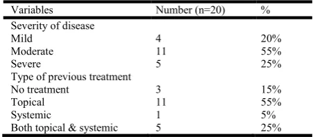
Disease severity showed that 20% (4 patients) had mild disease, 55% (11 patients) had moderate degree and 25% (5 patients) had severe degree as shown in(Table 3).
Table 1. Mean age of different study groups
Age (years)
Cases (n=20)
Controls
(n=20) p-value Sig. Mean SD Mean SD
33 20.9 32 8.9 0.9 NS
Table 2. Sex distribution among different study groups
Sex
Cases (n=20)
Controls
(n=20) p-value Sig. No. % No. %
Male 9 45% 13 65% 0.2 NS Female 11 55% 7 35%
[image:2.595.319.545.247.346.2]NS=no significance.
Table 3. Frequencies of disease characters among psoriatic cases
Variables Number (n=20) % Severity of disease
Mild 4 20%
Moderate 11 55%
Severe 5 25%
Type of previous treatment
No treatment 3 15%
Topical 11 55%
Systemic 1 5%
Both topical & systemic 5 25%
[image:2.595.308.556.437.480.2]Duration of disease ranged between 2 months and 30 years with a mean± SD of (4.7±7.1) years, the PASI score ranged between 1.2 and 34.8 with a mean± SD of (9.3 ± 8.9) as illustrated in (Table 4).
Table 4. Description of disease characters among psoriatic cases
Variables Minimum Maximum Mean SD Duration of disease
(years)
(2 months) 30 4.7 7.1
PASI score 1.2 34.8 9.3 8.9
IL-17 level in lesional area ranged between 1.5 and 14.4 with a mean± SD of (5.8 ± 3.8), and IL-17 level in non-lesional area ranged between1.1 and 3.3 with a mean± SD of (1.8±0.66) as shown in (Table 5).
Table 5. IL-17 level in psoriatic cases
Variables Minimum Maximum Mean SD Lesional IL-17 1.5 14.4 5.8 3.8 Non-Lesional IL-17 1.1 3.3 1.8 0.66
[image:2.595.306.562.698.741.2]There is a statistically significant difference between IL 17 level in lesional and non-lesional area among psoriatic cases with higher level of IL-17 in lesional area with p-value <0.05 as shown in (Table 6).
Table 6. Comparison of IL-17 level among psoriatic cases
Variables Cases (n=20) p-value Sig. Mean SD
Lesional IL-17 5.8 3.8 <0.001 HS Non-Lesional IL-17 1.8 0.66
HS=high significance.
5.8±3.8 and control group with a mean ±SD of 1.2±0.15 as shown in (Table 7).
Table 7. Comparison of IL 17 between cases (lesional) and control
Variable
(Lesional) Cases (n=20)
Controls
(n=20) p-value Sig. Mean SD Mean SD
IL-17 5.8 3.8 1.2 0.15 <0.001 HS
[image:3.595.311.559.55.266.2]
Also, there is statistically significant difference with p-value <0.05 between case group (non-lesional area) with a mean ±SD of 1.8±0.66 and control group with a mean ±SD of 1.2±0.15 (Table 8).
Table 8. Comparison of IL 17 between cases (non lesional) and control
Variable
(Non lesional) Cases (n=20)
Controls
(n=20) p-value Sig. Mean SD Mean SD
IL-17 1.8 0.66 1.2 0.15 0.001 HS
[image:3.595.38.286.111.154.2]There is a statistically significant difference with p-value <0.05 between different degrees of disease severity as regards to IL-17 level in lesional and non-lesional area among psoriatic cases with higher level among cases with severe degree of psoriasis(Table 9).
Table 9. Comparison of IL-17 level in lesional and non lesional skin between different degrees of disease severity among
psoriatic cases
Variables
Severity of disease
p-value Sig. Mild Moderate Severe
Mean ± SD Mean ± SD Mean ± SD Lesional
IL-17
2.2±0.50 4.5±1.7 11.4±2 <0.001 HS
Non-Lesional IL-17
1.3±0.18 1.6±0.60 2.5±0.46 <0.001 HS
There is no statistically significant correlation between disease duration and level of IL-17 in both lesional and non-lesional area among psoriatic patients (p-value > 0.05) as illustrated in (Table 10).
Table 10. Correlation between duration of disease and level of IL-17 among psoriatic cases
IL17 level Disease duration (years) r p-value Sig. Lesional IL-17 0.02 0.9 NS Non-Lesional IL-17 -0.05 0.8 NS
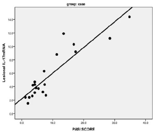
[image:3.595.310.557.303.513.2]This study showed that there is a statistically significant positive strong correlation with p-value <0.05 between PASI score and level of IL-17 in both lesional and non-lesional area among psoriatic patients as illustrated in (Table 11), (Fig. 1, 2).
Table 11. Correlation between PASI score and level of IL-17 among psoriatic cases
IL17 level PASI score
r p-value Sig. Lesional IL-17 0.90 <0.001 HS Non-Lesional IL-17 0.74 <0.001 HS
Fig.1. Correlation between PASI and IL-17 in lesional area among psoriatic patients
Fig. 2. Correlation between PASI score and level of IL-17 in non lesional area among psoriatic patients
DISCUSSION
[image:3.595.34.292.421.503.2] [image:3.595.36.287.752.797.2]skin and the difference was highly statistically significant with p-value <0.05. Both lesional and non lesional IL 17 was higher than control group and the difference was highly statistically significant with p-value <0.05, which is consistent with Johansen et al. (2009), Lowes et al. (2008), Zaba et al. (2007) whose researches showed increased expression of IL-17 ligands in lesional skin compared with both non lesional and normal skin. This was also in agreement with the results of Harper et al. (2009) Russell et al. (2011) who measured expression of IL-17A, IL-17C, and IL-17F which was elevated in psoriatic lesional tissue compared with non lesional tissue. The same with Wolk et al. in 2006 and Matsushita and Higashi in 2008 who observed increased IL-17A and IL-17F mRNA in psoriatic skin lesion comparing to non lesional or healthy skin. Moreover, many researchers have studied serum level of IL 17, not the tissue level, in psoriasis like Takahashi et al. (2010), Yilmaz et al. (2012) who detected that it is elevated and also significantly correlated with PASI score. Others have measured IL 17 cells level in both tissue and peripheral circulation of psoriatic patients as Zhang et al. (2010), Caproni
et al. (2009) who found that it was positively correlated with disease severity as measured by PASI score. This is in agreement with our results that documented positive strong correlation between IL 17 level and disease severity together with PASI score with high statistically significance. Kagami et al. (2010) found that circulating IL-17A+ cells were elevated in psoriatics compared with healthy individuals but -unlike our results-did not significantly correlate with skin disease severity. On the contrary, Kyriakou et al. (2014) documented that median serum levels of IL-17, using ELISA, did not differ significantly between the cases and the control group in his study and no significant correlations were found between PASI and the cytokine serum. In Egypt, to our knowledge, we are the first to study tissue gene expression of IL 17 in lesional and non lesional areas in psoriatic patients in relation to control group and correlating the results with disease severity through PASI score. Almost all studies were conducted on the serum level of IL 17, not the tissue level like our study. Almakhzangy and Gaballa (2009), Abdel Mawla et al. (2013), El-Moaty Zaher et al. (2013), Abo Elmajd et al. (2014) observed a statistically significant higher serum level of IL-17 (p< 0.05) in psoriatic patients versus control.
Conclusion and Recommendations
As we gain further insight into the immunopathogenesis of psoriasis, we hope it will provide the basis for the development of safer, more efficacious, and more durable therapeutics in the future. Given its enormous toll on patient health and quality of life, steps should be taken to prevent or decrease the risk of psoriasis associated comorbidities. In conclusion, there is no doubt that IL17 has a major role and importance in the orchestra played by the cytokines in order to create the psoriatic plaque in the end, evident by its high tissue gene expression in comparison to control in our study and the high significant correlation with PASI score. We suggest that, a use of an array of this cytokine and others related may be considered as a useful follow-up marker for monitoring of psoriatic patients and optimizing therapeutic strategies.
REFERENCES
Abdel Mawla MY, Abulmajd Y, Soliman M, Amer AM, Nasr M and Victor O 2013. Serum Interleukin (IL)-17 in Psoriasis. J Am Sci., 9(12):229-32.
Abo Elmajd Y, Ibrahim H, Raafat N and Abdel Mawla Y 2014. Evaluating the Interleukin 17 serum level in psoriatic patients from the Eastern Egyptian population. Ind J Appl Res., 4 (7): 537-9.
Almakhzangy I and Gaballa A 2009. Serum level of 17, IL-22, INF-γ in patient with psoriasis. Egypt Dermatol J., 5: 1-10.
Caproni M, Antiga E, Melani L, Volpi W, Del Bianco E and Fabbri P. 2009. Serum levels of IL-17 and IL-22 are reduced by etanercept, but not by acitretin, in patients with psoriasis: a randomized-controlled trial. J Clin Immunol.,
29: 210–4.
Carrier Y, Ma HL, Ramon HE, Napierata L, Small C, O’Toole M, Young DA, Fouser LA, Nickerson-Nutter C, Collins M, Dunussi-Joannopoulos K and Medley QG. 2011. Inter-Regulation of Th17 cytokines and the IL-36 cytokines in vitro and in vivo: implications in psoriasis pathogenesis. J Invest Dermatol., 131: 2428-37.
El-Darouti M and Abdel Hay R 2010. Psoriasis: highlights on pathogenesis, adjuvant therapy and treatment of resistant and problematic cases (part i). J Egypt Women Dermatol Soc., 7:64 - 70.
El-Moaty Zaher HA, El-Komy MH, Hegazy RA, Mohamed El Khashab HA, Ahmed HH. 2013. Assessment of interleukin-17 and vitamin D serum levels in psoriatic patients. J Am Acad Dermatol., 69(5): 840-2.
Harper EG, Guo C, Rizzo H, Lillis JV, Kurtz SE, Skorcheva I, Purdy D, Fitch E, Iordanov M and Blauvelt A. 2009. Th17 cytokines stimulate CCL20 expression in keratinocytes in vitro and in vivo: implications for psoriasis pathogenesis. J Invest Dermatol., 129: 2175–83.
Johansen C, Usher PA, Kjellerup RB, Lundsgaard D, Iversen L and Kragballe K. 2009. Characterization of the interleukin-17 isoforms and receptors in lesional psoriatic skin. Br J Dermatol., 160: 319–24.
Kagami S, Rizzo HL, Lee JJ, Koguchi Y, and Blauvelt A 2010. Circulating Th17, Th22, and Th1 cells are increased in psoriasis. J Invest Dermatol., 130(5): 1373-83.
Kuchekar AB, Pujari RR, Kuchekar SB, Dhole SN and Mule PM. 2011. Psoriasis: A comprehensive review. Int J of Pharm & Life Sci., 2(6): 857-77.
Kyriakou A, Patsatsi A, Vyzantiadis TA and Sotiriadis D 2014. Serum Levels of TNF- , IL-12/23p40 and IL-17 in Plaque Psoriasis and Their Correlation with Disease Severity. Scientific World J., 508178.
Lowes MA, Kikuchi T, Fuentes-Duculan J, Cardinale I, Zaba LC, Haider AS, Bowman EP and Krueger JG. 2008. Psoriasis vulgaris lesions contain discrete populations of Th1 and Th17 T cells. J Invest Dermatol., 128:1207–11. Matsushita S and Higashi T 2008. Human Th17 Cell Clones
and Natural Immune Responses. Allergol Int., 57: 135-40. Michalak-Stoma A, BartosiNska J, Kowal M,
Juszkiewicz-Borowiec M, Gerkowicz A and Chodorowska G. 2013. Serum levels of selected th17 and th22 cytokines in psoriatic patients. Disease Markers, 35 (6): 625-31. Monteleone G, Pallone F, Macdonald TT, Chimenti S and
Costanzo A. 2011. Psoriasis: from pathogenesis to novel therapeutic approaches. Clin Sci., 120: 1–11.
Peiser M. 2013. Role of Th17 Cells in Skin Inflammation of Allergic Contact Dermatits. Clin Dev Immunol., 1-10.
substantial pathway-specific effects within one week. J Invest Dermatol., 131:S11.
Skroza N, Proietti I, Pampena R, Viola GL, Bernardini N, Nicolucci F, Tolino E, Zuber S, Soccodato V, and Potenza C. 2013. Correlations between psoriasis and inflammatory bowel diseases. Biomed Res Int., 1-8.
Song X and Qian Y 2013. IL-17 family cytokines mediated signaling in the pathogenesis of inflammatory diseases.
Cellular Signalling, 25: 2335–47.
Takahashi H, Tsuji H, Hashimoto Y, Ishida-Yamamoto A and Iizuka H. 2010. Serum cytokines and growth factor levels in Japanese patients with psoriasis. Clin Exp Dermatol., 35: 645-9.
Van den Berg WB and McInnes IB. 2013. Th17 cells and
IL-17A- Focus on immunopathogenesis and
immunotherapeutics. Seminars in Arthritis and
Rheumatism, 43: 158–70.
Wolk K, Witte E, Wallace E, Döcke WD, Kunz S, Asadullah K, Volk HD, Sterry W and Sabat R. 2006. IL-22 regulates
the expression of genes responsible for antimicrobial
defense, cellular differentiation and mobility in
keratinocytes: a potential role in psoriasis. Eur J Immunol.,
36: 1309-23.
Yilmaz SB, Cicek N, Coskun M, Yegin O and Alpsoy E 2012. Serum and tissue levels of IL-17 in different clinical subtypes of psoriasis. Arch Dermatol Res., 304:465-9. Zaba LC, Cardinale I, Gilleaudeau P, Sullivan-Whalen M,
Suarez-Farinas M, Fuentes-Duculan J, Novitskaya I, Khatcherian A, Bluth MJ, Lowes MA and Krueger JG 2007. Amelioration of epidermal hyperplasia by TNF inhibition is associated with reduced Th17 responses. J Exp
Med., 204(13): 3183–94.
Zhang L, Yang XQ, Cheng J, Hui RS and Gao TW. 2010. Increased Th17 cells are accompanied by FoxP31 Treg cell accumulation and correlated with psoriasis disease severity.
Clin Immunol., 135:108-17.