R E S E A R C H P A P E R
Microarray analysis revealed different gene expression patterns
in HepG2 cells treated with low and high concentrations
of the extracts of
Anacardium occidentale
shoots
Shaghayegh Khaleghi•Azlina Abdul Aziz• Nurhanani Razali•Sarni Mat Junit
Received: 23 November 2010 / Accepted: 11 March 2011 / Published online: 29 March 2011
ÓThe Author(s) 2011. This article is published with open access at Springerlink.com
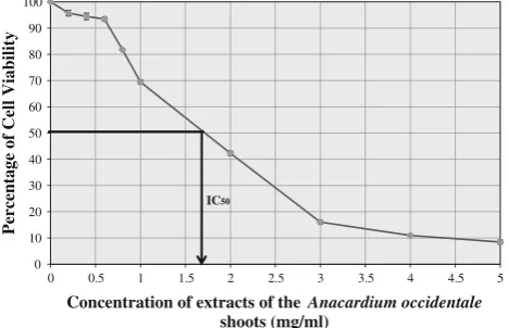
Abstract In this study, the effects of low and high con-centrations of the Anacardium occidentale shoot extracts on gene expression in liver HepG2 cells were investigated. From MTT assays, the concentration of the shoot extracts that maintained 50% cell viability (IC50) was 1.7 mg/ml. Cell viability was kept above 90% at both 0.4 mg/ml and 0.6 mg/ml of the extracts. The three concentrations were subsequently used for the gene expression analysis using Affymetrix Human Genome 1.0 S.T arrays. The microarray data were validated using real-time qRT–PCR. A total of 246, 696 and 4503 genes were significantly regulated (P\0.01) by at least 1.5-fold in response to 0.4, 0.6 and 1.7 mg/ml of the extracts, respectively. Mutually regulated genes in response to the three concentrations included
CDKN3, LOC100289612, DHFR, VRK1, CDC6, AURKB
and GABRE. Genes like CYP24A1, BRCA1, AURKA,
CDC2, CDK2, CDK4 and INSR were significantly
regu-lated at 0.6 mg/ml and 1.7 mg but not at 0.4 mg/ml. However, the expression of genes including LGR5,
IGFBP3, RB1, IDE, LDLR, MTTP, APOB, MTIX, SOD2
andSOD3 were exclusively regulated at the IC50 concen-tration. In conclusion, low concentrations of the extracts were able to significantly regulate a sizable number of genes. The type of genes that were expressed was highly dependent on the concentration of the extracts used. Keywords Anacardium occidentale shoots
Methanol extractsGene expression
cDNA microarray analysisHepG2 cells
Introduction
The cashew plant or Anacardium occidentale L(A. occi-dentale) has many medicinal properties that are beneficial to health. Various scientific evidences have linked the various parts of cashew plant to several biological activi-ties. The stem bark extract had been shown to have anti-bacterial [1], anti-viral [22], anti-diabetic [32] and anti-inflammatory [29] activities. Anti-tumour activity was detected in the cashew gum [30] and nut [42] while anti-ulcerogenic was reported in the cashew leaf extracts [19]. Antioxidant activities were also detected in the nut skin extracts [16]. In addition to the medicinal properties, the fruit of theA. occidentaleis a natural whitening agent that disrupts pigmentation through the inhibition of tyrosinase [21].
In Malaysia, the young leaves or shoots of theA. occi-dentale are widely consumed as salads, and the locals believed its benefits include diabetic control and preven-tion. The extracts of the shoots were found to have potent antioxidant activities [35], were able to inhibit the oxida-tion of LDL and up-regulated LDL receptor activity in cultured HepG2 cells [39]. The antioxidant activities observed in the A. occidentale shoot extracts were attrib-uted to the reported presence of phenolic compounds such as myricetin and quercetin [19, 27]. Intact quercetin glycosides, the most common flavonoids found in human diets, were shown to be absorbed at the small intestine probably through a sodium-dependent glucose transport pathway [9,14]. Once absorbed, quercetin circulates in the plasma in conjugated forms but its antioxidant properties were maintained [25].
Other active compounds found in the crude extracts of the
A. occidentaleleaves include catechin, epicatechin, tetramer of proanthocyanidin and biflavanoids amentoflavone [19] S. KhaleghiA. A. AzizN. RazaliS. M. Junit (&)
Department of Molecular Medicine, Faculty of Medicine, University of Malaya, 50603 Kuala Lumpur, Malaysia e-mail: [email protected]
and agathisflavone [20]. Agathisflavone was reported to be able to induce apoptosis in Jurkat cells (acute lymphoblastic leukaemia cell line) [20] as well as a potent, competitive inhibitor for the GABAA/benzodiazepine receptor [43].
Scientific molecular studies on the effects of the
A. occidentale shoot extracts on cells are still lacking despite its reported antioxidant properties and its use in traditional medicine. We had earlier reported that the methanol extracts of the A. occidentale contained the highest total phenolic content compared to ethyl acetate and hexane extracts [35]. In this study, we explored the effects of the methanol extracts of theA. occidentaleshoots on the expression of genes which could be associated with its antioxidant and medicinal properties.
Materials and methods
Chemicals
All reagents and chemicals used in the experiments were of analytical grade and obtained mostly from Sigma–Aldrich. Solvents used for extraction of plants were purchased from Fisher Scientific. Water used was of Millipore quality (ELGA Purelab Ultra Genetic system).
Preparation of methanol extract of the shoots ofAnacardium Occidentale
In our previous study [35], we reported that the methanol extract of theA. Occidentaleshoots possessed significantly higher antioxidant activities compared to those of the ethyl acetate and hexane extracts. The methanol extracts was subsequently used in this study. Briefly, the shoots were washed, air-dried followed by complete drying in an oven at 40°C. The dried shoots were then ground to powder and then extracted with methanol with a mass to volume ratio of 1:20 (g/mL), at room temperature for 24 h. The resulting extract was filtered and roto-evaporated (Rotavapor R-215) to dryness at 37°C, and the residues were then redissolved in dimethylsulfoxide (DMSO). For the subsequent cell culture experiments, the final concentration of DMSO was kept below 1% to avoid toxicity to the cells.
High-performance liquid chromatography
Acid hydrolyses was conducted on the dried powder of
A. occidentale[3]. Samples (20 mg) were mixed with 50% methanol containing 1.2 M HCl and 20 mM sodium diethyldithiocarbamate as an antioxidant, in reactive vials. The samples were hydrolysed for 2 h at 90°C. Following hydrolysis, samples were centrifuged at 50009gfor 5 min and diluted with distilled water (pH 2.5) prior to analysis
on the HPLC. The hydrolysed samples contained both free flavonoids and aglycones released from conjugated flavo-noids following acid hydrolysis.
The HPLC system used for the flavonoid analyses comprised a Shimadzu system consisting of a system controller, a binary pump (LC 20AC), a manual injector (Rheodyne 7725i manual injector), a column oven (CTO-10AS VP) and a dual channel UV detector (SPD-20A UV–VIS). Absorbance of the samples was monitored at a wavelength of 260 nm. Flavonoids in the samples were separated using a reversed-phase column (NovaPak C18, 150 93.0 mm, i.d 4 lm) (Waters, USA), at a temperature of 40°C. Separation of flavonoids was conducted using a gradient system containing 7–40% acetonitrile in water (pH 2.5) at a flow rate of 0.5 ml/min over 20 min. Standard solutions containing catechin, epicatechin, rutin, genistin, myricetin, morin, quercetin, genistein, kaempferol and isorhamnetin were prepared and injected on the HPLC under the same conditions.
Cell culture
The human hepatoblastoma HepG2 cell line (ATCC, Manassas, VA, USA) was grown in Dulbecco’s modified Eagle’s medium (DMEM) supplemented with 10% foetal bovine serum (Flowlab, Australia), 1% penicillin (Flowlab, Australia) and 1% streptomycin (Flowlab, Australia). Cells were maintained in humidified air with 5% CO2at 37°C. Cell viability analysis using the MTT assay
Cell viability of HepG2 cells in response to treatment with various concentration of the A. occidentale shoot extracts was analysed using an MTT assay as described by Mosmann, 1983 [28] with slight modifications [36]. Briefly, HepG2 cells at a density of 5000 cells per well were seeded in a 96-well ELISA microplate. The cells were incubated at 37°C in 5% CO2for 24 h. After 24 h, increasing concen-trations of the shoots extracts (0.2–5.0 mg/ml) were added into the wells, and the cells were further incubated for 48 h. Following this, MTT reagent (Merck) was added, and the mixture was incubated for 4 h. Next, the mixture in each well was removed, and formazan crystals formed were dissolved in 10ll of 75% isopropanol. Spectrophotometric measurement of the mixture was performed in a microplate-reader (Bio-Rad) at 590 and 620 nm wavelengths. A linear plot of cell viability (%) against the concentrations of plant extracts was constructed.
at 0.4, 0.6 and 1.7 mg/ml. The cells were then incubated at 37°C for 24 h. As a control, cells were incubated in fresh DMEM, in the absence of the extracts. All experiments were performed in triplicate. After 24 h, cells were trypsinized and then precipitated by centrifugation at 2619g for 5 min. Following this, cells were washed with PBS twice before total cellular RNA (tcRNA) was extracted from the treated and untreated cells.
Extraction of total cellular RNA (tcRNA)
tcRNA from both treated and untreated (control) HepG2 cells was isolated and then purified using RNAEasy kit and RNase-free DNAse set (Qiagen) according to the manufacturer’s instructions. The quality of the tcRNA was estimated by measuring the absorbance ratio of 260–280 nm while its integrity was analysed using dena-turing gel electrophoresis. An A260/A280 ratio above 1.8 indicated that the tcRNA was of good quality. The integrity of the tcRNA was indicated by the presence of two distinct bands corresponding to the ribosomal 28S and 18S sub-units, with the intensity of the larger, 28S band approxi-mately twice than that of the smaller, 18S band.
Microarray analysis
Affymetrix Human Gene 1.0 S.T (sense target) arrays were used for the gene expression analysis according to the con-ventional Affymetrix eukaryotic RNA labelling protocols (Affymetrix). Briefly, freshly extracted tcRNA (100 ng) isolated from the treated and untreated HepG2 cells was reversed transcribed to single-stranded sense strand DNA (cDNA) in two cycles using the Whole Transcript (WT) cDNA synthesis, amplification kit and sample clean-up module. The sense strand cDNA was then cleaved into small fragments using a mixture of UDP and apurinic/apyrimidinic endonuclease 1 or APE1. Following this, the fragments were end-labeled with biotinylated dideoxynucleotides using the WT Terminal Labeling kit. The biotinylated fragments (5.5lg) were then hybridized to the Affymetrix Human Gene 1.0 S.T array at 45°C for 16 h in hybridization Oven 640. After hybridization, the arrays were stained and then washed in the Affymetrix Fluidics Station 450 under stan-dard conditions. The stained arrays were scanned at 532 nm using an Affymetrix GeneChip Scanner 3000, and CEL files for each array were generated using the Affymetrix Gene-ChipÒ Operating Software (GCOS). The data were pre-analyzed using Affymetrix Expression Console software. Microarray data normalization and analysis
The CEL files generated were converted to text files and exported to Partek Genomic Suite software to get the whole
list of up-regulated and down-regulated genes. Probeset IDs without any annotation in the Partek software were filtered out. The filtered, whole gene list was then subjected to a one-way analysis of variance (ANOVA) in the Partek Genomic software, to determine significantly expressed sets of genes which was set according toPvalues less than 0.01 (P\0.01) instead ofP\0.05 to avoid false positive results. Significantly expressed genes were then re-filtered to include only those with fold change difference of equal to or greater than 1.5. Additional information on the bio-logical functions of the genes and the genes products was determined from the Gene Ontology (GO) Enrichment tool in the Partek Genomic Suite Software. Information on function of genes can be derived from the Gene Ontology database which provides a structured annotation of genes with respect to molecular function, biological process and cellular component. Further information on GO could be retrieved fromhttp://www.geneontology.org/.
Validation of the DNA microarray data using qRT–PCR
The microarray data were verified using real-time relative quantitative RT–PCR (qRT–PCR) which was performed in a StepOneTMReal-Time PCR System (Applied BioSystem). The same cDNA and primer pairs for the selected up-regulated and down-regulated genes as well as a housekeeping gene, GADPH, as listed in Table1 were used. The PCR amplification was carried out in 0.2 ml MicroAmpÒOptical 8-tube strips in a final volume of 20ll containing a mixture of cDNA (30 ng), reverse and for-ward primers (1lM), pre-prepared Power SYBRÒ Green PCR master mix containing SYBRÒGreen 1 dye,
Ampli-Taq GoldÒDNA Polymerase dNTPs, dUTP, Passive Ref-erence 1 and optimized buffer components. The PCR parameters consisted of 40 cycles of amplification with initial denaturation at 95°C for 15 s, annealing of primers and elongation of the newly synthesized strands at 60°C for 60 s. The PCR mixture was initially held for 10 min at 95°C for AmpliTaq GoldÒ DNA polymerase activation. The Comparative CT Method (DDCT) was chosen for the relative quantitation of gene expression. Each sample type was run in triplicate. mRNA levels of the selected genes were normalized against that of GADPH.
Results
Cell viability analysis
the viability of HepG2 cells in response to the treatment with different concentrations of the methanol extract of
A. occidentaleshoots is shown in Fig.1. The graph shows a tri-phasal response. In the first phase, cell viability was maintained above 90% until 0.6 mg/ml of the extract concentration was reached. In the second phase, cell via-bility decreased steeply to below 20% from 0.6 to 3 mg/ml extract concentrations. In the third phase, the cells barely survived (viability below 10%) beyond 5 mg/ml extract concentration. The concentration of the shoot extract that reduced cell viability by 50% (IC50) was 1.7 mg/ml. Low concentrations of 0.4 and 0.6 mg/ml as well as the IC50 concentration were subsequently chosen for the gene expression analysis.
HPLC analysis
HPLC analyses of the hydrolysed samples revealed the presence of quercetin and kaempferol (Fig.2a) and the absence of catechin, epicatechin, rutin, genistin, genistein, myricetin, morin and isorhamnetin. The quercetin and kaempferol peaks were confirmed by comparing retention times of the peaks with the standards (Fig. 2b) which was run under the same conditions as the samples. The presence of kaempferol has not been reported previously.
Microarray analysis: normalization and visualization of data
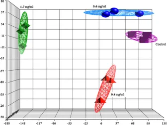
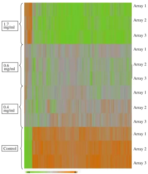
Figure3 shows the principal component analysis (PCA) plot of the microarray data for each of the array for the control and samples treated with 0.4, 0.6 and 1.7 mg/ml of the shoot extracts. The three arrays for the control samples were grouped together but separately from those of the treated samples. Figure4 shows hierarchical clustering of the microarray data generated from each of the arrays where the reproducibility pattern correlates with that shown by the PCA plot.
Venn diagram in Fig. 5shows the number of genes that were significantly regulated (P\0.01) by at least 1.5-fold. The total number of genes regulated by 0.4, 0.6 and 1.7 mg/ml of the methanol extracts ofA. occidentalewere 246, 696 and 4,503, respectively. An increase in concen-tration by 1.5-fold from 0.4 to 0.6 mg/ml was able to increase the number of regulated genes by threefold (65%). In addition, increasing the concentration from 0.4 to 1.7 mg/ml increased the number of regulated genes by 18-fold (95%). Figure5also shows that a total of 94 genes were mutually regulated in response to the three
Table 1 Primer sequences for the selected genes used for validation of the microarray data using real-time relative quantitative PCR (qRT–PCR)
Gene name (Genebank ID) Primer sequences Product size (bp)
DHFR(NM_000791) Forward: 50CATGGTCTGGATAGTTGGTGGC 30
Reverse: 50GTGTCACTTTCAAAGTCTTGCATG 30 108
TYMS(NM_001071) Forward: 50ATCAAGGGATCCACAAATGA 30
Reverse: 50GGTCAACTCCCTGTCCTGAA 30
205
LIPC(NM_000236) Forward: 50CAAGTGCCCTTGGACAAAGC 30
Reverse: 50TGACAGCCCTGATTGGTTTCT 30
130
CYP24A1(NM_000782) Forward: 50CTCATGCTAAATACCCAGGTG 30
Reverse: 50TCGCTGGCAAAACGCGATGGG 30
300
PLAUR(NM_002659) Forward: 50TGCGGTGCATGCAGTGTAAGAC 30
Reverse: 50TCAAGCCAGTCCGATAGCTCAG 30
183
PLCXD1(NM_018390) Forward: 50ACGAGTACCTGGTCGCCTGTAT 30
Reverse: 50CATAGGAGACGATGACCTGTTGG 30 117
SQSTM1(NM_003900) Forward: 50CCAGTGACGAGGAATTGACAA 30
Reverse: 50CATCGCAGATCACATTGGGG 30
156
0 10 20 30 40 50 60 70 80 90 100
0 0.5 1 1.5 2 2.5 3 3.5 4 4.5 5
Percentage of Cell Viability
Concentration of extracts of the Anacardium occidentale
shoots (mg/ml) IC50
concentrations where 5 were up-regulated and 89 were down-regulated. Ninety-eight genes were mutually regu-lated in response to the 0.4 and 0.6 mg/ml extracts. On the other hand, 178 were mutually regulated in response to the 0.4 and 1.7 mg/ml extracts. A larger number of genes, with a total of 571, were regulated when cells were treated with 0.6 and 1.7 mg/ml of the extracts.
Mutually regulated genes in response to the three con-centrations included CDKN3, LOC100289612, DHFR,
VRK1, CDC6, AURKB, CYP2S1 and GABRE (Table2).
Genes like CYP24A1, CDH2, E2F5, BRCA1, BRCA2,
AURKA, CDC2 (CDK1), CDK2, CDK4, CHECK1,
CCNA2, ACAT, IGFBP1, DUSP5 andINSR were
signifi-cantly regulated at 0.6 and 1.7 mg/ml but not at 0.4 mg/ml. In addition, the expressions of genes such as the LGR5,
IGFBP3, RB1, IDE, LDLR, MTTP, APOB, SCP2, MTIX, SODandSOD3were exclusively regulated at the 1.7 mg/ ml dose. Amongst the highly significantly suppressed genes were CYP24A1, LGR5,CDH2andDHFRby 27.8-, 16.4-, 15.5-, 10.0-fold, respectively. On the other hand, amongst the highly induced genes were the DUSP5,
IGFBP 3, IGFBP1, LDLR and INSR by 9.1-, 8.0-, 4.2-, 3.6- and 2.6-fold, respectively (Table2).
Other genes that were being significantly regulated in response to the 1.7 mg/ml shoot extracts were those asso-ciated with cell cycle check points either directly or indi-rectly. These included the CDK5, CDK6, CCNB1 and 2,
CCNE1 and 2, CCNH, CDKN1A (p21/Cip1), CDKN1C
(p57/Kip2), CDKN2B (p15), CDKN2D (p19), CDKN3,
RBL1, RBL2, NSUN6, NOP2, DAPK1, PAK2, HDAC2and
G2E3. Genes coding for other ubiquitin ligase isoforms,
UBE2C and UBE3B, were also aberrantly expressed. In
addition, genes associated with the Wnt/b-catenin signal-ling pathway including theWnt6,FZDs 1, 4,6, 7, 8, 9and
10, CDH1, CTNNA2, DKK1, APC, NUCKS1, CSNK1G2,
CSNK1G3 and TCF were all aberrantly expressed. In
addition, genes associated with cancers, BRIP1, BAP, BRCC3, RAS, SOS1, STAT2,were all down-regulated (data not shown).
1
2
20.0 21.0 22.0 23.0 24.0
mins (a)
20.0 22.5
1
2
mins (b)
Fig. 2 Gradient reverse phase HPLC analysis of flavonoids in the shoots ofA. occidentaleand the flavonoid standards. HPLC analysis was performed on the hydrolysed samples ofA. occidentale(Fig.2a) and the flavonoid standards (Fig.2b). Flavonoids were separated on a NovaPak C18 reversed-phase column (15093.0 mm i.d, 4lm), using a linear gradient system of 7–40% acetonitrile in water (pH 2.5), at a flow rate of 0.5 ml/min. Absorbance was measured at a wavelength of 260 nm. 1:quercetin; 2:kaempferol
Gene ontology (GO): biological interpretation
Gene ontology analysis of the products of the significantly regulated genes in response to the 3 concentrations of the
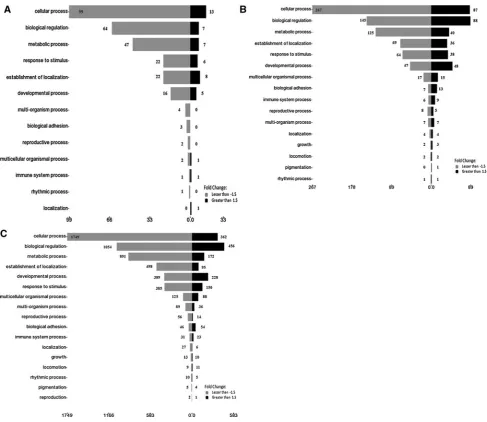
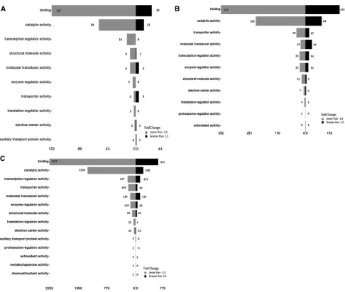
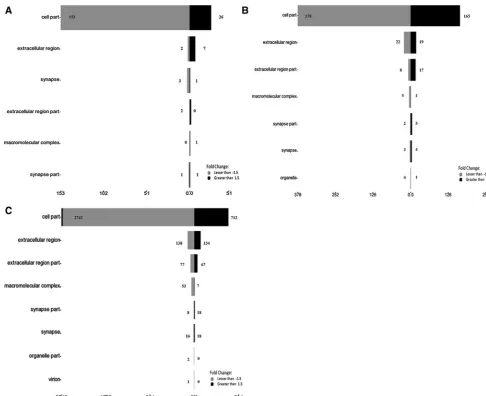
A. occidentaleshoot extracts is shown in Figs.6,7and8. The data are presented according to the following catego-ries; biological process (Fig.6), molecular function (Fig.7) and cell component (Fig.8). Selected significantly down-regulated and up-regulated genes in each of the category are shown in Table3.
Figures6,7and8show that most of the genes (and the subsequent gene products) are involved in cellular pro-cesses, biological regulations and metabolic processes in response to the three concentrations of the extracts. A total of 18 genes that are involved in localization, growth, locomo-tion and pigmentalocomo-tion were regulated in response to the 0.6 mg/ml but not the 0.4 mg/ml extracts. As the concen-tration was increased from 0.6 to 1.7 mg/ml, the number of genes rose from 18 to 88 in the same 3 subcategories. In addition, 3 genes that are involved in reproduction were regulated only in the presence of 1.7 mg/ml of the extracts. Genes likeCYP24A1andDHFRare involved in cellular as well as metabolic processes (Table3A). In addition, under the ‘‘Molecular function’’ category, the majority of genes that were regulated in response to the 3 concentrations were involved in binding, followed by catalytic activity and transcriptional regulation activity (Fig.7). Table3B lists
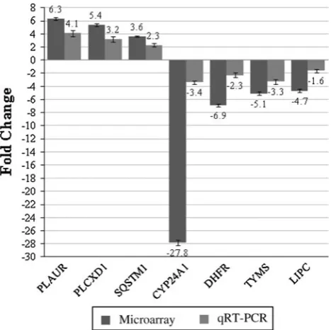
selected genes such asINSRandLDLRthat are involved in binding, MAOB in catalytic activity andRB1in transcription regulator activity. The majority of the gene products were found as cell part, in extracellular region/part and in syn-apses (Fig. 8), and the selected genes are listed in Table3C. Validation of the microarray data using qRT–PCR The microarray data were validated by quantitating selec-ted significantly regulaselec-ted genes, PLAUR, PLCXD1,
SQSTM1, CYP24A1, DHFR, TYMS and LIPC using
real-time RT–PCR (qRT–PCR). All data were normalized to the reference gene, GAPDH. As shown in Fig.9, the expres-sions of thePLAUR, PLCXD1andSQSTM1genes were all up-regulated while those of CYP24A1, DHFR, TYMS and
LIPC were down-regulated. The expression patterns obtained through qRT–PCR analysis were consistent with those of the microarray results.
Discussion and conclusion
In Malaysia, the young leaves or shoots of the A.occiden-taleare widely consumed as salads and the locals believed Control
0.6 mg/ml 1.7 mg/ml
0.4 mg/ml
Array 1
Array 2
Array 3
Array 1
Array 2
Array 3
Array 1
Array 2
Array 3
Array 1
Array 2
Array 3
Fig. 4 Hierarchical cluster analysis of genes showing the differential expression of genes in HepG2 cells in response to the treatment with 0.4, 0.6 and 1.7 mg/ml of extracts ofA. occidentaleshoots
1.7 mg/ml vs control
0.4 mg/ml vs control 0.6 mg/ml vs control
its benefits include diabetes control or prevention. The methanol extracts of the shoots were found to have potent antioxidant activities [35]. Bioactive compounds found in the A. occidentale shoot extracts that could be linked to the antioxidant activities and other medicinal properties included myricetin and quercetin [19, 27], amentoflavone [19] and agathisflavone [20]. Dietary antioxidants could be absorbed through the intestine, albeit in small quantities. Once in the circulation, they are quickly metabolized
[14,44] but the antioxidant properties were retained [25]. Our group had reported that low concentration of crude extract of antioxidant-rich T. indica(0.3 mg/ml) was able to significantly regulate a sizable number of genes in HepG2 cells [36]. In this study, based on the MTT assays, cells showed more than 90% viability at 0.4 and 0.6 mg/ml. The IC50 concentration was found to be 1.7 mg/ml. This study was aimed to (1) investigate the effects of low and high concentration of theA. occidentale shoot extracts on
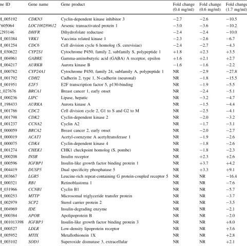
Table 2 Selected significantly expressed genes in HepG2 cells in response to treatment with 0.4, 0.6 and 1.7 mg/ml of the extracts of A. occidentaleshoots
Gene ID Gene name Gene product Fold change
(0.4 mg/ml)
Fold change (0.6 mg/ml)
Fold change (1.7 mg/ml)
NM_005192 CDKN3 Cyclin-dependent kinase inhibitor 3 -2.7 -2.6 -10.5
AY605064 LOC100289612 Arsenic transactivated protein 1 -3.0 -3.6 -10.2
AK293146 DHFR Dihydrofolate reductase -2.4 -2.4 -10.0
NM_003384 VRK1 Vaccinia related kinase 1 -2.3 -2.6 -6.7
NM_001254 CDC6 Cell division cycle 6 homolog (S. cerevisiae) -2.4 -2.7 -4.3
NM_030622 CYP2S1 Cytochrome P450, family 2, subfamily S, polypeptide 1 ?1.8 ?2.3 ?3.5
NM_004961 GABRE Gamma-aminobutyric acid (GABA) A receptor, epsilon ?1.6 ?2.1 ?2.7
NM_004217 AURKB Aurora kinase B -1.6 -1.6 -2.2
NM_000782 CYP24A1 Cytochrome P450, family 24, subfamily A, polypeptide 1 NR -2.9 -27.8
NM_001792 CDH2 Cadherin 2, type 1, N-cadherin (neuronal) NR -1.8 -15.5
NM_001951 E2F5 E2F transcription factor 5, p130-binding NR -1.9 -5.5
NR_027676 BRCA1 Breast cancer 1, early onset NR -2.4 -5.1
NM_000236 LIPC Lipase, hepatic NR -3.2 -4.7
NM_198433 AURKA Aurora kinase A NR -1.5 -4.4
NM_001786 CDC2 Cell division cycle 2, G1 to S and G2 to M NR -2.5 -4.1
NM_001798 CDK2 Cyclin-dependent kinase 2 NR -2.0 -3.2
NM_001237 CCNA2 Cyclin A2 NR -1.7 -3.1
NM_000059 BRCA2 Breast cancer 2, early onset NR -2.0 -2.7
NM_000019 ACAT1 Acetyl-coenzyme A acetyltransferase 1 NR -1.9 -2.6
NM_000075 CDK4 Cyclin-dependent kinase 4 NR -1.8 -2.6
NM_001274 CHEK1 CHK1 checkpoint homolog (S. pombe) NR -1.8 -2.3
NM_000208 INSR Insulin receptor NR ?2.3 ?2.6
NM_000596 IGFBP1 Insulin-like growth factor binding protein 1 NR ?3.7 ?4.2
NM_004419 DUSP5 Dual specificity phosphatase 5 NR ?3.3 ?9.1
NM_003667 LGR5 Leucine-rich repeat-containing G protein-coupled receptor 5 NR NR -16.4
NM_000321 RB1 Retinoblastoma 1 NR NR -7.6
NM_031966 CCNB1 Cyclin B1 NR NR -5.7
NM_000253 MTTP Microsomal triglyceride transfer protein NR NR -3.7
NM_002979 SCP2 Sterol carrier protein 2 NR NR -3.5
NM_004969 IDE Insulin-degrading enzyme NR NR -2.1
NM_000384 APOB Apolipoprotein B NR NR -2.0
NM_001013398 IGFBP3 Insulin-like growth factor binding protein 3 NR NR ?8.0
NM_000527 LDLR Low-density lipoprotein receptor NR NR ?3.6
NM_005952 MTIX Metallothionein 1X NR NR ?2.8
NM_003102 SOD3 Superoxide dismutase 3, extracellular NR NR ?2.1
the expression of genes in liver HepC2 cells and (2) identify genes that could be associated with the medicinal properties of the shoot extracts.
HPLC analyses showed the presence of quercetin in the extracts of theA. occidentaleshoots that confirmed earlier findings by other researchers [19,27]. In addition, we also detected the presence of kaempferol which has not been reported previously. Kaempferol possessed significant antioxidant activities [34] and has cancer chemopreventive properties towards several tumour cell lines including lung and leukaemic cell lines [8,31].
In this study, cDNA microarray analysis showed that the extracts of theA. occidentaleshoots at a low concentration of 0.4 mg/ml was able to significantly up-regulated (P\0.01) a total of 248 genes by at least 1.5-fold. Amongst the down-regulated genes were those encoding
arsenic transactivated protein (LOC100289612), vaccinia-related kinase 1 (VRK1),dihydrofolate reductase (DHFR),
cell division cycle 6 homolog (S.cerevisiae) (CDC6),
cyclin-dependent kinase inhibitor 3 (CDKN3) and aurora kinase B (AURKB). Increasing the concentrations from 0.4 to 0.6 mg/ml led to an increase in the number of regulated genes from 248 to 696. Amongst the 696 genes were
CYP24A1, CDH2, E2F5, BRCA1, BRCA2, AURKA, CDC2
(CDK1), CDK2, CDK4, CHECK1, CCNA2, ACAT,
IGFBP1, DUSP5 and INSR. These genes were also
expressed in response to the IC50 concentration, but as expected, the fold change was much larger compared to that of the 0.6 mg/ml. At an IC50concentration of 1.7 mg/ml, a total of 4286 genes were significantly regulated. In addition, the expressions of genes such as theLGR5,IGFBP3,RB1,
IDE,LDLR, MTTP,APOB, SCP2,MTIX, SODandSOD3
were exclusively regulated at the 1.7 mg/ml dose. Inter-estingly, for all three concentrations, the down-regulated genes were three times as many as those that were up-regulated (full data are not shown) as indicated in the hierarchical analysis. Mutually regulated genes in response to the three concentrations includedCDKN3, LOC100289612,
DHFR, VRK1, CDC6, AURKB, CYP2S1 and GABRE
(Table2).Amongst the highly significantly suppressed genes
wereCYP24A1, LGR5, CDH2and DHFR by 27.8-, 16.4-,
15.5-, 10.0-fold, respectively. On the other hand, amongst the highly induced genes were the DUSP5, IGFBP 3, IGFBP1, LDLR and INSR by 9.1-, 8.0-, 4.2-, 3.6- and 2.6-fold, respectively.
CYP24A1gene codes for the hepatic enzyme, CYP24A1 or 24-hydroxylase which is the rate limiting enzyme in the catabolism of the active form of vitamin D, 1a,25-(OH)2D3 or calcitriol [18, 23]. 1a, 25-(OH)2D3, synthesized in the
kidney by the CYP27B1, promotes dietary absorption of calcium and phosphate as well as maintain the levels of the two minerals. There are increasing evidences that indi-viduals with low serum vitamin D have a higher risk of developing various types of cancers [18,23] and myocar-dial diseases [33]. In addition, 1a, 25-(OH)2 D3 is also important in regulating cell cycle check points as well as controlling multiple signalling pathways including those of the MAPK/ERK, PI3 K/AKT, Wnt and TGF-b [5]. The
CYP24A1gene has been reported to act as an oncogene and its overexpression was detected in cancers of the colon, ovary and lung [2,18,23]. In vivo studies have indicated that exposing cancer cells to a high concentration of the active metabolites of vitamin D stopped the cells from progressing. This occurred via a mechanism affecting cell cycle and increasing apoptosis, ultimately slowing or stopping growth of the tumour [45]. The shoot extracts of
the A. Occidentale were able to highly suppress the
CYP24A1 gene; hence, it has the potential to be used synergistically with vitamin D to maintain the bioavail-ability and bioactivity of the latter.
Apart from theCYP24A1gene, LGR5andCDH2 were also highly suppressed, by 16- and 15-fold respectively at the IC50concentration of the shoot extracts. The leucine-rich repeat-containing G-protein-coupled receptor 5 (LGR5) belongs to the G-protein-coupled receptor (GPCR) superfamily [15]. LGR5 had been reported to be a marker of adult stem cells where the LGR5 gene transcription is under the control of the canonical or beta-catenin Wnt signalling pathway [11]. This pathway, which is involved in embryogenesis and normal physiological processes, is critical in the regulation of adult stem cells [11, 37]. Dysregulation of this pathway is linked to cancers [37]. An overexpression of LGR5 was observed in many types of
cancers including those of the colon [26,46], oesophagus [4], ovary [26] and the hepatocytes [47]. In this study, genes associated with the Wnt/b-catenin signalling path-way were also regulated at the IC50 concentration, albeit modestly. These included the Wnt6,FZDs 1, 4,6, 7, 8, 9
and 10, CDH1, CDH2, CTNNA2, DKK1, APC, NUCKS1,
CSNK1G2,CSNK1G3andTCF(data not shown).
One of the most interesting observations of this study was the fact that the shoot extracts were able to directly regulate a spectrum of genes involved in the G1 as well as G2 cell cycle check points. These included the CDK1 (CDC2), CDK2, CDK4, CDK5, CDK6, CCNA2, CCNB1
and 2, CCNE1 and 2, CCNH, CDKN1A (p21/Cip1),
CDKN1C (p57/Kip2), CDKN2B (p15), CDKN2D (p19),
CDKN3, RB1, RBL1, RBL2, NSUN6, NOP2, AURKA,
AURKB, DAPK1, PAK2, E2F5, HDAC2, VRK1, CREBand
G2E3. Genes coding for other ubiquitin ligase isoforms,
Table 3 Gene ontology analysis of selected significantly regulated genes (A) Gene ontology
(Biological process)
Selected down-regulated genes Selected up-regulated genes
Cellular process CYP24A1, DHFR, CDH1, CDH2, CDC2, CDK2, CDK4, CDK5, CDK6, CDKN3, CCNA2, CCNB1, CCNB2, CCNE1, CCNE2, CHEK1, RB1, AURKA, AURKB, BRCA1, BRCA2, IDE, LIPC, MTTP, SCP2, APOB, ACAT1
DUSP5, INSR, IRS2, SOD2, SOD3, LDLR
Biological regulation
CDH1, CDC2, CDK2, CDK4, CDK5, CDK6, CDKN3, CHEK1, CCNA2, CCNB1, CCNB2, CCNE1, CCNE2, E2F4, AURKA, RB1, CKS1B, PAK2, BRCA1, BRCA2, CSNK1G3, FZD4, FZD6, LIPC, APOB, APOBEC3F, APOH, MTTP, IDE
CDH4, CDKN2B, CDKN1A, APC2, IRS2, WNT6, FZD1, FZD7, FZD8, FZD9, FZD10, IGFBP1, IGFBP2, IGFBP3, IGFBP6, INSR, LDLR, SOD2
Metabolic process CDH1, CCNH, CHEK1, CDK5, E2F4, CYP24A1, RB1, DHFR, BRCA1, LIPC, MAOB, MTTP, SCP2, ACAT1, APOB
APC2, SOD2, SOD3, INSR, LDLR
Response to stimuli
BRCA1, BRCA2, CHEK1, CDK5 IRS2, CDKN2D, SOD2, SOD3, CDKN2B, CDKN1A, MT1X
Establishment of localization
LIPC, MTTP, SCP2, APOB LDLR
Developmental process
CDH2, RB1, CCNB2, BRCA1, SCP2, FZD6, CDK6, BRCA2, CCNF, DKK1, CDK5 E2F4, IDE, APOB, CDH1, FZD4
FZD7, CDK5R1, FZD9, IRS2, IGF2, CDKN2D, FZD10, CDKN1C, SOD2, FZD8, WNT6, FZD1, INSR
Multi-organism process
RB1 INSR, LDLR
Biological adhesion
CDH2, FZD6, CDK5, CDH1 CDH4
Reproductive process
SCP2, BRCA2, CHEK1, APOB
Multicellular organismal process
APOH, ACAT1, CDK5, E2F4 FZD9, LDLR
Rythmic process CDK4 –
Growth CCNB2, BRCA2 –
Pigmentation – SOD2
(B) Gene ontology (Molecular function)
Selected down-regulated genes Selected up-regulated genes
Binding CYP24A1, CDH2, CDKN3, RB1, DHFR, CCNB1, CCNB2, BRCA1, LIPC, AURKA, CDC2, BRCC3, MTTP, SCP2, CDK2,CCNA2, APOH, CDK6, RBL2, BRCA2, ACAT1, CDK4, CHEK1, CCNE1, AURKB, CDK5, E2F4, IDE, APOB, CDH1, FZD4, CSNK1G2, CCNE2
CDH4, IGFBP2, IRS2, FZD10, SOD2, FZD8, WNT6, SOD3, IGFBP6, FOXO3, FZD1, INSR, MT1X, LDLR, IGFBP3
Catalytic activity CYP24A1, CDKN3, DHFR, BRCA1, LIPC, AURKA, CDC2, MAOB, SCP2, CDK2, CDK6, BRCA2, ACAT1, CCNH, CDK4, CHEK1, AURKB, CDK5, IDE
SOD2, SOD3, INSR, DUSP5
Transcription regulator activity RB1, BRCA1, BRCA2, E2F4, CDH1 FOXO3
Structural molecular activity – WNT6
Molecular transducer activity FZD6, CDK5, IDE, FZD4 FZD7, FZD9, IRS2, FZD10, FZD8, WNT6, FZD1, INSR, MT1X, LDLR
Enzyme regulator activity CDH1, CCNE1 IGFBP3
Transporter activity LIPC, MTTP, APOB LDLR
Electron carrier activity CYP24A1, MAOB –
UBE2CandUBE3B,was also aberrantly expressed. VRK1 is involved in the regulation of DNA replication through the phosphorylation of CREB leading to the regulation of
CCND1gene expression [17].
Aurora kinases comprise three members, Aurora A, Aurora B and Aurora C. Aurora-A is transcriptionally regulated by E2F3 during a cell cycle. E2F3 induces
Aurora-A expression by binding directly toAurora-A pro-moter and subsequently stimulates the propro-moter activity [12]. Both could thus be an important target for cancer intervention [12]. In addition, aurora-B has been shown to be overexpressed in many cancers including breast cancers [10]. The methanol extracts of theA. occidentaleshoots at the IC50concentration, suppressed theAurora Aand Aur-ora B, but notAurora C, by fourfold and twofold respec-tively. An RNA methyltransferase, NSUN2 had been shown to be a novel substrate for Aurora B, which con-tained a NOL/NOP/sun domain [38]. In this study, the extracts were able to suppress bothAurora AandAurora B
as well asNOP2andNSUN6,suggesting its potential as an anti-cancer agent.
Insulin-like growth factor binding protein-3 (IGFBP3) inhibits the growth of non-small cell lung cancer (NSCLC) cells. IGFBP3 overexpression inhibits the phosphorylation of Akt and glycogen synthase kinase-3 beta and the activity of MAPK, all three are activated by IGF-mediated sig-nalling pathways that have mitogenic and anti-apoptotic properties and have been implicated in the development of
Fig. 9 Validation of the microarray data using semi-quantitative RT–PCR (qRT–PCR). A few genes that were significantly regulated in HepG2 cells in response to treatment with extracts of the A. occidentale shoots were selected namely PLAUR, PLCXD1, SQSTM1, CYP24A1, DHFR, TYMS and LIPC using real-time RT–PCR. All data were normalized to the reference gene,GAPDH. The expressions of thePLAUR, PLCXD1andSQSTM1genes were all up-regulated while those ofCYP24A1, DHFR, TYMSandLIPCwere down-regulated. The expression patterns obtained through real-time RT–PCR were consistent with the microarray results
Table 3 continued (C) Gene ontology (Cellular component)
Selected down-regulated genes Selected up-regulated genes
Cell part CYP24A1, LGR5, CDH2, RB1, CCNB1, CCNB2, E2F5, TYMS, BRCA1, LIPC, ATG4C, SOS1, AURKA, G2E3, CDC2, CHEK2, BRCC3, MAOB, MTTP, SCP2, GAS2, MTHFD1, CDK2, CCNA2, APOH, CKS1B, RBL1, UBE2C, FZD6, BARD1, PAK2, CDK6, RBL2, BRCA2, CCNF, ACAT1, CCNH, CDK4, PAK1IP1, NUCKS1, DKK1, STAT2, CHEK1, CCNE1, AURKB, CDK5, E2F4, DAPK1, IDE, TOP2B, BCCIP, CSNK1G3, APOB, CDH1, TET1, APOBEC3F, FZD4, ACAT2, CSNK1G2, CCNE2, CDK7, LRP1
FZD7, CTNNA2, CDK5R1, CDH4, APC2, FZD9, IRS2, CDKN2D, FZD10, CDKN1C, AQP6, SOD2, FOLR3, FZD8, AQP12A, SOD3, FOXO3, FZD1, CDKN2B, CDKN1A, LRP12, INSR, LDLR, AQP3, EGLN3, IGFBP3, DUSP5
Extracellular region LIPC, ATG4C, APOH, DKK1, APOB APC2, IGFBP2, IGF2, FOLR3, WNT6, SOD3, IGFBP6, IGFBP4, IGFBP1, IGFBP3
Synapse CDH2, CDK5 CDK5R1
Extracellular region part LIPC, APOH, IDE, APOB APC2, IGFBP2, IGF2, WNT6, SOD3, IGFBP4, IGFBP1, IGFBP3
Macromolecular complex – APC2
Synapse part SOS1 –
lung cancer [24]. Nuclear IGFBP3 induces apoptosis and is targeted to ubiquitin/proteosome-dependent proteolysis. IGFBP3 degradation is dependent on active ubiquitin-E1 ligase [41].
The shoot extracts also up-regulatedLDLR gene which correlated with findings by Salleh et al.[39] who reported that the cashew shoots were able to increase LDLR activity in cultured HepG2 cells. Other genes associated with lipid metabolism including LIPC, ACAT1, MTTP, APOB and SCP2 were down-regulated. LDL-R is responsible for the internalization of cholesterol-rich lipoprotein, LDL, from the blood circulation through the recognition of its ApoB by the receptor [6]. SCP2 gene expression was reported to be enhanced by oxidized LDL [13] which could be ingested by macrophages leading to the formation of foam cells, a critical step in atheroscle-rosis. ACAT is responsible to convert free cholesterol to cholesteryl ester in tissues. ACAT1 is the main isoenzyme in the neuronal brain [7], and its presence is associated with certain forms of Alzheimer disease. MTTP encodes microsomal triacylglycerol transfer protein (MTP) which is required for the assembly of nascent chylomicrons and VLDL while hepatic lipase encoded by LIPC does not only hydrolyses triacylglycerols and phospholipids in cir-culating plasma lipoproteins, it also regulates lipoprotein uptake by cells [40]. Except for theACAT1, the expression of the LDLR, MTTP, SCP2 and APOB genes was only observed in response to the IC50 concentration, not those of 0.4 and 0.6 mg/ml suggesting a concentration-depen-dent expression of the genes.
The anti-diabetic properties of the shoot extracts could be linked to the up-regulation of the genes coding for the insulin receptor and the down-regulation of the insulin-degrading enzyme (IDE).
The hypolipidaemic, anti-diabetic and anti-cancer properties could be attributed to the presence of quercetin and kaempferol in the shoot extracts of theA. occidentale. Quercetin and kaempferol are normally present in nat-ure as glycosides. Free and glycoside forms of the two flavonoids are absorbed in the intestine (9, 14, 48) and are found conjugated with glucuronides or sulphates in the blood circulation. The concentrations of the extracts selected in the study were based on MTT assays whereby cell viability was maintained above 90% at the extract concentrations of 0.3 and 0.4 mg/ml. The concentrations used were probably not a true reflection of the flavonoids levels in the plasma as it was reported that the levels of free and conjugated flavonoids such as kaempferol in blood was in the range of 1.7–6.1lg/ml after oral inges-tion of 25–50 mg/kg body weight [48]. However, when using an in vitro model like HepG2 cells, sometimes it is necessary to use higher concentrations of extracts to see significant changes in gene expression [36]. Nevertheless,
this in vitro study is still useful to provide preliminary information on the possible molecular mechanisms in relation to the medicinal properties of the shoot extracts of
A. occidentale. Further confirmation using an in vivo model would need to be carried out to corroborate the in vitro findings.
Conclusion
In conclusion, low concentrations of the shoot extracts of
A. occidentalewere able to significantly regulate a sizable number of genes in HepG2 cells. However, the type of the expressed genes was highly dependent on the concentration of the extracts used. Amongst the genes that were signifi-cantly regulated were those involved in regulating cell cycle check points, apoptosis and cell proliferation, lipo-protein metabolism and insulin signalling suggesting the potential of the plant to act as anti-cancer, hypolipidaemic and anti-diabetic agents.
Acknowledgments This study was funded by the following research grants: E-Science Fund (12-03-02-2061) from the Ministry of Science, Technology and Innovation Malaysia (MOSTI), FRGS (FP004/2003C) from the Ministry of Higher Education Malaysia (MOHE), the UMRG (RG014/09AFR) and Research University Grants (SF076-2007A & FR113/2007A) from the University of Malaya. We would like to thank Mr Siah Eng Tian for the technical assistance on the microarray analysis.
Open Access This article is distributed under the terms of the Creative Commons Attribution Noncommercial License which per-mits any noncommercial use, distribution, and reproduction in any medium, provided the original author(s) and source are credited.
References
1. Akinpelu DA (2001) Antimicrobial activity of Anacardium occidentale bark. Fitoterapia 72:286–287
2. Anderson MG, Nakane M, Ruan X, Kroeger PE, Wu-Wong JR (2006) Expression of VDR and CYP24A1 mRNA in human tumors. Cancer Chemother Pharmacol 57:234–240
3. Aziz AA, Edwards CA, Lean ME, Crozier A (1998) Absorption and excretion of conjugated flavonols, including quercetin-4’-O-beta-glucoside and isorhamnetin-4’-O-quercetin-4’-O-beta-glucoside by human volunteers after the consumption of onions. Free Radic Res 29:257–269
4. Becker L, Huang Q, Mashimo H (2010) Lgr5, an intestinal stem cell marker, is abnormally expressed in Barrett’s esophagus and esophageal adenocarcinoma. Dis Esophagus 23:168–174 5. Brown AJ, Slatopolsky E (2008) Vitamin D analogs: therapeutic
applications and mechanisms for selectivity. Mol Aspects Med 29:433–452
6. Brown MS, Goldstein JL (1986) A receptor-mediated pathway for cholesterol homeostasis. Science 232:34–47
8. Dimas K, Demetzos C, Mitaku S, Marselos M et al (2000) Cytotoxic activity of kaempferol glycosides against human leu-kaemic cell lines in vitro. Pharmacol Res 41:83–86
9. Gee JM, DuPont S, Rhodes MJC, John IT (1998) Quercetin glucosides interact with the intestinal glucose transport pathway. Free Radic Biol Med 25:19–25
10. Gully CP, Zhang F, Chen J, Yeung JA et al (2010) Antineoplastic effects of an Aurora B kinase inhibitor in breast cancer. Mol Cancer 9:42
11. Haegebarth A, Clevers H (2009) Wnt signaling, lgr5, and stem cells in the intestine and skin. Am J Pathol 174:715–721 12. He L, Yang H, Ma Y, Pledger WJ et al (2008) Identification of
Aurora-A as a direct target of E2F3 during G2/M cell cycle progression. J Biol Chem 283:31012–31020
13. Hirai A, Kino T, Tokinaga K, Tahara K et al (1994) Regulation of sterol carrier protein 2 (SCP2) gene expression in rat peritoneal macrophages during foam cell formation. A key role for free cholesterol content. J Clin Invest 94:2215–2223
14. Hollman PC, de Vries JH, van Leeuwen SD, Mengelers MJ, Katan MB (1995) Absorption of dietary quercetin glycosides and quercetin in healthy ileostomy volunteers. Am J Clin Nutr 62:1276–1282
15. Hsu SY, Liang SG, Hsueh AJ (1998) Characterization of two LGR genes homologous to gonadotropin and thyrotropin receptors with extracellular leucine-rich repeats and a G protein-coupled, seven-transmembrane region. Mol Endocrinol 12:1830–1845
16. Kamath V, Rajini PS (2007) The efficacy of cashew nut (Ana-cardium occidentale L.) skin extract as a free radical scavenger. Food Chem 103:428–433
17. Kang TH, Park DY, Kim W, Kim KT (2008) VRK1 phosphor-ylates CREB and mediates CCND1 expression. J Cell Sci 121:3035–3041
18. King AN, Beer DG, Christensen PJ, Simpson RU, Ramnath N (2010) The vitamin D/CYP24A1 story in cancer. Anticancer Agents Med Chem 10:213–224
19. Konan NA, Bacchi EM (2007) Antiulcerogenic effect and acute toxicity of a hydroethanolic extract from the cashew (Anacardium occidentaleL.) leaves. J Ethnopharmacol 112:237–242 20. Konan NA, Lincopan N, Collantes Dı´az IE, de Fa´tima Jacysyn J,
Tanae Tiba MM, Pessini Amarante Mendes JG, Bacchi EM, Spira B (in press) Cytotoxicity of cashew flavonoids towards malignant cell lines. Exp Toxicol Pathol Article. doi:10.1016/j.etp.2010. 10.010
21. Kubo I, Kinst-Hori I, Yokokawa Y (1994) Tyrosinase inhibitors from Anacardium occidentale fruits. J Nat Prod 57:545–551 22. Kudi AC, Myint SH (1999) Antiviral activity of some Nigerian
medicinal plant extracts. J Ethnopharmacol 68:289–294 23. Lechner D, Kallay E, Cross HS (2007) 1alpha,
25-dihydroxyvi-tamin D3 downregulates CYP27B1 and induces CYP24A1 in colon cells. Mol Cell Endocrinol 263:55–64
24. Lee HY, Chun KH, Liu B, Wiehle SA et al (2002) Insulin-like growth factor binding protein-3 inhibits the growth of non-small cell lung cancer. Cancer Res 62:3530–3537
25. Manacha C, Moranda C, Crespya V, Demigne´a C, Texierb O, Re´ge´ratb F, Re´me´sya C (1998) Quercetin is recovered in human plasma as conjugated derivatives which retain antioxidant prop-erties. Febs Lett 426:331–336
26. McClanahan T, Koseoglu S, Smith K, Grein J et al (2006) Identification of overexpression of orphan G protein-coupled receptor GPR49 in human colon and ovarian primary tumors. Cancer Biol Ther 5:419–426
27. Miean KH, Mohamed S (2001) Flavonoid (myricetin, quercetin, kaempferol, luteolin, and apigenin) content of edible tropical plants. J Agric Food Chem 49:3106–3112
28. Mosmann T (1983) Rapid colorimetric assay for cellular growth and survival: application to proliferation and cytotoxicity assays. J Immunol Methods 65:55–63
29. Mota ML, Thomas G, Barbosa Filho JM (1985) Anti-inflamma-tory actions of tannins isolated from the bark of Anacardium occidentaleL. J Ethnopharmacol 13:289–300
30. Mothe´ CG, de Souza IA, Calazans GMT (2008) Antitumor activity of cashew gum fromAnacardium OccidentaleL. Agro Food Ind Hi Tech 19:50–52
31. Nguyen TTT, Tran E, Ong CK, Lee SK et al (2003) Kaempferol-induced growth inhibition and apoptosis in A549 lung cancer cells is mediated by activation of MEK-MAPK. J Cell Physiol 197:110–121
32. Ojewole JA (2003) Laboratory evaluation of the hypoglycemic effect of Anacardium occidentale Linn (Anacardiaceae) stem-bark extracts in rats. Methods Find Exp Clin Pharmacol 25:199–204
33. Pilz S, Tomaschitz A, Drechsler C, Dekker JM, Marz W (2010) Vitamin D deficiency and myocardial diseases. Mol Nutr Food Res 54:1103–1113
34. Rao YK, Geethangili M, Fang SH, Yew-Min Tzeng YM (2007) Antioxidant and cytotoxic activities of naturally occurring phe-nolic and related compounds: a comparative study. Food Chem Toxicol 45:1770–1776
35. Razali NH, Razab R, Mat Junit S, Abdul Aziz A (2008) Radical scavenging and reducing properties of extracts of cashew shoots (Anacardium occidentale). Food Chem 111:38–44
36. Razali N, Abdul Aziz A, Mat Junit S (2010) Gene expression profiles in human HepG2 cells treated with extracts of the Tamarindus indica fruit pulp. Genes Nutr 5:331–341
37. Reya T, Clevers H (2005) Wnt signalling in stem cells and cancer. Nature 434:843–850
38. Sakita-Suto S, Kanda A, Suzuki F, Sato S et al (2007) Aurora-B regulates RNA methyltransferase NSUN2. Mol Biol Cell 18:1107–1117
39. Salleh MN, Runnie I, Roach PD, Mohamed S, Abeywardena MY (2002) Inhibition of low-density lipoprotein oxidation and up-regulation of low-density lipoprotein receptor in HepG2 cells by tropical plant extracts. J Agric Food Chem 50:3693–3697 40. Santamarina-Fojo S, Gonzalez-Navarro H, Freeman L, Wagner
E, Nong Z (2004) Hepatic lipase, lipoprotein metabolism, and atherogenesis. Arterioscler Thromb Vasc Biol 24:1750–1754 41. Santer FR, Bacher N, Moser B, Morandell D et al (2006) Nuclear
insulin-like growth factor binding protein-3 induces apoptosis and is targeted to ubiquitin/proteasome-dependent proteolysis. Cancer Res 66:3024–3033
42. Sung B, Pandey MK, Ahn KS, Yi T et al (2008) Anacardic acid (6-nonadecyl salicylic acid), an inhibitor of histone acetyltrans-ferase, suppresses expression of nuclear factor-kappaB-regulated gene products involved in cell survival, proliferation, invasion, and inflammation through inhibition of the inhibitory subunit of nuclear factor-kappaBalpha kinase, leading to potentiation of apoptosis. Blood 111:4880–4891
43. Svenningsen AB, Madsen KD, Liljefors T, Stafford GI et al (2006) Biflavones from Rhus species with affinity for the GABAA/benzodiazepine receptor. J Ethnopharmacol 103:2276– 2280
44. Tesoriere L, Allegra M, Butera D, Livrea MA (2004) Absorption, excretion, and distribution of dietary antioxidant betalains in LDLs: potential health effects of betalains in humans. Am J Clin Nutr 80:941–945
46. Uchida H, Yamazaki K, Fukuma M, Yamada T et al (2010) Overexpression of leucine-rich repeat-containing G protein-cou-pled receptor 5 in colorectal cancer. Cancer Sci 101:1731–1737 47. Yamamoto Y, Sakamoto M, Fujii G, Tsuiji H et al (2003)
Overexpression of orphan G-protein-coupled receptor, Gpr49, in human hepatocellular carcinomas with beta-catenin mutations. Hepatology 37:528–533