92
MR Brain Image Segmentation Using Region
Based Active Contour Model
Phyo Thant Thant Aung, Aung Soe Khaing, Hla Myo Tun
Abstract: Various image segmentation methods are widely used for finding diseases and illness. Detection of any kind of brain tumors from magnetic resonance imaging (MRI) is very important for radiologists and image processing researchers. This paper described a segmentation method based on region based active contour model using level set approach to be useful for region of interest (ROI) based image compression system. The brain tumors (ROI) may be anywhere in MR brain images. The aim of this paper is to segment an image into non-intersecting regions, region of interest and other than region of interest and background for region based medical image compression system. In this system, the initial mask is firstly created. The initial curve can be anywhere in the images and interior contours are automatically detected. This method performs two main steps, curve evolution and segmenting process. Curve evolution is done by using level set method and active contour model segments the region. The proposed method is applied on the different weighted MR brain images and this method is found to be very convenient for segmenting the region around ROI is wanted.
Index Terms: Image segmentation, Image Compression, Magnetic resonance imaging, Region based active contour model, Level set method
I. INTRODUCTION
Image segmentation is the process of partitioning a digital image into multiple regions. These regions are sometimes called region of interest (ROI). The goal of the image segmentation is to simplify and/or change the representation of an image into something that is more meaningful and easier to analyze [1]. Medical image segmentation is an important task for identification and location of tumors, diagnosis, and computer guided surgery etc. Thus an effective image segmentation is utmost important not only in detecting the location of diseases in medical images but also equally imperative to observe the extent to which diseases spread across the ROI. Researchers have developed the methods for image segmentation like watershed segmentation, region based segmentation, clustering based segmentation, histogram based segmentation, segmentation based on edge detection and segmentation based on partial differential equation, but all of these methods deal with specific category of images [2]. Imaging of the tumors, cancers and other pathologies can be done by computed tomography (CT) scans, X-rays, ultrasound images, magnetic resonance imaging (MRI) and so on. MRI imaging method is the best due to its higher resolution [3]. MRI is an advanced medical imaging technique used to produce high quality images of the parts contained in the human body. From high resolution images, detailed anatomical information can be derived to examine human brain development and discover abnormalities. Different views MRI consists of T1 weighted, T2 weighted, FLAIR weighted and so on. And the details of these views and weighted images are expressed in [4]. In this system, region based active contour model using level set approach is used for segmenting ROI. This system is very useful for region based image compression system. Firstly the color image is converted to gray scale image and then mark the initial mask (ROI) in MR brain images. Curve evolution and segmentation process are done by proposed method.
II. DESCRIPTION OF THE PROPOSED METHOD
Two active contour models are widely used in medical image segmentation. They are region based active contour model and edge based active contour model. The edge based active contour is more suitable for the exact region of disease. If the region around ROI is wanted, region based active contour model is useful. Region based active contour model detects blurred effects contained in the images. Initial mask can be placed anywhere. It is less sensitive to noise. It can segmentimages with very weak edges and without edges. It can detect the exterior and interior contour boundaries simultaneously. Segmentation result is less dependent on the location of the initial mask [4].
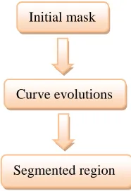
Fig. 1. Block diagram of proposed method
Fig. 1 shows the block diagram of the proposed method. In this active contour model, the basic idea is to evolve a curve C in a given image, and to stop the evolution when the curve reaches the desired iteration step. Then, region based active contour model is for segmenting the region of interest. The benefits of this algorithm can be summarized in: automatically detecting interior contours, robust with respect to noise, ability to detect and represent complex boundaries or segments, and finally, extraction of geometric measurements, such as length, area, volume, intensity of a detected contour, surface or region, respectively. The information can be later used to study the evolution in time of a disease (a growing tumor), or to compare two different subjects, usually a normal one and abnormal one.
Initial mask
Curve evolutions
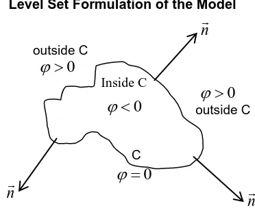
A. Level Set Formulation of the Model outside C outside C C
Fig. 2. Curve C propagating in normal direction
Where,
C = the boundary of an evolving curve = the level set function [5]
In the level set method, C is represented by the zero level set of a Lipschitz function φ: , such that
{
*( ) ( ) + ( ) *( ) ( ) + ( ) ̅⁄ *( ) ( )
Recall that is open, and C = . This is illustrated in Fig. 2, the above assumptions and notations on the level set function φ, defining the evolving curve C [6].
Step-1.Original image is denoted by and define the maximum iteration manually.
Step-2. Create the initial mask at the region of interest.
Step-3. Initialize from the initial mask at n=0
Step-4. Compute ( ) by (1) if ∫ ( ( )) > 0 (i.e. if the curve has a nonempty interior in Ω) and ( ) by (2) if ∫ ( ( ( ))) > 0 (i.e. if the curve has a nonempty exterior in Ω)
( ) ∫ ( ) ( ( )) ∫ ( ( )) ( ) ( ) ∫ ( ) ( ( ( ))) ∫ ( ( ( ))) ( ) ( ) {
Where, = average intensity of inside C = average intensity of outside C = input image
H(φ)= Heaviside function
Step-5. Solve the partial differential equation (PDE) in φ by (3) to obtain by (4)
( ) [ (| | ) ( ) ( ) ] ( ) ( ) ( ) ( ) ( )
Fix λ = 1, ν = 0 and μ < 1
Where, δ (φ) = one dimensional Dirac measure μ, λ = positive parameters
ν = a force pushing the curve towards the object
The iterative equation;
( ), ( √( )⁄( ) ( )⁄( ) ) ( √ ( ) ( ) ⁄ ( ) ( ) ⁄ ) ( ( )) ( ( )) ( )
The finite differences are;
Step-6. Re-initialize φ to the signed distance function to the curve by (5),
( ) ( )
( ) ( )
G = {√ ( ) ( ) ( ) √ ( ) ( ) ( )
( ) ( ) ( ) ( )
( ) ( ) ( ) ( )
Fix T = 1 and h = 1,
Where, a and c are backward differences
94 h = the step space
T= time to reach final segmentation
Step-7. Check whether the final contour reaches 100 iterations. If not, n = n+1 and repeat.
B. Region based active contour model for segmentation
This model is the minimization of an energy based segmentation. The image is formed by two regions of approximately piecewise-constant intensities, of distinct values and . The object to be detected is represented by the region with the value . Let denote its boundary by . Now we have inside the object and outside the object. The energy fitting term of this model (6),
( ) ( ) ∫| ( ) |
∫| ( ) | ( )
Where, C is any other variable curve, and the constants and are the averages of inside C and respectively outside C.
C. Relation with the Mumford-Shah Active Contour Model
The Mumford-Shah functional for segmentation is (7).
( ) ( )
∫| ( ) ( )|
∫| ( )| ( )
Where, C ,
μ and λ are positive parameters. u = average inside C and outside C
The case of minimal partition problem can be formulated and solved using level set method [7].
III. IMPLEMENTATION OF THE SYSTEM
Image acquisition is the first step of the system. Different weighted or sequences of MR brain images are obtained from the Department of Radiology of Mandalay General Hospital. Then convert the color MR images into gray scale images. And the initial mask was created at the region of interest (tumor region) in the MR brain images. Curve evolution processes are accomplished from this step. In this system, iteration step is defined by user manually. Level set function is stopped at the desired iteration step. After iterating process, the final segmented region is detected. Fig. 3 shows the overall flow diagram of the proposed system.
Fig. 3. Overall flow diagram of the system
IV. EXPERIMENTAL RESULTS
This proposed method can work well for different views of MR brain tumor images such as axial view, sagittal view and coronal view. These tumor regions are remarked by radiologists from Mandalay General Hospital. Figure.5 shows the segmentation results of different views of a patient.
(a)
(b)
MR brain images
Convert RGB to gray
Initial mask
Curve evolutions
(c)
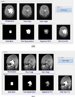
Fig. 5. Segmentation results of different views. (a). Axial view FLAIR weighted, (b). Sagittal view FLAIR weighted, (c).
Coronal view FLAIR weighted.
In this system, axial view MR brain tumor images with FLAIR weighted, T1 weighted and T2 weighted images of three patients who were suffered from diseases are tested because most pathologies and diseases can easily find from axial view and this view contains the central nervous system of the human brain. Patient 1’s tumor region is in the left of the brain. But the region of tumor is not distinct in T2 weighted sequence of Patient 1.
(a)
(b)
Fig. 6. Segmentation results of Patient 1. (a). FLAIR weighted, (b). T1 weighted.
(a)
(b)
(c)
Patient 2’s tumor region is in the midline of the brain.
96 (a)
(b)
(c)
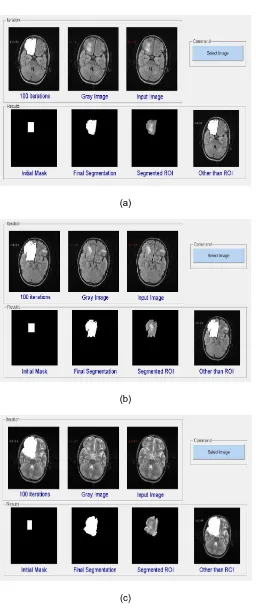
Patient 3’s tumor region is in the right of the brain.
Fig. 8. Segmentation results of Patient 3. (a). FLAIR weighted, (b). T1 weighted, (c). T2 weighted.
V. DISCUSSIONS AND CONCLUSION
In this system, iteration steps can be controlled for the segmenting process. The more iteration steps there are, the wider and larger the region of interest (region of tumor). The desired regions for all views of magnetic resonance (MR) brain images have been obtained at 100 iterations. In this system, re-initialization has been used for maintaining stable curve evolutions and ensuring the desirable results. The region based active contour model can get better results when the region around ROI is wanted. In this proposed system, the interior contours are automatically detected. Edge based active contour can be used if the exact area is wanted. For segmentation process, it does not need to find the exact area of disease because this system is not calculating the tumor area and volume. Therefore, region based active contour model is more suitable than edge based active contour model for this system. Since the aim of this system is for the region based medical image compression, the ROI and non-ROI are separated out independently. This segmentation method can segment all views of MR brain images such as axial view, sagittal view and coronal view. But in this system, axial view different weighted MR brain images are used because radiologist can find more pathologies from this view than other views. This region based active contour model works well for all views and sequences of MR brain images.
ACKNOWLEDGEMENTS
The author would like to express her gratitude to her supervisor, Dr. Aung Soe Khaing, Associate Professor for his guidance and her teachers from Department of Electronic Engineering, Mandalay Technological University. The author specially thanks to her family and her friends for their supports and encouragement. And she also thanks to the doctors from Department of Radiology, Mandalay General Hospital for giving the MR brain tumor images to test for her research.
REFERENCES
[1] Manish Khare and Rajneesh Kumar Srivastava, “Medical image segmentation using Level Set Method without Re-initialization,” International Conference on Signal, Image and Video Processing (ICSIVP), 2012.
[2] Sajith A.G and Hariharan.S, “Medical Image Segmentation Using CT Scans- A Level Set Approach ” International Journal of Innovation Technology and Exploring Engineering (IJITEE), ISSN: 2278-3075, Volume-2, Issue-6, May 2013.
[3] B.S Saini and G.Sethi, “Comparative Analysis of Edge Based and Region Based Active Contour Using Level Set and its Application on CT Images,” International Journal of Research in Engineering and Technology (IJRET), ISSN: 2319-1163, Volume: 02, Issue: 04, Apr-2013.
[5] Tony F. Chan, Member, IEEE, and Luminita A. Vese, “Active Contours Without Edges,” IEEE Transactions on Image processing, Vol. 10, No. 2, February 2001.
[6] S. Osher and J. A. Sethian, “Fronts propagating with curvature-dependent speed: Algorithms based on Hamilton-Jacobi Formulation,” J. Comput. Phys., Vol.79, PP. 12-49, 1998.