Zuevaet al. Frontiers in Zoology (2018) 15:1
DOI 10.1186/s12983-017-0247-4
R E S E A R C H
Open Access
The complex simplicity of the brittle star
nervous system
Olga Zueva
1, Maleana Khoury
1, Thomas Heinzeller
2, Daria Mashanova
3and Vladimir Mashanov
1*Abstract
Background: Brittle stars (Ophiuroidea, Echinodermata) have been increasingly used in studies of animal behavior, locomotion, regeneration, physiology, and bioluminescence. The success of these studies directly depends on good working knowledge of the ophiuroid nervous system.
Results: Here, we describe the arm nervous system at different levels of organization, including the microanatomy of the radial nerve cord and peripheral nerves, ultrastructure of the neural tissue, and localization of different cell types using specific antibody markers. We standardize the nomenclature of nerves and ganglia, and provide an anatomically accurate digital 3D model of the arm nervous system as a reference for future studies. Our results helped identify several general features characteristic to the adult echinoderm nervous system, including the extensive anatomical interconnections between the ectoneural and hyponeural components, neuroepithelial organization of the central nervous system, and the supporting scaffold of the neuroepithelium formed by radial glial cells. In addition, we provide further support to the notion that the echinoderm radial glia is a complex and diverse cell population. We also tested the suitability of a range of specific cell-type markers for studies of the brittle star nervous system and
established that the radial glial cells are reliably labeled with the ERG1 antibodies, whereas the best neuronal markers are acetylated tubulin, ELAV, and synaptotagmin B. The transcription factor Brn1/2/4 – a marker of neuronal
progenitors – is expressed not only in neurons, but also in a subpopulation of radial glia. For the first time, we describe putative ophiuroid proprioceptors associated with the hyponeural part of the central nervous system.
Conclusions: Together, our data help establish both the general principles of neural architecture common to the phylum Echinodermata and the specific ophiuroid features.
Keywords: Echinodermata, Brittle star, Nervous system, Glia, Neurons
Background
Brittle stars are emerging model organisms in modern biology. They have been increasingly used to address a wide range of fundamental questions, including post-traumatic regeneration of lost body appendages [1–3], organization and physiology of the mutable connective tissue [4], and bioluminescence [5]. Brittle stars are also among the fastest-moving echinoderms capable of coor-dinated complex locomotory behaviors. The neurobiology of ophiuroid locomotion has been receiving attention in the contexts of body plan evolution, neurobiology, and robotics. Unlike many other members of the phylum,
*Correspondence: [email protected] 1University of North Florida, Jacksonville, FL, USA
Full list of author information is available at the end of the article
they do not use their numerous small podia for move-ment. Instead, they quickly propel their body over the substratum via rapid large-scale rowing-like movements of highly motile segmented body appendages called arms. The behavior of the five individual arms is centrally con-trolled to produce a true bilateral movement pattern [6, 7]. To achieve this level of coordination, the nervous system must be able to synchronize muscular activity between different segments within each individual arm, as well as large-scale movements across the five arms.
All the above phenomena are controlled by or depend on the nervous system. Nevertheless, the ophiuroid nervous system has never been comprehensively studied with modern techniques. The last and only complete
microanatomical description dates back to the 19th century [8] and remains the best reference to date. The pioneering neurophysiological studies that were per-formed in 1970s and 1980s [9, 10] provided valuable initial insights into the role of one brittle star neuronal type – giant neurons. They established the role of these cells in propagation of neural impulses throughout the nervous system and in integration of sensory inputs to coordinate contraction and relaxation of the arm muscles. These early works however have never been followed up by more detailed and comprehensive studies of other cell types and of the overall structure and function of neural circuits. Most modern reports focus on individual aspects of echinoderm neurobiology (e.g., immunostaining with one or a few cell type markers, ultrastructure of certain regions of the nervous system). These isolated studies, although each valuable in itself, do not assemble together into a general cohesive picture. The scope of this paper is to provide a comprehensive view of the organization of the nervous system in the brittle star arm that can serve as a reference for future studies in the biology of ophiuroids and echinoderms in general. We use a synthesis of different techniques to characterize various aspect of the neural architecture. These experimental approaches include three-dimensional (3D) modeling,
transmission electron microscopy, and
immunos-taining with a series of cell-type specific glial and neuronal markers coupled with laser scanning confocal microscopy.
Here, we:
1. Present an anatomically precise digital 3D model of the nervous system in an arm segment. We made an effort to trace the origin and targets of all major peripheral nerves and propose standardized terminology for them.
2. Provide a detailed description of the
neurohistological organization of the radial nerve cord, peripheral nerves and ganglia. We also discuss both the features that ophiuroids share with other echinoderms and unique characteristics of the brittle star nervous system
3. Describe the immunoreactivity of the cells of the nervous tissue with cell type-specific antibody markers.
Together, our data help establish both the general prin-ciples of neural architecture common to the phylum Echinodermata and the features that are specific to the class Ophiuroidea. We also confirmed and expanded on earlier observations [11] of complex and molecularly het-erogeneous organization of echinoderm glia. This study also describes for the first time putative proprioceptors embedded in the CNS.
Methods
Animal collection and maintenance
Adult Amphipholis kochii Lütken, 1872 were collected from Vostok Bay, Sea of Japan (Russia). Adult individu-als ofOphioderma brevispinumSay, 1825 were purchased from Gulf Specimen Marine Laboratories, Inc. (Panacea, FL). The animals were kept in glass aquaria with aerated sea water.
Electron microscopy
For transmission electron microscopy (TEM), arms ofA. kochiiwere fixed in 2.5% glutaraldehyde dissolved in 0.05 M cacodylate buffer (pH 7.6) for 24 h at 4 °C. After fix-ation, the specimens were rinsed in the same buffer and postfixed in 1% OsO4in cacodylate buffer for 1 h. The
tis-sue samples were then decalcified in several changes of a solution containing 1% ascorbic acid and 0.15 M NaCl, dehydrated in a graded series of ethanol and acetone and embedded in the Araldite epoxy resin. Sections were cut with glass knives on Ultracut E (Reichert, Vienna, Aus-tria) and UC6 (Leica) ultratomes. Ultrathin (50 – 70 nm) sections were stained with aqueous uranyl acetate and lead citrate and then examined and photographed with a Zeiss EM 10 transmission electron microscope.
For scanning electron microscopy (SEM), specimens were fixed in 2.5% glutaraldehyde in 0.05 M cacodylate buffer at pH 7.6, dehydrated in ethanol followed by an ace-tone series, critical point dried, and then sputter coated with carbon and gold. Specimens were examined with a Jeol JSM-IC848 scanning electron microscope (JEOL Ltd., Tokyo, Japan).
3D surface reconstruction
Zuevaet al. Frontiers in Zoology (2018) 15:1 Page 3 of 26
The original stack of images, saved as a video file, is available in Additional file 1. The final 3D model in vari-ous interactive and non-interactive (video) formats can be accessed in Additional files 2, 3, 4, 5, 6, 7, 8, 9, 10 and 11.
Immunohistochemistry
Fluorescent immunohistochemistry on frozen sections was performed as described elsewhere [12]. Briefly, ani-mals were anesthetized in 0.2% chlorobutanol (Sigma) and then portions of the arm, approximately 5 segments long, were fixed in 4% paraformaldehyde prepared in 0.01 M PBS (pH 7.4) overnight at 4 °C. For immunos-taining with anti-synaptotagmin antibodies, the samples were fixed in ice-cold methanol for 30 min. The samples were then washed in PBS, decalcified in 10% EDTA, cry-oprotected in sucrose and embedded in the Tissue-Tek O.C.T. Compound (Sakura). Cryosections (10μm thick) were cut using a Leica CM1860 cryostat, collected on gelatin-covered slides and incubated at 42 °C overnight. The slides were then washed in PBS incubated in 0.1 M glycine for 1 h to quench autofluorescence. After another wash in PBS (3×10 min), the sections were blocked using 2% goat serum for 1 h. The first antibodies (see Table 1) were applied at 4 °C overnight. After extensive washing in PBS (4×10 min), the sections were incubated in the sec-ondary antibodies (Table 1) for 1 h at room temperature. Unbound antibodies were removed in four changes of PBS (10 min each) and nuclei were stained with 5μM DRAQ5 (Thermo Scientific) for 30 min. After the final round of washes (3×5 min), the slides were coverslipped in an anti-fading medium containing 2.5% DABCO and 10% Mowiol 4-88 in 25% buffered glycerol (0.2M Tris-HCL, pH 8.5).
Whole-mount staining was performed in a similar way with the following modifications. After the fixation and decalcification steps, the tissues were bleached in increas-ing concentrations of hydrogen peroxide (0.3%, 1%, and
3%) and then permeabilized by Proteinase K digestion (2.5μg/ml, 15 min at room temperature). All wash buffers contained 0.5% Triton X-100. The incubation steps in the first and second antibodies were increased to 2 days each and performed at 4 °C.
Stacks of optical sections were taken using the Olympus confocal laser scanning microscope FV1000. Maximum intensity Z-projections were generated in the Fiji image processing software [13].
Unless indicated otherwise, images of whole-mount specimens and micrographs of longitudinal sections are oriented with the distal side to the right.
Results
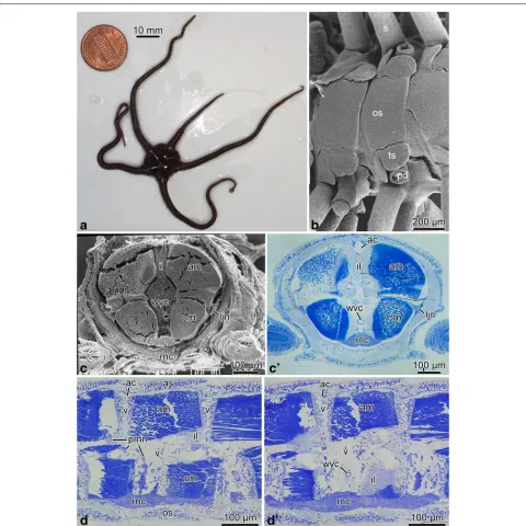
Anatomical organization of the nervous system in the arm Nerve ring and radial nerve cord. The brittle star body is composed of a flattened disk and five long unbranched segmented arms (Fig. 1a, b). Each segment contains a set of calcareous skeletal elements, including the cen-tral vertebral ossicle surrounded by four peripheral arm shields or plates: a dorsal, a ventral, and two lateral ones (Fig. 1b, d, d’). Each lateral arm shield bears a vertical row of arm spines (Fig. 1b). On the ventral surfaces of the arm segment, a pair of podia emerges through pores adjacent to bases of the ventral arm spines. Each podium is protected by two tentacle scales (Fig. 1b). The verte-brae of adjacent segments are joined together by paired aboral and oral intervertebral muscles and the interver-tebral ligament that has a complex geometry (Figs. 1c–d’ and 4c, e).
The main components of the central nervous system in brittle stars are organized into a pentaradial pattern and thus correspond to the overall layout of the general body plan. In each arm, the radial nerve cord (RNC) lies beneath the water-vascular canal (Figs. 1c–d’, 3 and 4a, e) and is protected by the oral skeletal shield and oral
Table 1Antibodies used in this study
Antibodies used Source Host species Dilution
First antibodies
Acetylated tubulin Sigma (T6793) Mouse 1:1,000
Brn1/2/4 [22] Rat 1:1,000
ELAV [22] Rabbit 1:3,000
ERG1 [18] Mouse 1:1
GFSKLYFamide [20] Rabbit 1:1,000-1:5,000
Second antibodies
Goat anti-mouse Cy3 Jackson Immunoresearch (115-165-146) Goat 1:2,000
Goat anti-rabbit Cy3 Jackson Immunoresearch (115-165-144) Goat 1:1,000
Goat anti-rabbit FITC ThermoFisher Scientific (65-6111) Goat 1:200
Goat anti-rat FITC GenWay (GWB-7B4D70) Goat 1:100
Fig. 1Brittle star arm anatomy.aAn individual ofO. brevispinum.bScanning electron micrograph of the oral arm surface ofA. kochii.candc’ Cross section of the arm ofA. kochii.cThe arm was embedded into epoxy resin and sectioned transversely. The resin was then removed, and the cut surface was imaged with a scanning electron microscope.c’ The corresponding plastic section stained with methylene blue.dandd’ Longitudinal paragsagittal sections through the arm inA. kochiistained with methylene blue. The skeletal elements are not visible in any of the micrographs, because they were removed during tissue processing. Abbreviations:ac– arm coelom;am– aboral intervertebral muscle;as– aboral shield;il– intervertebral ligament;ljn– lateral juxtaligamental node;om– oral intervertebral muscle;os– oral shield;pd– podium;pmn– hyponeural proximal muscle nerve;rnc– radial nerve cord;s– spine;ts– tentacle scale;wvc– water-vascular canal;v– vertebral ossicle
ligament. At the attachment of the arm to the disk, each of the the RNC approaches the centrally located esophagus, ascends aborally and bifurcates (Fig. 2). The side branches of adjacent RNCs fuse together to form a continuous nerve ring (Fig. 2b). Both the
Zuevaet al. Frontiers in Zoology (2018) 15:1 Page 5 of 26
Fig. 2Transition between the radial nerve cord and the circumoral nerve ring inA. kochii. Semi-thin sections stained with methylene blue.aRadial section (parallel to the long axis of the arm).bHorizontal section (i.e., orthogonal with respect to the oral-aboral axis) through the nerve ring. Abbreviations:nr– nerve ring;rnc– radial nerve cord
The radial nerve cord in brittle stars has clear metameric organization and corresponds to segmented arrangement of other components of the arm, including the skeleton, ligaments, muscles, and water-vascular system. At the level of each vertebral ossicle, both the ectoneural and hyponeural layers of the RNC are thickened to form gan-glionic swellings (Fig. 1d, d’). In each segment, the radial nerve cords give off a complex system of peripheral nerves that innervate different metameric anatomical structures of the arm (Figs. 3 and 4). This peripheral nervous system is described below.
As in the previously studied CNS of sea cucumbers [14, 15], the ectoneural and hyponeural components of the RNC have organization of tubular cords. The aboral wall of the ectoneural cord is very thick and is formed by a tall
ectoneural epithelium (Figs. 5a and 6a). The opposite oral wall is composed of a very thin epineural epithelium. The cavity that separates the ectoneural neuroepithelium and the epineural epithelium constitutes the epineural canal (Figs. 6a and 7a, b).
Ectoneural peripheral nerves and ganglia. At the level of the podia, the ectoneural part of the RNC gives off a pair of short and thick podial nerves (Figs. 3, 4a, b, e and 5a, j). Shortly after emerging from the RNC, the podial nerve forms a thick ring ganglion at the base of the podium. A sleeve-like extension of the podial gan-glion surrounds the hydrocoelic lining of the podium and descends down to the distal tip. The lateral side of the podial ganglion further gives off thick spine nerves to
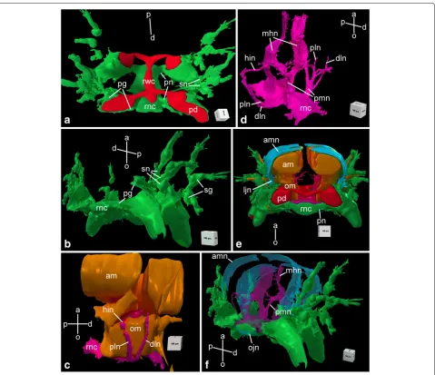
Fig. 4Three-dimensional reconstruction of the radial nerve cord and peripheral nerves in the arm segment inA. kochii. Other anatomical structures are provided for reference, when indicated.aAboral view of ectoneural system(green). Components of the water-vascular system are shown inred. bDistal view of the ectoneural system.cSide view of muscles(brown)and the hyponeural system(magenta).dOblique side view of the hyponeural system(magenta).eProximal view of the complete nervous system. The muscles(brown)and the water-vascular system(red)are also shown. fDistal view of the complete nervous system. The orientation of the projections is indicated by axes on each image:a– aboral;d– distal;o– oral; p– proximal. Abbreviations:am– aboral intervertebral muscle;amn– aboral mixed nerve;dln– distal lateral hyponeural nerve;hin– horizontal intermuscular hyponeural nerve;ljn– lateral juxtaligametal node;mhn– median hyponeural nerve;ojn– oral juxtaligamental node;om– oral intervertebral muscle;pd– podium;pg– podial ganglion;pln– proximal lateral hyponeural nerve;pmn– proximal muscle nerve;pn– podial nerve; rnc– radial nerve cord;rwc– radial water-vascular canal;sg– spine ganglion;sn– spine nerve
each spine (Figs. 3, 4a, b and 5a, j). As they penetrate the lateral arm shields, these nerves pass throughspine ganglia. The podial ganglia also give off small nerves that innervate tentacle scales (Fig. 5j).
Hyponeural peripheral nerves. The hyponeural part of the RNC gives off an extensive system of peripheral nerves (Figs. 3 and 4c, d). Although the hyponeural system by itself forms no purely hyponeural peripheral ganglia, it contributes to the formation of the mixed ganglia, known
Zuevaet al. Frontiers in Zoology (2018) 15:1 Page 7 of 26
Fig. 6Radial glia in the ectoneural neuroepithelium of the radial nerve cord inA. kochii. Transmission electron microscopy.aLow magnification view of the ectoneural neuroepithelium.bIntercelluar junctions between apicolateral surfaces of two adjacent radial glial cells.cBasal endfoot of a radial glial cell.dAttachment of the apical surface of a radial glial cell to the epineural cuticle. Abbreviations:bl– basal lamina;bp– basal process of a radial glial cell;c– epineural cuticle;ec– epineural canal;if– intermediate filaments;ne– neuropil;rg– radial glia.Arrowheadsshow hemidesmosomes
– both aboral and oral – and the intervertebral ligament and fuses with its oral margin with the hyponeural part of the RNC (Figs. 3, 4d and 5h, i). It gives off numerous short branches on its surface that innervate both the interver-tebral muscles and the mutable collagenous tissue of the intervertebral ligament in the central region of the arm (Figs. 4d, f and 5d–g).
On either side of the arm, a horizontal intermuscu-lar nerveconnects the median hyponeural nerves to the lateral juxtaligamental node (see below). This nerve runs between the intervertebral and lateral ligaments in the space between the oral and aboral muscles (Figs. 3, 4c, d and 5f, g).
Immediately behind the point of origin of the proxi-mal muscle nerves, the hyponeural part of the RNC gives off two pairs of lateral nerves that both ascend aborally along the lateral surface of the oral intervertebral muscle (Figs. 4c, d and 5c–e, g). One of them – thedistal lateral nerve– innervates the lateral arm shield, whereas the sec-ond one – theproximal lateral nerve– directly connects the radial nerve cord to thelateral juxtaligamental node
(see below) (Fig. 5c–g).
Zuevaet al. Frontiers in Zoology (2018) 15:1 Page 9 of 26
Fig. 7Neurons in the ectoneural neuroepithelium of the radial nerve cord inA. kochii. Transmission electron microscopy.aSub-apical neuron. a’ Cilium in a subapical neuron(n).bNeuron(n, colored)that reaches the lumen of the epineural canal(ec).c–f’ Ectoneural neuropil.canddshow the transversal and longitudinal sections, respectively, of the neuropil area containing processes of giant neurons(gn).eProcess of the
either ectoneural nor hyponeural, as these structures are formed by contribution of both. All components of the mixed peripheral nervous system are embedded into the outer wall of the arm coelom. The most prominent structures of this mixed system are the paired large
lateral juxtaligamental nodesthat lie one on each side of the arm, midway between the oral and aboral surfaces (Figs. 3, 4e and 5f, g). Each lateral node is formed by the fusion of three neural components: the lateral end of the horizontal intermuscular nerve, the aboral end of the proximal lateral nerve, and the distal margin of the podial ganglion (Figs. 3, 4c–e and 5f, g). The first two structures originate from the hyponeural system, whereas the podial ganglion is ectoneural (Figs. 4a, b and 5a, d–g, j). The lateral juxtaligamental nodes give off numerous short branching protrusions that enter the adjacent collagenous tissue of the lateral ligament (Fig. 5f, g). They also give rise to two pairs of larger nerves, both thin and wide, which run in opposite directions in the lateral wall of the arm coelom (Figs. 3, 4e, f and 5f, i). One pair – the abo-ral mixed nerves – ascend towards the aboral midline (Figs. 3 and 5f, i). The two oral mixed nerves, one on each side of the arm, descend towards the oral midline and fuse to form theoral juxtaligamental node, which innervates the oral ligament (Figs. 3 and 5h).
Cellular architecture of the nervous tissue Radial nerve cord
The ectoneural neuroepithelium of the RNC contains prominent radial glial cells (Fig. 6). The brittle star radial glia is very similar to their counterparts previously described in starfish and sea cucumbers [12, 14, 17, 18]. These cells are robustly labeled by the ERG1 antibody (Figs. 15a, 16e, f, 17b, c and 18b, c”’), which was raised against a sea cucumber glial antigen [18]. They stretch throughout the height of the neuroepithelium between the apical and basal surfaces (Figs. 6a, 16e, f and 17c) and show clear epithelial cell features. Their cell bodies are often located at the apical surface of the neuroepithe-lium (Figs. 6a and 7a, b) and give off a long basal process that crosses the underlying neuropil (Figs. 6a and 7a, c). The distal end of the glial process forms a flattened end-foot that attaches to the basal lamina by hemidesmosomes (Fig. 6c). The cell bodies of adjacent glial cells are con-nected by apicolateral junctional complexes composed of zonula adherens and septate junctions (Fig. 6b). A typical intracellular characteristic of radial glia is the presence of thick bundles of intermediate filaments which run along the long axis of the cell (Fig. 6c, d). The epineural canal contains a dense accumulation of fibrous extracellular material that forms a flat cuticle-like structure overlay-ing the apical surface of the ectoneural neuroepithelium (Fig. 6a, d). This cuticle varies in thickness and some-times shows a mesh-like organization of interconnected
layers. The apical surface of the radial glial cells is attached to the cuticle via hemidesmosome-like contacts (Fig. 6d).
Most neuronal cell bodies are localized to the sub-apical region of the ectoneural neuroepithelium beneath the layer of glial cell bodies (Fig. 7a). Some neuronal perikarya, however, reach the lumen of the epineural canal (Fig. 7b). These neuronal cells are flanked by radial glia and joined to them by intercellular junctions. The region of the neuroepithelium located between the apical layer of the neuronal and glial cell bodies and the basal lam-ina is occupied by an extensive neuropil (Figs. 6a and 7c – f’). The neuropil is composed of densely packed neuronal processes filling all the space between the basal processes of radial glia (Fig. 7c). By their size, the neuronal processes can be clearly classified into “regular” (measur-ing 130 nm – 600 nm across) and “giant” (up to∼5μm across) (Fig. 7c, d). Occasionally, the neuropil contains processes of neurosecretory-like cells (Fig. 7e) filled with large dense granules, which are morphologically identical to those described in juxtaligamental cells [16]. Well-defined chemical synapses are often seen in the neuropil (Fig. 7f, f’).
The roof of the epineural canal is formed by a thin epineural epithelium, a simple epithelial monolayer com-posed of flattened glial cells (Fig. 8a). Unlike the radial glia in the neuroepithelium, these glial cells mostly lack inter-mediate filaments, except those that are associated with the hemidesmosomes that anchor the cells to the basal lamina and to the cuticle in the epineural canal (Fig. 8b). Very occasionally, this epithelium contains basiepithelial nerve processes, but never neuronal perikarya (Fig. 8c).
Zuevaet al. Frontiers in Zoology (2018) 15:1 Page 11 of 26
Fig. 8Roof of the epineural canal (epineural epithelium) inA. kochii. Transmission electron microscopy.aFlattened glial cell(gc)of the epineural epithelium.bHemidesmosomes(arrowheads)anchoring a glial cell of the epineural epithelium to the basal lamina(bl)and to the epineural cuticle (c).cThe epineural epithelium occasionally contains basiepithelial neural processes(np), but not neuronal perikarya. Abbreviations:bl– basal lamina;c– epineural cuticle;ec– epineural canal;gc– flattened glial cell;if– intermediate filaments;ne– ectoneural neuroepithelium;np– neuronal processes
bundles of intermediate filaments in their cytoplasm. All glial cells in the hyponeural system are secretory. Their cytoplasm contains vacuoles filled with material of mod-erate electron density, which is released into the lumen of the hyponeural canal via exocytosis (Fig. 9b, c).
The hyponeural part of the brittle star RNC is also associated with two non-neural anatomical components. The first one is the radial hemal lacuna, which is a local expansion of the otherwise thin extracellular space that separates the ectoneural and hyponeural parts of the RNC (Fig. 9a). The lumen of the lacuna, therefore, does not have epithelial lining, but is instead surrounded by the basal lamina of the hyponeural neuroepithelium. The second non-neural component of the hyponeural system are two compact bundles of muscle cells immersed into the oral wall of the hyponeural cord on either side of the midline (Fig. 9a, e, g). These muscle bundles run longitudinally throughout the length of the radial nerve cord.
As has been previously documented in studies of the sea cucumber CNS [14, 19], the ectoneural and hyponeural neuroepithelia of the RNC form extensive direct anatom-ical connections with each other. Even though the basal surfaces of these two neuroepithelia are separated from each other by a dense basal lamina, this separation is never complete. Frequent gaps in this basal lamina allow
for the passage of neuronal processed from one neuroep-ithelium into another (Fig. 10a, a’). The basal lamina is also absent in the regions where the hyponeural part of the radial nerve cord comes in close contact with the oral intervertebral muscle (Fig. 10b, b’).
Peripheral nerves and ganglia
All peripheral nerves in the arm are organized as densely packed bundles of neuronal processes. They either run as anatomically distinct neural tracts or enter the wall of the arm coelom and form a basiepithelial nerve plexus there. Specific details on individual components of the peripheral nervous system are provided below.
Fig. 9Organization of the hyponeural part of the radial nerve cord (RNC) inA. kochii. Transmission electron microscopy.aandbHyponeural system in the interganglionic region of the RNC.aLow-magnification view of the hyponeural cord.bPart of the oral wall (floor) of the interganglionic hyponeural cord formed by flattened glial cells with no neuronal elements.c–gHyponeural system in the ganglionic swelling of the RNC. CSecretory radial glia.dNeurosecretory-like cell in the aboral wall (roof) of the hyponeural cord.eandfAbundant neurons in the oral wall (floor) of the hyponeural cord.fGiant neuron.gHigh-magnification view of a muscle bundle integrated in the lateral region of the hyponeural neuroepithelium. Abbreviations:bp– basal process of radial glial cell;en– ectoneural neuroepithelium;gc– flattened glial cell;gn– “giant” neuron; hc– lumen of the hyponeural canal;hl– hemal lacuna;hn– hyponeural neuroepithelium;m– bundle of muscle cells;np– neuronal processes;nsc– neurosecretory-like cell;rg– radial glia
cells (Fig. 11c). At the periphery, the spine ganglia are covered by a sheath of flattened glial cells and a basal lamina (Fig. 11e).
Zuevaet al. Frontiers in Zoology (2018) 15:1 Page 13 of 26
Fig. 10The hyponeural neuroepithelium makes direct contacts with the ectoneural epithelium (aanda’) and with the intervertebral muscles (bandb’) inA. kochii. Transmission electron microscopy. The colorized neuron inaanda’ has its cell body within the hyponeural neuroepithelium and sends a process into the ectoneural neuropithelium.a’ andb’ show detail views of the boxed areas inaandb, respectively. Abbreviations: en– ectoneural neuroepithelium;hn– hyponeural neuroepithelium;n– neuron;np– neural processes;om– oral intervertebral muscle.White arrowheadsindicate the basal lamina
nerve contains mostly thick processes of “giant” neu-rons, less abundant processes of the “regular” diame-ter and occasional neuronal perikarya (Fig. 12a). The nerve is surrounded by a sheath of flattened glial cells and a continuous basal lamina. In contrast, the median hyponeural nerve lacks continuous glial envelope and has basal lamina only on the median side, which faces the intervertebral ligament (Fig. 12b, c). On its lat-eral side, the nerve directly abuts the intervertebral muscle. The nerve processes are often seen to pene-trate into the muscle and form synaptic contacts with myocytes (Fig. 12b). The median nerves are particularly
rich in cell bodies and processes of neurosecretory-like cells. The bundles of these processes frequently leave the nerve through the gaps in the basal lamina and branch in the collagenous connective tissue of the ligament (Fig. 12c).
Fig. 11Organization of the spine ganglion inA. kochii. Transmission electron microscopy.aPeripheral part of the spine ganglion with cell bodies of neurons and two types of neurosecretory-like cells.bThe central neuropil area of the spine ganglion with nerve processes of spine nerve passing through.cSynapses(arrows)between a neural process and processes of neurosecretory-like cells.dBasal body of a cilium in a neurosecretory cells. eGlial cells at the periphery of the ganglion. Abbreviations:gc– glial cell;n– neuron;nsc I– neurosecretory-like cell type I;nsc II– neurosecretory-like cell type II
located between the cell bodies of the coelomic epithe-lial cells and the basal lamina of the coelomic epithelium (Fig. 13a, b). The ganglia – the oral and lateral juxtaliga-mental nodes – are local expansions of the basiepithelial plexus (Fig. 13c–h). They contain dense accumulations of cell bodies and extensive neuropil regions. The latter con-tain frequent synapses between neuronal processes and neurosecretory-like cells (Fig. 13e, h). Bundles of neurose-cretory processes leave the ganglia through perforations in the basal lamina and enter the adjacent collagenous connective tissue (Fig. 13g).
Immunohistochemistry
We used a number of cell-type specific markers to study localization of different cell types in the brittle star nervous system (Table 1). These include antibodies against general neuronal antigens, such as synaptotagmin
B (SynB), ELAV, and acetylated tubulin; Brn1/2/4 – an antigen specific to neuronal progenitors, and the neu-ropeptide GFSKLYFamide [20–22]. To label glial cells, we used the ERG1 monoclonal antibody that stains radial glia in the echinoderm CNS [18].
Zuevaet al. Frontiers in Zoology (2018) 15:1 Page 15 of 26
Fig. 12Organization of the hyponeural peripheral nerves inA. kochii. Transmission electron microscopy.aProximal muscle nerve.b, cMedian hyponeural nerve.bBundles of neuronal processes(asterisk)branching off the median hyponeural nerve and entering the oral intervertebral muscle. The inset inbshows a neuromuscular synapse(arrow).cProcesses of neurosecretory-like cells passing through an opening in the basal lamina. Abbreviations:bl– basal lamina;gc– glial cell;il– intervertebral ligament;m– myocyte;np– neuronal processes
as a fine net. Among other immunopositive structures in the peripheral nervous system are the spine nerves, as well as the cell bodies and fibers in the median hyponeu-ral nerve that innervate the intervertebhyponeu-ral muscles and ligaments (Figs. 14a, 15b–d). In the muscles, the SynB-containing processes form two parallel tracts: one near the proximal and one near the distal end. Individual fibers branch off those bundles and run parallel to the
myocytes (Fig. 15b, c). The intervertebral ligament also contains a dense array of SynB-positive neural elements. These cells have small perikarya and give off long processes running parallel to the long axis of the ligament (Fig. 15d).
Fig. 13Organization of the mixed peripheral nerves and ganglia inA. kochii. Transmission electron microscopy.a, bAboral mixed nerve.c–eLateral juxtaligamental node.f–hOral juxtaligamental node.aNeuronal cell body in the wall of the arm coelom.bNeurosecretory-like cell in the wall of the arm coelom.cLow magnification view of the lateral juxtaligamental node.dCoelomic epithelial cell separating neurosecretory cells from the lumen of the coelom.eSynapse(arrow)between an axon and a process of a neurosecretory cell in the neuropil area of the lateral juxtaligamental node. fGeneral view of the oral juxtaligamental node.gProcesses of neurosecretory-like cells leaving the oral juxtaligamental node and entering the collagenous connective tissue.hSynapse(arrow)between an axon and a process of a neurosecretory-like cell. Abbreviations:ce– coelomic epithelial cell;cl– lumen of the coelom;ec– epineural canal;ee– epineural epithelium;en– ectoneural neuroepithelium;n– neuron;np– neural processes;nsc– neurosecretory cell
positioned on either side of the midline and run contin-uously throughout the length of the arm. More loosely organized longitudinal processes also run on either side of the longitudinal tracts, but they do not form well-defined
Zuevaet al. Frontiers in Zoology (2018) 15:1 Page 17 of 26
Fig. 14Synaptotagmin B (SynB) in the nervous system of the arm inO. brevispinum. Whole-mount preparation. Maximum intensity Z-projection of a confocal stack.aLow-magnification view of two segments of the radial nerve cord and associated peripheral nerves.a’ anda” show high-magnification views of the corresponding boxed areas ina. Abbreviations:om– oral intervertebral muscle;pg– podial ganglion;rnc– radial nerve cord;sn– spine nerve.Arrowheadsina” show neuronal cell bodies
arm segment, the longitudinal GFSKLYFamide-positive tracts give off side branches that contribute to the podial nerves and podial ganglion (Fig. 16d, d’). The immunopos-itive neuronal cell bodies are not very numerous and are scattered in the ectoneural neuroepithelium with-out forming any noticeable clusters (Fig. 16a–c, e, e’). They lie at the apical region of the neuroepithelium. Some are clearly bipolar with an apical process reach-ing towards the lumen of the epineural canal and the basal axon descending in to the neuropil. Staining with the anti-GFSKLYFamide antibodies also reveals that at least some neurons in the RNC occupy stereotyped positions in different arm segments. For example, we identified a pair of large ectoneural unipolar neurons, which were always precisely localized to the distal region of the ganglionic swelling and projected their axons
into the longitudinal tracts in the ectoneural neuropil (Fig. 16b, c).
The neuron-specific RNA-binding protein ELAV is expressed in the majority of (or possibly all) neurons in both the ectoneural and hyponeural neuroepithelia of the RNC (Fig. 17). The anti-ELAV antibody also labels neu-rons in peripheral nerves, such as the podial nerve and the proximal muscle nerve (Fig. 17b, b’). The immunoreac-tivity appears to be specific to neurons, as ELAV-positive cells are never co-labeled by the radial glial marker ERG1 (Fig. 17d–d”’).
The transcription factor Brn1/2/4 is expressed
in numerous cells of the ectoneural epithelium in the RNC. The expression is however restricted to cells of the ganglionic swelling (Fig. 18). Double
Fig. 15Synaptotagmin B (SynB) in the nervous system of the arm inO. brevispinum. Longitudinal sections.aRadial nerve cord stained with the anti-SynB antibody and with the glial marker ERG1. The SynB staining is shown by itself ina’. The inset inashows a low-magnification view of the radial nerve cord.bAboral intervertebral muscle and ligament. Note the immunopositive bundles of nerve fibers in the muscle(arrows).cDetailed view of innervation of the aboral muscle by SynB-immunopositive neurons.dInnervation of the intervertebral ligament. Abbreviations:am– aboral muscle;en– ectoneural neuroepithelium;hn– hyponeural neuroepithelium;il– intervertebral ligament.Arrowheadsina,a’ anddhow the cell bodies of SynB-immunopositive neurons
and the glial maker ERG1 shows that Brn1/2/4 is produced in both radial glial cells and neurons (Fig. 18c–c”’). Although all neurons (i.e., ERG1-negative cells) in the ganglionic swelling appear to express this transcription factor, there are two pop-ulations of glial cells: Brn1/2/4-positive glia and Brn1/2/4-negative glia. In the ganglionic swelling these two types of glia are intermixed, whereas
all glia in the interganglionic regions appear to be Brn1/2/4-negative.
Zuevaet al. Frontiers in Zoology (2018) 15:1 Page 19 of 26
Fig. 16Distribution of the neuropeptide GFSKLYFamide (GFS) in the arm nervous system ofO. brevispinum.aanda’ Low magnification view of a RNC segment. Whole-mount specimen, Z-projection of a confocal stack.bandcA pair of stereotypically positioned neurons in two different arm segments. Whole-mount specimen, volume-rendered confocal stack.dandd’ A branch of the longitudinal immunopositive tract contributing to the podial nerve.eande’ Longitudinal section through the ectoneural neuroepithelium of the RNC.eshows triple staining with the anti-GFS antibodies, the ERG1 glial marker and the DRAQ5 nuclear stain, wherease’ shows only the GFS-positive cells in a separate channel.fandf’ Cross section through the ectoneural epithelium of the RNC.fshows staining with the anti-GFS antibodies, ERG1 antibodies, and DRAQ5 nuclear dye, whereasf’ shows only anti-GFS immunostaining in a separate channel. Abbreviations:ec– epineural canal;pg– podial ganglion;open arrowhead– longitudinal immunopositive tracts;asterisk– median network of immunopositive fibers between the longitudinal tracts;filled arrowheads– a pair of stereotypically positioned neurons;filled arrow– a bundle of processes contributing to the podial nerve;open arrow– a bipolar neuron
apically positioned neuronal perikarya. They descend into the neuropil and form mostly longitudinal bundles (Fig. 19c). Some fibers, however, are assembled into com-missural tracts instead and cross into the contralateral
Fig. 17ELAV in the arm nervous system ofO. brevispinum.aanda’ Low magnification view of a RNC segment. Whole-mount specimen.
Zuevaet al. Frontiers in Zoology (2018) 15:1 Page 21 of 26
Fig. 18Expression of the transcription factor Brn1/2/4 in the radial nerve cord (RNC) ofO. brevispinum.aanda’ Whole-mount preparation showing the oral view of two RNC segments. Maximum intensity Z-projection of a confocal stack.bA low-magnification view of a sagittal section through the RNC.c–c”’ High-magnification view of a longitudinal section of the RNC co-labeled with anti-Brn1/2/4 antibody and the ERG1 glial marker
axon, but no staining in the cell body. On the other had, “giant” neurons have both their cell bodies and processes strongly labeled with this antibody (Fig. 19c).
Acetylated tubulin is also a reliable marker of the peripheral nervous system, as it marks both large nerves and their finest branches (Fig. 20). For example, it strongly stains the lateral and oral juxtaligamental nodes and the processes that these ganglia give off to innervate the adjacent regions of the collagenous connective tissue (Fig. 20a, b). It also marks the hyponeural proximal mus-cle nerve throughout its course, including the fibers that contribute to the intermusclular nerve and those that join the median hyponeural nerve to innervate the interverte-bral muscles (Fig. 20c). Other peripheral nerves strongly marked with acetylated tubulin include the ectoneural spine nerves (Fig. 20d).
Discussion
Fig. 19Acetylated tubulin in the radial nerve cord (RNC) ofO. brevispinum.aLow-magnification aboral view. Maximum intensity Z-projection of a confocal image stack.bHigh-magnification view of neuronal fibers in the RNC.Arrowheadsindicate commissural bundles crossing into the contralateral regions of the RNC.cSagittal section through the ganglionic swelling of the RNC. Dual labeling with anti-acetylated tubulin(magenta) and anti-ELAV(green)antibodies. The nuclei are stained with DRAQ. Note a “giant” neuron(gn)in the distal region of the ganglionic region co-labeled with ELAV and acetylated tubulin. Abbreviations:gn– “giant” neuron;pmn– proximal muscle nerve;rnc– radial nerve cord;sn– spine nerve
two components of the nervous system originate from the same source in development [26]. Second, the neu-rons were found to extensively communicate via “classical” chemical synapses [14], which were previously considered to be absent in echinoderms. Another critical finding was that echinoderm CNS has neuroepithelial architecture with the scaffold composed of radial glial cells. These glial cells are similar to the chordate radial glia in a number of morphological and functional properties, including their function as neuronal progenitors in adult neurogenesis and neural generation [11, 12, 14, 17, 18, 27, 28].
All these new significant findings, however, mostly emerged from studies of the sea cucumber CNS, one of the five existing classes in the phylum Echinodermata. It is therefore unclear whether or not these newly discov-ered principles are applicable to other echinoderm classes and whether they characterize the nervous system of the phylum in general. In this study, we establish that all general features mentioned above (radial glia scaffold in the neuroepithelium, frequent chemical synapses in the neuropil regions in the neuroepithelium, direct anatom-ical connections between the ectoneural and hyponeural
systems) are also seen in the nervous system of brittle stars. However, there are some distinct features too that are present in ophuiroids, but not in the sea cucumber CNS. One such characteristic is the clearly defined seg-mental organization of the arm nervous system at the anatomical and cellular levels, as has been suggested in some earlier studies [29, 30]. At the anatomical level, the RNC is subdivided into ganglionic swellings separated by narrower interganglionic regions. In each segment, the peripheral nerves emerge from the same regions of the RNC and innervate the same effectors. At the cellular level, one can identify the stereotypically posi-tioned individual cell bodies of giant neurons that con-tribute their axons to certain areas of the neuropil. This observation has several implications: (1) an existence of a patterning mechanism responsible for the segmenta-tion in development/arm regenerasegmenta-tion; (2) a degree of functional autonomy of the nervous system within each segment.
Zuevaet al. Frontiers in Zoology (2018) 15:1 Page 23 of 26
Fig. 20Acetylated tubulin in the peripheral nerves of the arm inO.brevispinum.aCross section showing the radial nerve cord, horizontal
intermuscular hyponeural nerve, and lateral juxtaligamental node.bLongitudinal section showing the oral juxtaligamental node and the bundles of immunopositive processes that originate from this ganglion and innervate the adjacent collagenous connective tissue(arrowheads).cLongitudinal section showing immunopositive neural processes(arrows)innervating the aboral muscle.dCross section through the podial ganglion and spine nerves.sn. Abbreviations:am– aboral muscle;hin– horizontal intermuscular hyponeural nerve;ljn– lateral juxtaligamental node;ojn– oral juxtaligamental node;om– oral muscle;pd– podium;pg– podial ganglion;pmn– proximal muscle nerve;rnc– radial nerve cord;sn– spine nerves
within the class Ophiuroidea appears remarkably con-served. For example, the arrangement of the peripheral nerves does not change across the three species stud-ied so far, which represent three different ophiuroid families. Except for the minor differences, such as the number of spine nerves, the two species described in the present study –O. brevispinumandA. kochii– showed the same neuroanatomical architecture, which also matched Hamann’s detailed descriptions forOphioglypha albida[8].
Another interesting feature of the brittle star nervous system is its intimate association with effectors, including the arm muscles and the collagenous connective tissue structures. The median hyponeural nerve is particu-larly remarkable in this regard, as it innervates both large intervertebral muscles and the adjacent interverte-bral ligament. Not only does it give off numerous side branches penetrating the muscles, the nerve itself lacks
glial covering and is not separated from the muscle by a basal lamina. This intimate association between the nervous system and effectors probably provides the anatomical basis for the distinct locomotory behavior of brittle stars. Unlike in other echinoderms, tube feet play a relatively minor role in the whole-body movement of brittle starts. Instead, they extensively use bending of their jointed arms to crawl over the substratum [6]. These movements can be unusually rapid (by echinoderm standards) and are highly coordinated across the individual arms.
and posture control, while irreversible loss of tensile strength in ligaments and tendons is the main mecha-nism of CNS-controlled autotomy in echinoderms. The changes in the properties of the extracellular matrix are mediated by substances released by juxtaligamental cells. These neurosecretory cells, in turn, are believed to be directly innervated by the central nervous system. Here, we provide direct support to this model, as we consistently see direct chemical synapses formed by neuronal ter-minals on juxtaligamental cells. Previously, cell bodies of juxtaligamental cells were mainly found in peripheral ganglia (juxtaligamental nodes) in the direct vicinity of the mutable collagenous structures they control [16, 31]. Here, we found cells of identical ultrastructural appear-ance in the RNC. This suggests that there are two cohorts of juxtaligamental cells: peripheral juxtaligamental cells localized in the vicinity of the structures they control and the central juxtaligamental cells with the cell bod-ies localized in the RNC and processes contributed to peripheral nerves. The respective physiological roles of these two populations of neurosecretory cells remain to be established.
A pair of small bundles of muscles cells are also incor-porated into the oral wall of the hyponeural part of the RNC and run throughout the length of the nerve cord. These bundles of myocytes within the RNC are unique features of the brittle star nervous system, as they have never been observed in other echinoderms. The purpose of these cells is not known, but their position and organi-zation allows us to formulate some preliminary thoughts. These bundles are very small, especially in comparison with the powerful intervertebral muscles. The cytoskele-tal components of their contractile apparatus are weakly developed. Finally, unlike other muscles in the arm, these myocytes are never connected to any of the skeletal ele-ments in the arm. Instead, they are completely immersed into the nervous tissue and fully surrounded by glial and neuronal cells. Taken together, these three observations suggest that these bundles of myocytes are highly unlikely to generate any significant contractile force. We hypoth-esize that they instead may function as stretch receptors (proprioceptors) immersed into the CNS. It would be interesting to experimentally probe into the function of these cells and to trace their origin in development and regeneration.
Another important finding that emerged from this study is that it contributes evidence in support of the idea that the glia in echinoderms are diverse and heterogeneous. Previous studies of the sea cucumber CNS demonstrated that the radial glial cells in the RNC, in spite of being all morphologically alike, fell into two distinct subpop-ulations: some of the glia expressed the transcription factor Myc, while in others it remained transcription-ally silent [11]. Here, we show that in the brittle star
RNC, radial glial cells also differ in their expression of the Brn1/2/4 transcription factor and thus form distinct Brn1/2/4+and Brn1/2/4−subpopulations. These two glial subtypes are intermixed within the ganglionic swellings of the RNC, but the interganglionic regions contain only the Brn1/2/4− glia. Besides glia, Brn1/2/4 is also expressed in all neurons within the ganglionic swellings and thus its expression is not turned off in fully mature brittle star neurons. Brn proteins are a subgroup of the POU family transcription factors. In vertebrates, they have been implicated in neurogenesis and specification of the neu-ronal fate [32, 33]. The neurogenic function appears to be evolutionary conserved, as Brn1/2/4 was expressed in post-mitotic differentiating neuronal progenitors in the developing larval nervous system of a sea urchin [22]. The functional significance of Brn1/2/4 in a subset of glial cells remains unclear. One possibility is that this tran-scription factor marks the neurogenic population of radial glia. It has been previously shown that radial glia in sea cucumbers give rise to both neurons and new glial cells in neural regeneration, but also in the uninjured adult CNS [11, 12]. Even though neurogenesis is negligible in the brittle star RNC, radial glia undergoes rapid activation fol-lowed by extensive cell proliferation after arm autotomy (Mashanov et al., in preparation). It therefore remains to be established if the differences in gene expression is related to the potency of radial glial cells in post-traumatic neurogenesis.
An additional level of echinoderm glial complexity involves the fact that glial cells are not restricted to the CNS only. Here, we confirm the existence of the peripheral glial cells, previously reported by Byrne [34], which are associated with some peripheral nerves (e.g. the hyponeural proximal muscle nerve) and ganglia (e.g. spine ganglia).
Zuevaet al. Frontiers in Zoology (2018) 15:1 Page 25 of 26
Conclusions
• Our results in combination with already available data on the sea cucumber nervous system enhance our understanding of general principles of
echinoderm nervous system organization including:
– The ectoneural and hyponeural components of the nervous system are extensively interconnected.
– The CNS has neuroepithelial organization with the supporting scaffold formed by radial glial cells.
– Radial glial cells in the CNS are molecularly and probably functionally diverse.
• The brittle star CNS is highly metameric. The same pattern of peripheral nerves/ganglia and precisely positioned cell bodies of at least some neurons are repeated in all arm segments.
• For the first time, we have described a system of putative proprioceptors that are associated with the CNS and embedded into the hyponeural
neuroepithelium.
• We tested the suitability of glial and neuronal markers for studies of the brittle star CNS. As expected, the radial glial cells reliably marked with the ERG1 antibody, whereas the best neuronal markers are acetylated tubulin, ELAV, and synaptotagmin B. The transcription factor Brn1/2/4, a marker of neuronal progenitors, is expressed in all neurons within the ganglionic swellings of the RNC, but also in a subset of glial cells.
Additional files
Additional file 1:Aligned stack (saved as a video.avifile) that was used to generate the 3D model. The movie progresses in the proximal-to-distal direction. (AVI 17,818 kb)
Additional file 2:The 3D model of the arm nervous system (one segment is shown only) saved as a.blendfile. The file can be opened and
manipulated in Blender, a free open-source 3D editor (https://www. blender.org/). The following anatomical structures are represented:green– ectoneural system;magenta– hyponeural system;light blue– mixed peripheral nerves;brown– intervertebral muscles;red– water-vascular system (hydrocoel). A side of the scale cube measures 100μm. (BLEND 32,768 kb)
Additional file 3:Ectoneural system. 3D animation. (AVI 3840 kb) Additional file 4:Hyponeural system. 3D animation. (AVI 19,149 kb) Additional file 5:All components of the arm nervous system. 3D animation. (AVI 19,558 kb)
Additional file 6:All components of the arm nervous system (green– ectoneural system;magenta– hyponeural system;light blue– mixed peripheral nerves) plus the intervertebral muscles (brown) and hydrocoel (red). This is an interactive 3D model generated from the original.blendfile (Additional file 2) using the Blend4Web tool (https://www.blend4web. com). The model can be opened in any modern web browser. Upon
opening, zoom out using the scroll wheel. To rotate, press and hold the left mouse button. To pan the view, press and hold the right mouse button. (HTML 23,040 kb)
Additional file 7:Ectoneural system. Interactive 3D model. See caption to Additional file 6 for instructions. (HTML 6502 kb)
Additional file 8:Hyponeural system. Interactive 3D model. See caption to Additional file 6 for instructions. (HTML 4680 kb)
Additional file 9:Mixed nerves. Interactive 3D model. See caption to Additional file 6 for instructions. (HTML 5079 kb)
Additional file 10:Arm hydrocoel. Interactive 3D model. See caption to Additional file 6 for instructions. (HTML 2837 kb)
Additional file 11:Intervertebral muscles of the arm. Interactive 3D model. See caption to Additional file 6 for instructions. (HTML 5222 kb)
Acknowledgments
The authors thank Dr. Robert Burke (University of Victoria, Canada) and Dr. José García-Arrarás (University of Puerto Rico) for their gift of the antibodies. We are also grateful to Ms. Beate Aschauer (LMU, Munich) for her technical assistance and to all members of the Mashanov lab at UNF for critical discussion and inspiring comments.
Funding
The study was supported by the Alexander von Humboldt Foundation and University of North Florida.
Availability of data and materials
All data are reported in the article and contained in additional files submitted along with the manuscript.
Authors’ contributions
VM, OZ, and TH conceived the study. OZ and MK performed
immunohistochemical analysis. OZ and VM collected light and electron microscopy data. OZ, VM, and DM reconstructed the 3D model of the nervous system. All authors analyzed and interpreted the data. VM drafted the manuscript. All authors edited the draft and prepared it for submission. All authors read and approved the final manuscript.
Ethics approval and consent to participate
No human subjects were involved in the study. All experiments with brittle stars were carried out in compliance with the NSF and NIH guideline. The brittle starsAmphipholis kochiiandOphioderma brevispinumare not regulated or endangered species.
Consent for publication Not applicable
Competing interests
The authors declare that they have no competing interests.
Publisher’s Note
Springer Nature remains neutral with regard to jurisdictional claims in published maps and institutional affiliations.
Author details
1University of North Florida, Jacksonville, FL, USA.
2Ludwig-Maximilians-Universität München, Munich, Germany.3Mandarin
High School, Jacksonville, FL, USA.
Received: 30 September 2017 Accepted: 11 December 2017
References
1. Bannister R, McGonnell IM, Graham A, Thorndyke MC, Beesley PW. Coelomic expression of a novel bone morphogenetic protein in regenerating arms of the brittle starAmphiura filiformis. Dev Genes Evol. 2007;218(1):33. doi:10.1007/s00427-007-0193-9.
3. Czarkwiani A, Ferrario C, Dylus DV, Sugni M, Oliveri P. Skeletal regeneration in the brittle starAmphiura filiformis. Front Zool. 2016;13:18. doi:10.1186/s12983-016-0149-x.
4. Wilkie IC. Functional morphology of the arm spine joint and adjacent structures of the brittlestarOphiocomina nigra(Echinodermata: Ophiuroidea). PLoS ONE. 2016;11(12):1–36. doi:10.1371/journal.pone. 0167533.
5. Delroisse J, Ullrich-Lüter E, Blaue S, Ortega-Martinez O, Eeckhaut I, Flammang P, Mallefet J. A puzzling homology: a brittle star using a putative cnidarian-type luciferase for bioluminescence. Open Biol. 2017;7(4). doi:10.1098/rsob.160300. http://rsob.royalsocietypublishing. org/content/7/4/160300.full.pdf.
6. Astley HC. Getting around when you’re round: quantitative analysis of the locomotion of the blunt-spined brittle star,Ophiocoma echinata. J Exp Biol. 2012;215(11):1923–9. doi:10.1242/jeb.068460. http://jeb.biologists. org/content/215/11/1923.full.pdf.
7. Matsuzaka Y, Sato E, Kano T, Aonuma H, Ishiguro A. Non-centralized and functionally localized nervous system of ophiuroids: evidence from topical anesthetic experiments. Biol Open. 2017;6(4):425–38.
doi:10.1242/bio.019836. http://bio.biologists.org/content/6/4/425.full.pdf. 8. Hamman O. Anatomie der ophiuren und crinoiden. Ztschrft f Naturw
(Jena). 1889;43:233–384.
9. Brehm P. Electrophysiology and luminescence of an ophiuroid radial nerve. J Exp Biol. 1977;71(1):213–27. http://jeb.biologists.org/content/71/ 1/213.full.pdf.
10. Cobb JLS. Enigmas of Echinoderm Nervous Systems. In: Anderson PAV, editor. Evolution of the First Nervous Systems. NATO ASI Series (Series A: Life Sciences), vol 188. Boston: Springer. 1989. p. 329–37.
11. Mashanov VS, Zueva OR, García-Arrarás JE. Heterogeneous generation of new cells in the adult echinoderm nervous system. Front Neuroanat. 2015;9:123. doi:10.3389/fnana.2015.00123.
12. Mashanov VS, Zueva OR, García-Arrarás JE. Radial glial cells play a key role in echinoderm neural regeneration. BMC Biol. 2013;11(1):49. 13. Schindelin J, Arganda-Carreras I, Frise E, Kaynig V, Longair M, Pietzsch T,
Preibisch S, Rueden C, Saalfeld S, Schmid B, Tinevez J-Y, White DJ, Hartenstein V, Eliceiri K, Tomancak P, Cardona A. Fiji: an open-source platform for biological-image analysis. Nat Methods. 2012;9(7):676–82. doi:10.1038/nmeth.2019.
14. Mashanov VS, Zueva O, Heinzeller T, Dolmatov I. Ultrastructure of the circumoral nerve ring and the radial nerve cords in holothurians (Echinodermata). Zoomorphology. 2006;125(1):27–38. doi:10.1007/s00435-005-0010-9.
15. Mashanov V, Zueva O, Rubilar T, Epherra L, García-Arrarás JE. In: Schmidt-Rhaesa A, Harzsch S, Purschke G, editors. Structure and Evolution of Invertebrate Nervous Systems. Oxford and New York: Oxford University Press; 2016. Chap. 51 Echinodermata.
16. Wilkie IC. The juxtaligamental cells ofOphiocomina nigra(Abildgaard) (Echinodermata: Ophiuroidea) and their possible role in
mechano-effector function of collagenous tissue. Cell Tissue Res. 1979;197(3):515–30. doi:10.1007/BF00233574.
17. Viehweg J, Naumann WW, Olsson R. Secretory radial glia in the ectoneural system of the sea starAsterias rubens(Echinodermata). Acta Zool. 1998;79(2):119–31. doi:10.1111/j.1463-6395.1998.tb01151.x. 18. Mashanov VS, Zueva OR, Garcia-Arraras JE. Organization of glial cells in
the adult sea cucumber central nervous system. Glia. 2010;58(13): 1581–93. doi:10.1002/glia.21031.
19. Hoekstra LA, Moroz LL, Heyland A. Novel insights into the echinoderm nervous system from histaminergic and FMRFaminergic-like cells in the sea cucumberLeptosynapta clarki. PLoS ONE. 2012;7(9):44220. doi:10.1371/journal.pone.0044220.
20. Díaz-Miranda L, Blanco RE, García-Arrarás JE. Localization of the heptapeptide GFSKLYFamide in the sea cucumberHolothuria glaberrima
(Echinodermata): a light and electron microscopic study. J Comp Neurol. 1995;352(4):626–40. doi:10.1002/cne.903520410.
21. Nakajima Y, Kaneko H, Murray G, Burke RD. Divergent patterns of neural development in larval echinoids and asteroids. Evol Dev. 2004;6(2): 95–104. doi:10.1111/j.1525-142X.2004.04011.x.
22. Garner S, Zysk I, Byrne G, Kramer M, Moller D, Taylor V, Burke RD. Neurogenesis in sea urchin embryos and the diversity of deuterostome neurogenic mechanisms. Development. 2016;143(2):286–97.
doi:10.1242/dev.124503. http://dev.biologists.org/content/143/2/286.full. pdf.
23. Díaz-Balzac CA, Lázaro-Peña MI, Vázquez-Figueroa LD, Díaz-Balzac RJ, García-Arrarás JE. Holothurian nervous system diversity revealed by neuroanatomical analysis. PLoS ONE. 2016;11(3):1–22.
doi:10.1371/journal.pone.0151129.
24. Cobb JLS. Neurobiology of the Echinodermata. In: Ali MA, editor. Nervous Systems in Invertebrates. Boston: Springer US. 1987. p. 483–525. 25. Cobb JLS. The nervous systems of Echinodermata: Recent results and new
approaches. In: Breidbach O, Kutsch W, editors. The Nervous Systems of Invertebrates: An Evolutionary and Comparative Approach: With a Coda written by T.H. Bullock. Basel: Birkhäuser Basel. 1995. p. 407–24. 26. Mashanov VS, Zueva OR, Heinzeller T, Aschauer B, Dolmatov IY.
Developmental origin of the adult nervous system in a holothurian: an attempt to unravel the enigma of neurogenesis in echinoderms. Evol Dev. 2007;9(3):244–56. doi:10.1111/j.1525-142X.2007.00157.x. 27. Mashanov VS, Zueva OR, Heinzeller T, Aschauer B, Naumann WW,
Grondona JM, Cifuentes M, Garcia-Arraras JE. The central nervous system of sea cucumbers (echinodermata: Holothuroidea) shows positive immunostaining for a chordate glial secretion. Front Zool. 2009;6:11. doi:10.1186/1742-9994-6-11.
28. Mashanov VS, Zueva OR, García-Arrarás JE. Myc regulates programmed cell death and radial glia dedifferentiation after neural injury in an echinoderm. BMC Dev Biol. 2015;15(1):24. doi:10.1186/s12861-015-0071-z. 29. Cobb JLS, Stubbs TR. The giant neurone system in ophiuroids. Cell Tissue
Res. 1981;219(1):197–207. doi:10.1007/BF00210028.
30. Bremaeker ND, Deheyn D, Thorndyke MC, Baguet F, Mallefet J. Localization of S1– and S2–like immunoreactivity in the nervous system of the brittle starAmphipholis squamata(Delle Chiaje 1828). Proc R Soc Lond B Biol Sci. 1997;264(1382):667–74. doi:10.1098/rspb.1997.0095. http://rspb.royalsocietypublishing.org/content/264/1382/667.full.pdf. 31. Wilkie IC. Mutable collagenous tissue: overview and biotechnological perspective. In: Matranga V, editor. Echinodermata. Berlin, Heidelberg: Springer-Verlag. 2005. p. 219–248.
32. Vierbuchen T, Ostermeier A, Pang ZP, Kokubu Y, Südhof TC, Wernig M. Direct conversion of fibroblasts to functional neurons by defined factors. Nature. 2010;463(7284):1035–41.
33. Brombin A, Grossier J-P, Heuzé A, Radev Z, Bourrat F, Joly J-S, Jamen F. Genome-wide analysis of the pou genes in medaka, focusing on expression in the optic tectum. Dev Dyn. 2011;240(10):2354–63. doi:10.1002/dvdy.22727.
34. Byrne M. Ophiuroidea. In: Microscopic Anatomy of Invertebrates. New York: Wiley-Liss. 1994. p. 247–343.
35. Long KA, Nossa CW, Sewell MA, Putnam NH, Ryan JF. Low coverage sequencing of three echinoderm genomes: the brittle starOphionereis fasciata, the sea starPatiriella regularis, and the sea cucumber
Australostichopus mollis. GigaScience. 2016;5(1):1–4. doi:10.1186/s13742-016-0125-6.