EVALUATION OF A CT HEAD RADIATION DOSE REDUCTION PROTOCOL IN CHILDREN USING
ANATOMICAL DELINEATION, DIAGNOSTIC CONFIDENCE AND IMAGING QUALITY OF CT BRAIN
*
Anurag Bhushan, Sarita Magu, Nitin Yadav, Seema, Perdeep Singh Gahlot and Rohtas K Yadav
Department of Radiodiagnosis, Pt. B D Sharma PGIMS, Rohtak, Haryana, India
ARTICLE INFO ABSTRACT
Objective:
delineation, diagnostic confidence and imaging quality Method:
and 50 with reduced radiation protocol. Image quality was assessed using five point grading scale based on anatomical delineation,
Results:
approximately 15%. There was no statistically significant difference (p>0.05) in overall image quality by both observers.
Conclusion:
anatomical delineation, diagnostic confidence and overall image quality.
Copyright © 2018, Anurag Bhushan et al. This is an open use, distribution, and reproduction in any medium, provided
INTRODUCTION
Since the advent of clinical computerized technology (CT) in the early 1970s by Sir Godfrey Hounsfield its use in the investigation of both adult and pediatric pathology has continued to grow (Rao et al., 2013). The last three decades have seen major advances in CT. Over the recent years there has been increasing concern about the long term effects of exposure to ionizing radiation particularly in the pediatric population. Children’s less mature, rapidly dividing tissues are more sensitive to the effects of ionizing radiation. In addition, children’s longer life expectancy means they have a much longer latent period of oncogenic effects of ionizing radiation compared with adults. Several studies have tried to estimate the risk of radiation induced cancer from pediatric CT (Brenner et al., 2001). They estimated the lifetime cancer mortality risks attributable to radiation exposure from CT head in a one-year-old to be 0.07% and from CT abdomen to be 0.18%. More recent data suggest that the brain is significantly more radiosensitive than was previously thought, the risk increasing with decreasing age. The estimated risk of cancer death for those undergoing CT is 12.5/10000 population for each pass of the CT scan through the abdomen
2010).
*Corresponding author: Anurag Bhushan,
Department of Radiodiagnosis, Pt. B D Sharma PGIMS, Rohtak, Haryana, India.
ISSN: 0975-833X
Vol.
Article History:
Received 13th November, 2017
Received in revised form 27th December, 2017
Accepted 08th January, 2018
Published online 28th February, 2018
Citation: Anurag Bhushan, Sarita Magu, Nitin Yadav, Seema, Perdeep Singh Gahlot and Rohtas K Yadav
dose reduction protocol in children using anatomical delineation, diagnostic confidence and imaging quality of CT brain as pa
of Current Research, 10, (02), 66017-66021. Key words:
CT Dose Reduction, Pediatric CT, CT Head.
ORIGINAL RESEARCH
EVALUATION OF A CT HEAD RADIATION DOSE REDUCTION PROTOCOL IN CHILDREN USING
ANATOMICAL DELINEATION, DIAGNOSTIC CONFIDENCE AND IMAGING QUALITY OF CT BRAIN
AS PARAMETERS
Bhushan, Sarita Magu, Nitin Yadav, Seema, Perdeep Singh Gahlot and Rohtas K Yadav
Department of Radiodiagnosis, Pt. B D Sharma PGIMS, Rohtak, Haryana, India
ABSTRACT
Objective: To evaluate a CT head radiation dose reduction protocol in children using anatomical delineation, diagnostic confidence and imaging quality of CT brain as parameters.
Method: The study prospectively reviewed head CT studies of 100 children, 50 with normal protocol and 50 with reduced radiation protocol. Image quality was assessed using five point grading scale based on anatomical delineation, diagnostic confidence and overall image quality.
Results: The relative dose reduction between normal and reduced radiation protocol was approximately 15%. There was no statistically significant difference (p>0.05) in overall image quality by both observers.
Conclusion: Pediatric CT dose can be reduced by approximately 15% while still maintaining anatomical delineation, diagnostic confidence and overall image quality.
open access article distributed under the Creative Commons Attribution provided the original work is properly cited.
Since the advent of clinical computerized technology (CT) in the early 1970s by Sir Godfrey Hounsfield its use in the investigation of both adult and pediatric pathology has . The last three decades have seen major advances in CT. Over the recent years there has been increasing concern about the long term effects of exposure to ionizing radiation particularly in the pediatric population. Children’s less mature, rapidly dividing tissues are ive to the effects of ionizing radiation. In addition, children’s longer life expectancy means they have a much longer latent period of oncogenic effects of ionizing radiation compared with adults. Several studies have tried to estimate on induced cancer from pediatric CT . They estimated the lifetime cancer mortality risks attributable to radiation exposure from CT head old to be 0.07% and from CT abdomen to be brain is significantly more radiosensitive than was previously thought, the risk The estimated risk of cancer death for those undergoing CT is 12.5/10000 population for each pass of the CT scan through the abdomen (Tsapaki et al.,
Department of Radiodiagnosis, Pt. B D Sharma PGIMS, Rohtak,
Therefore, concerns regarding a reduction in radiation dose have been recently raised during CT acquisitions
2012). Although decreasing tube current is the most common means of reducing CT radiation dose
Hamberg et al., 2003; Frush et al
this alteration also reduces the contrast to noise ratio (CNR) which may affect the diagnostic outcome of the examination. Although CT comprises a relatively small fraction (4%) of all radiological examinations, it contributes to as much as 35% of the collective radiation dose to the population from radiological examinations, since the radiation dose for each CT examination is relatively high
authors have focused on reducing the radiation dose to the patient by decreasing the
mAs-1994; Verdun, 1996). Doing this increase
the diagnostic accuracy is still acceptable, it is a profitable way of reducing the radiation dose.
there is no consensus over optimal tube current
currents from 200 to 533 mAs for chest CT have been reported. Tube currents have been chosen arbitrarily without assessing impact on image quality and lesion detectability. Appropriate tube current is more difficult to define for CT than for conventional radiography because CT is a digital technique in which acquisition and display are not related. Therefore when tube current is excessive, the CT image does not become too dark but merely improves because of decreased image noise.
International Journal of Current Research Vol. 10, Issue, 02, pp.66017-66021, February, 2018
Anurag Bhushan, Sarita Magu, Nitin Yadav, Seema, Perdeep Singh Gahlot and Rohtas K Yadav, 2018
dose reduction protocol in children using anatomical delineation, diagnostic confidence and imaging quality of CT brain as pa
EVALUATION OF A CT HEAD RADIATION DOSE REDUCTION PROTOCOL IN CHILDREN USING
ANATOMICAL DELINEATION, DIAGNOSTIC CONFIDENCE AND IMAGING QUALITY OF CT BRAIN
Bhushan, Sarita Magu, Nitin Yadav, Seema, Perdeep Singh Gahlot and Rohtas K Yadav
Department of Radiodiagnosis, Pt. B D Sharma PGIMS, Rohtak, Haryana, India
evaluate a CT head radiation dose reduction protocol in children using anatomical of CT brain as parameters.
The study prospectively reviewed head CT studies of 100 children, 50 with normal protocol and 50 with reduced radiation protocol. Image quality was assessed using five point grading scale
diagnostic confidence and overall image quality.
The relative dose reduction between normal and reduced radiation protocol was approximately 15%. There was no statistically significant difference (p>0.05) in overall image quality
mately 15% while still maintaining anatomical delineation, diagnostic confidence and overall image quality.
ribution License, which permits unrestricted
Therefore, concerns regarding a reduction in radiation dose have been recently raised during CT acquisitions(Tang et al., . Although decreasing tube current is the most common means of reducing CT radiation dose (Sohaib et al., 2001;
et al., 2002; Kopp et al., 2002),
this alteration also reduces the contrast to noise ratio (CNR) affect the diagnostic outcome of the examination. Although CT comprises a relatively small fraction (4%) of all radiological examinations, it contributes to as much as 35% of the collective radiation dose to the population from ince the radiation dose for each CT examination is relatively high (Jangland, 2004). Several authors have focused on reducing the radiation dose to the -value (Jurik, 1997; Kamel et al., . Doing this increases image noise, and if the diagnostic accuracy is still acceptable, it is a profitable way of reducing the radiation dose. Most centers use 120 kVp, but there is no consensus over optimal tube current13. Tube currents from 200 to 533 mAs for chest CT have been reported. Tube currents have been chosen arbitrarily without assessing impact on image quality and lesion detectability. Appropriate tube current is more difficult to define for CT than ional radiography because CT is a digital technique in which acquisition and display are not related. Therefore when tube current is excessive, the CT image does not become too dark but merely improves because of decreased image
INTERNATIONAL JOURNAL OF CURRENT RESEARCH
Because radiation dose is linearly related to amperage at a fixed kilovoltage, reduction in the miliamperage or tube current used is equivalent to dose reduction. Thus optimal CT tube current is an appropriate balance between image quality and radiation (Nishizawa, 1991). Image quality in CT, as in all medical imaging, depends on 4 basic factors: image contrast, spatial resolution, image noise, and artifacts (International Atomic Energy Agency, 2009). Depending on the diagnostic task, these factors interact to determine sensitivity (the ability to perceive low-contrast structures) and the visibility of detail. In radiography, image noise is related to the numbers of X-ray photons contributing to each small area of the image (e.g., to each pixel of a direct digital radiograph) (Goldman, 2007). In the brain, use of CT has to be balanced against the need to minimize radiation exposure and the increased availability and use of MRI to manage intracranial pathology in some settings. MRI gives superior differentiation of the tissues but the long scan times inevitably means that patient motion becomes an issue resulting in increased need for sedation or GA.
MATERIALS AND METHODS
This study population included 100 patients referred from different outpatient and inpatient departments of Pt.B.D.Sharma PGIMS, Rohtak. Patients less than 14 years of age with suspected brain pathology referred from different outpatient and inpatient departments were included in the study. CT images with poor image quality from uncertain protocol, technical error, severe motion or streak artefacts, resulting in the radiologists being unable to interpret the imaging study, were excluded from the study.
Details of imaging techniques used
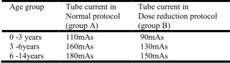
[image:2.595.48.273.573.635.2]All patients with suspected brain pathology, categorized randomly into two groups by lottery system (group A and group B), were subjected to CT Brain examination on spiral CT machine: one group with normal protocol and other group with dose reduction protocol. The data included age; sex; indications for scan categorized into four items: trauma, tumor, congenital and infection; and scan parameters: kVp, mA. The dose reduction was done as per Table 1.
Table 1. Dose reduction criteria
Age group Tube current in Normal protocol (group A)
Tube current in Dose reduction protocol (group B)
0 -3 years 110mAs 90mAs
3 -6years 160mAs 130mAs
6 -14years 180mAs 150mAs
Assessment of image quality
Qualitative assessment: All imaging studies were reviewed in
blind random order and independently by two experienced radiologists. The guidelines for image assessment were adapted from the standard international atomic energy agency(IAEA) protocol. The quality of each image were assessed in terms of anatomical criteria (Table 3), diagnostic confidence (Table 4) and overall quality using a five point scale grade for each element (Tables 2). If there was a greater than two grading score discrepancy by the two assessors, the image was reviewed again until an agreement was reached.
Diagnostic confidence was assessed in terms of clinical disease or pathological lesions in head (Kritsaneepaiboon et al., 2014).
Table 2. Five point grading scale
Score Anatomical criteria
Diagnostic confidence
Overall quality
1 Not possible Non- diagnostic Non-acceptable,
repeat scan
2 Difficult Low Low
3 Acceptable Acceptable Acceptable
4 Good Good Good
5 Excellent Excellent Excellent
Table 3. Anatomical criteria
Grey-white differentiation Delineation of ventricular contours Delineation of basal ganglia Definition of gyri
Table 4. Diagnostic confidence
Pathologies assessed Bleeding
Infarction Mass lesion
Statistical analysis
Different findings between the normal protocol and reduced radiation protocol images were compared to check for any significant differences by using Independent sample t-test. The statistical significance level was set at 0.05.
OBSERVATIONS AND RESULTS
Hence, it was concluded that both observers found no significant difference in the anatomic delineation, diagnostic confidence and over all image quality between normal protocol and reduced radiation protocol.
DISCUSSION
Medical imaging has become a significant source of radiation exposure. In the developed nations, the contribution from medical imaging can be greater (Kubo, 2019). In the United States, medical radiation exposure is reported to have exceeded natural back ground radiation from environment. If we leave this upward trend unchecked, we will take a risk of increase in malignant disease in the future. Prevention of further expansion of medical radiation exposure is necessary. Growth of medical radiation exposure is largely attributable to the increase in the number of CT examinations (O’Connor, 2012). Since children are more sensitive to radiation and at a relatively greater risk of carcinogenesis than are adults, it is even more important to avoid unnecessary radiation exposure in this group than in the adult population (Kritsaneepaiboon, 2014). An optimal CT radiation dose in MDCT studies can be achieved by modifying the acquisition parameters, using the automatic exposure control, and adjusting acquisition parameters for patient size or iterative reconstruction. Kritsaneepaiboon et al. (2014) retrospectively did a study to compare the multidetector CT(MDCT) radiation doses between default settings(phase 1) and a revised dose reduction
protocol (phase 2) and to determine whether the diagnostic confidence can be maintained with imaging quality made under the revised protocol in paediatric head, chest and abdominal CT studies. They found that paediatric CT radiation doses can be significantly reduced from manufacturer’s default protocol while still maintaining anatomical delineation, diagnostic confidence and overall imaging quality. Ledenius21
et al used computer-simulated images that were based on
[image:3.595.88.503.81.151.2]existing patient examinations (retrospective material) and studied several different tube currents per patient, i.e. paired data. They did a study to investigate the effect of tube current on diagnostic image quality in paediatric cerebral multidetector CT (MDCT) images in order to identify the minimum radiation dose required to reproduce acceptable levels of different diagnostic image qualities. Their ages ranged from newborn to 15 years. Three experienced radiologists blindly and randomly assessed the resulting images from two different levels of the brain with regard to reproduction of structures and overall image quality. Final data were evaluated using the non-parametric statistical approach of inter-scale concordance. The minimum value of tube current–time product (mAs) required to reproduce an image of sufficient diagnostic quality was established in relation to the age of the patient. The corresponding CT dose index values by volume (CTDIvol (mGy)) were also established. In conclusion, acceptable reproduction of low contrast structures was possible at CTDIvol values down to 20 mGy (patients 1–5 years old). We studied 100 patients divided in three age groups.
Table 5. Mean of findings of evaluation of films by observer 1
mAs NUMBER MEAN STD.DEVIATION
Observer 1 anatomical delineation Decreasd 50 3.98 0.845
Normal 50 4.10 0.814
Observer 1 diagnostic confidence Decreased 50 4.50 0.580
Normal 50 4.48 0.614
Observer 1 quality confidence Decreased 50 4.20 0.700
[image:3.595.60.535.190.262.2]Normal 50 4.24 0.687
Table 6. Levene’s test for equality of variances of observer 1
mAs F Sig t df Sig.(2-tailed)
Observer 1 anatomical delineation Equal variances assumed 0.034 0.854 -0.723 98 0.471
Equal variances not assumed -0.723 97.868 0.471
Observer 1 diagnostic confidence Equal variances assumed 0.247 0.620 0.167 98 0.867
Equal variances not assumed 0.167 97.687 0.867
Observer 1 quality confidence Equal variances assumed 0.000 0.983 -0.288 98 0.774
Equal variances not assumed -0.288 97.966 0.774
Table 7. Mean of findings of evaluation of films by observer 2
mAs Number Mean std.Deviation
Observer 2 anatomical delineation Decreasd 50 3.42 0.950
Normal 50 3.76 0.771
Observer 2 diagnostic confidence Decreased 50 4.34 0.688
Normal 50 4.48 0.707
Observer 2 quality confidence Decreased 50 4.24 0.744
Normal 50 4.44 0.705
Table 8. Levene’s test for equality of variances of observer 2
mAs F Sig t df Sig.(2-tailed)
Observer 2 anatomical delineation Equal variances assumed 5.249 0.024 -1.966 98 0.520
Equal variances not assumed -1.966 94.031 0.520
Observer 2 diagnostic confidence Equal variances assumed 0.097 0.756 -1.003 98 0.318
Equal variances not assumed -1.003 97.932 0.318
Observer 2 quality confidence Equal variances assumed 0.002 0.967 -1.380 98 0.171
[image:3.595.133.460.299.369.2] [image:3.595.81.516.401.472.2]
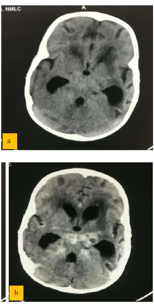
Figure 1. NCCT(a) AND CECT(b) head in a 4 year old child with decreased mAs protocol in a patient of meningitis showing acceptable image quality at reduced radiation dose protocol
It is important to use patients of similar size and developmental stage when assessing the effect of a dose reduction, as the image quality at a constant radiation dose is dependent on the patient attenuation. This study was limited by only using two observers who were experienced radiologists in the field. The results may have been different if less experienced radiologists had participated in the study. Reductions in radiation dose should therefore be implemented with care, using a safety margin and supervision for a period of time. There is a risk of bias in assessing the overall impression of image quality, as radiologists tend to recognise and favour their old settings. The age distribution of patients in this study was representative of patients within our paediatric department and surgery department. The majority of patients (between 5 years and 10 years old) are at an active age, resulting in an increased number of accident-related injuries. Among the younger children (under 1 year old), pathology is often suspected at birth, resulting in a scarcity of patients between 6 months and 1
year old. Follow-up MDCT examinations of shunt-treated hydrocephalus patients are common in paediatric patients, with the ventricles being of special interest. These patients are repeatedly scanned during an extended period of time, leading to relatively high accumulated patient doses. Protocols especially designed for hydrocephalus follow-up examinations were already in use prior to this study, with the radiation doses being up to 50% lower than those in the standard protocols. The images produced at these radiation doses agree relatively well with the results of this study as regards the minimum tube current–time product required to reproduce the ventricles acceptably for patients older than 1 year. In our study tube current was reduced approximately by 15%. The anatomic delineation, diagnostic confidence and overall image quality showed no significant difference between normal radiation protocol and reduced radiation protocol (Figure 1a & b). Based on the results of this study, the tube currents in our standard protocols for patients aged between 1 and 10 years of age can be lowered by approximately 15%.
Conclusion
This was a prospective study to assess the radiation dose reduction protocol and evaluate anatomical delineation, diagnostic confidence and imaging quality of CT brain in children using this protocol. 100 patients of pediatric age group were subjected to CT scan head examination on spiral CT machine. 50 of them were subjected to normal protocol and 50 of them were subjected to decreased radiation protocol (decreased tube current) randomly. All of 100 CT films were assessed randomly by the two observers and scores were given on the basis of anatomic delineation, diagnostic confidence and overall quality. After assessment by the two observers we applied student t test to know whether there is significant difference between the images of normal protocol and reduced radiation protocol. After applying it, we observed that there was no significant difference between the images of normal protocol and reduced radiation protocol. We conclude that tube current can be reduced up to certain level (in our study: 15%) without significant decrease in image quality and diagnostic confidence.
REFERENCES
Brenner DJ, Elliston CD, Hall EJ, Berdon WE. 2001. Estimated risks of radiation-induced fatal cancer from pediatric CT. Am J Roentgenol.176(2):289–96.
Frush DP, Slack CC, Hollingsworth CL, Bisset GS, Donnelly LF, Hsieh J, et al. 2002. Computer-simulated radiation dose reduction for abdominal multidetector CT of pediatric patients. Am J Roentgenol.179(5):1107–13.
Goldman LW. 2007. Principles of CT: radiation dose and image quality. J Nucl Med Technol., 35(4):213-25.
Hamberg LM, Rhea JT, Hunter GJ, Thrall JH. 2003. Multi-detector row CT: Radiation dose characteristics. Radiology. 226(3):762–72
Holmedal LJ, Friberg EG, Børretzen I, Olerud H, Lægreid L, Rosendahl K. 2007. Radiation doses to children with shunt-treated hydrocephalus. Pediatr Radiol.37(12):1209–15. International Atomic Energy Agency. Dose reduction in CT
Jangland L, Sanner E, Persliden J. 2004. Dose reduction in computed tomography by individualized scan protocols.
Acta radiol., 45(3):301–7.
Jurik a G, Jessen K a, Hansen J. 1997. Image quality and dose in computed tomography. Eur Radiol [Internet]. 7(1):77– 81.
Kamel IR, Hernandez RJ, Martin JE, Schlesinger AE, Niklason LT, Guire KE. 1994. Radiation dose reduction in CT of the pediatric pelvis. Radiology [Internet].190(3):683–7. Kopp AF, Heuschmid M, Claussen CD. 2002. Multidetector
helical CT of the liver for tumor detection and characterization. Eur Radiol.12(4):745–52.
Kritsaneepaiboon S, Siriwanarangsun P, Tanaanantarak P, Krisanachinda A. 2014. Can a revised paediatric radiation dose reduction CT protocol be applied and still maintain anatomical delineation, diagnostic confidence and overall imaging quality? Br J Radiol., 87:20140032
Kubo T, Ohno Y, Nishino M, Lin PJ, Gautam S, Kauczor HU,
et al. 2016. Low dose chest CT protocol (50 mAs) as a
routine protocol for comprehensive assessment of intrathoracic abnormality. Eur J Radiol Open., 3:86–94. Ledenius K, Gustavsson M, Johansson S, Stålhammar F,
Wiklund LM, Thilander-Klang A. 2009. Effect of tube current on diagnostic image quality in paediatric cerebral multidetector CT images. Br J Radiol., 82(976):313–20. Mayo JR, Hartman TE, Kyung Soo Lee, Primack SL, Vedal S,
Muller NL. 1995. CT of the chest: Minimal tube current required for good image quality with the least radiation dose. Am J Roentgenol.164(3):603–7.
Mullins ME, Lev MH, Bove P, O’Reilly CE, Saini S, Rhea JT,
et al. 2004. Comparison of Image Quality between
Conventional and Low-Dose Nonenhanced Head CT. Am J
Neuroradiol. 25(4):533–8.
Nishizawa K, Maruyama T, Takayama M, Okada M, Hachiya J, Furuya Y. 1991. Determinations of organ doses and effective dose equivalents from computed tomographic examination. Br J Radiol., 64(757):20–8.
O’Connor Gt HH. 2012. LUng cancer screening, radiation, risks, benefits, and uncertainty. JAMA J Am Med Assoc [Internet].307(22):2434–5.
Rao P, Bekhit E, Ramanauskas F, Kumbla S. 2013. CT head in children. Eur J Radiol. 82(7):1050–8.
Sohaib SA, Peppercorn PD, Horrocks JA, Keene MH, Kenyon GS, Reznek RH, 2001. “The effect of decreasing mAs on image quality and patient dose in sinus CT,” Br J Radiol.,
74(878):157-61
Tang K, Wang L, Li R, Lin J, Zheng X, Cao G. 2012. Effect of low tube voltage on image quality, radiation dose, and low-contrast detectability at abdominal multidetector CT: Phantom study. J Biomed Biotechnol. Article ID 130169, 6 pages.
Tsapaki V, Rehani M, Saini S. 2010. Radiation Safety in Abdominal Computed Tomography. Semin Ultrasound, CT MRI [Internet]. 31(1):29–38.
Verdun FR, Meuli RA, Bochud FO, Imsand C, Raimondi S, Schnyder P, et al. 1996. Image quality and dose in spiral computed tomography. Eur Radiol.6(4):485–8