Original Article
Screening and identification of antisense accessible
sites of the human telomerase reverse transcriptase
gene in prostate cancer cell lines
Fangmin Chen1,3, Bo Yan2, Jiaqi Shi3, Yu Chen4, Dengbao Li3, Xinan Jiang3, Deshuai Ren5
Departments of 1Urology, 4Library, Tianjin Third Central Hospital, Tianjin 300171, China; 2Department of
Urol-ogy, Guizhou Provincial People’s Hospital, Guiyang 550004, China; 3Department of Urology, Affiliated Hospital of
Guizhou Medical University, Guiyang 550004, China; 5Department of Neurosurgery, The Third Affiliated Hospital
of Qiqiha’er Medical College, Qiqiha’er, China
Received March 2, 2017; Accepted March 25, 2017; Epub May 1, 2017; Published May 15, 2017
Abstract: Selecting optimal antisense binding sites remains difficult during application of antisense oligonucle -otide techniques. A random oligonucle-otide library/RNase H cutting method combined with software analysis has emerged as an effective strategy. Here, we used the method to explore therapy targeted to the human telomerase reverse transcriptase (hTERT) gene in prostate cancer. A 20 mer random oligonucleotide library was synthesized and then hybridized with total hTERT complementary RNA (cRNA) that had been transcribed in vitro. The RNA in the specific hybrid double-stranded duplex was hydrolyzed by RNase H, so the total hTERT cRNA was cut into fragments. After primer extension and autoradiography, the antisense accessible sites (AAS) of hTERT were selected. Optimal AAS with obvious stem-loop structures were identified after RNA structure software analysis. The complementary antisense oligonucleotides (AS-ODNs) of these AAS were synthesized and transferred into cells expressing high hTERT levels and the effects upon cell growth, apoptosis and expression of hTERT mRNA were determined. There were twenty-six AAS of the hTERT gene screened, and seven AAS, which had obvious stem-loop structures were selected. After transfection of complementary antisense oligonucleotides of these optimal AAS, the hTERT mRNA expression levels in the cells were significantly decreased. Cellular growth was significantly inhibited and apoptosis was detected. Screening antisense accessible sites of a target gene through random oligonucleotide library/RNase H cutting in combination with computer analysis was effective. The resulting complementary antisense oligonucle-otides efficiently blocked the biological function of the hTERT gene suggesting their potential for cancer treatment.
Keywords: hTERT, random oligonucleotide library, screening, antisense accessible sites, targeted blocking, stem-loop structure
Introduction
The human telomerase is mainly composed of human telomerase reverse transcriptase (hTE- RT) and human telomerase RNA (hTR). The hTR acts as the reverse transcription template and is complementary to telomeric repeat sequenc-es. The hTERT is the catalytic subunit that cata-lyzes the reverse transcription reaction. These two components work together to maintain the length of telomeres. The expression of hTERT mRNA is related to the level of telomerase ac- tivity and this is considered to be the rate-limit-ing factor [1, 2]. The expression of hTERT is also associated with cellular aging and
tumorigene-sis, and high telomerase activity is observed in over 85% of human cancer cells [1-3], whereas normal tissues and cells have minimal expres- sion.
(PSA). DU145 cells have moderate metastatic potential. In both these prostate cancer cell lines telomerase activity is likely to be high because telomerase can be used as a tumor marker in the diagnosis of prostatic intraepithe -lial neoplasia and prostate cancer [6].
Targeting hTERT in prostate cancer cells may; therefore, provide an effective method of can-cer therapy. At present research strategies to inhibit a targeted gene’s mRNA expression include antisense oligodeoxynucleotide tech-niques, RNA interference technology, ribozyme technology, small molecule compounds, and immune therapy, but the first two techniques are the most researched and applied in the area (7). Antisense oligonucleotides have been used as effective methods of studying gene function and gene expression regulation in addition to targeted biological treatments, and telomerase has been successfully targeted with antisense by many investigations [7, 8]. These studies show the potential for anti- sense therapy in cancer treatment, but the ef- ficacy of the treatment could be improved by selection of antisense oligonucleotides target-ed to the optimum antisense accessible sites that most effectively shut down the target mRNA. Although antisense oligodeoxynucleo-tide techniques have been used for 32 years [9], selection of the most effective antisense binding site is often difficult. Early methods of designing antisense oligonucleotides were mainly based on the primary structure of RNA molecules, such as the translation initiation site. However, this leads to reduced specificity of antisense oligonucleotides due to the high homology of translation initiation sites in a wide range of genes. The effectiveness of anti-sense accessible site screening by the “Ran- dom shotgun” method is only 2% to 5% be- cause the secondary structure of RNA mole-cules is ignored [10]. Currently, computer-as- sisted folding programs can forecast the sec-ondary structure of RNA molecules using the free energy principle. From a structural molecu-lar biology perspective, sites without intramo-lecular base pairing in the loop region are anti-sense active accessible sites, while the stems are non-active accessible sites. The acknowl -edged five antisense accessible sites of the hTERT gene are screened out using only a com-puter-assisted folding program [1]. However, for long-chain RNA, computer-assisted folding programs will form many different structures with similar free energies, which make judging
the correct folding conformation very difficult. Using computer folding programs to predict antisense binding sites seems to be ineffective without also considering screening and identifi -cation in vitro.
In this study, a random oligonucleotide library/ RNase H cutting method was combined with computer software analysis that has been suc-cessfully used in other studies to screen the antisense binding sites of target sequences in vitro [11]. Twenty-six antisense accessible sites of the hTERT gene were screened to select the optimal ones [10, 12, 13]. Stem-loop structures were selected as these have been shown to have higher activity than other common mRNA structures, and have been shown to have bet-ter accessibility than other mRNA structures [14, 15]. The complementary phosphorothioate antisense oligonucleotides of these antisense accessible sites were then synthesized and transferred into the prostate cancer cell lines PC-3 and DU145 that express high levels of hTERT, in order to study their influence on the biological behavior of prostate cancer cells. To our knowledge, this is the first report using the random oligonucleotide library/RNase H cut-ting method combined with software analy- sis to explore targeted therapy of the human telomerase reverse transcriptase (hTERT) gene in prostate cancer.
Materials and methods
Design and synthesis of the random oligo-nucleotide library
The random oligonucleotide library was synthe-sized according to previously described meth-ods [16, 17]. Specific requirements were as follows: (1) the length was 20 bp; (2) the con-tent ratio of A:C:G:T was 1.5:1.25:1.15:1.0; (3) the sequence was 5’-MMMNNNNNNNNNNN- NNNNMM-3’. G, A, T, C represents the four dif-ferent deoxidizing nucleotides, M represents methylation deoxycytidine, N represents ran-dom oligodeoxynucleotides. The ranran-dom oligo-nucleotide library was synthesized and puri- fied by the Nanjing Shengxing Biological Engi-neering Company (China).
In vitro transcription of the target gene
tran-scription of the gene was performed by the fol-lowing steps. The plasmid was digested by the restriction enzyme Xba I (Fermentas company) to linearize it, and then transcribed to comple-mentary RNA (cRNA) using the T7 transcription kit (Fermentas company) in vitro [18]. The total
Design of the extension primers
[image:3.612.91.368.97.634.2]According to the mRNA sequence of the hTERT gene contained in the pcDNA-3.1 plasmid (+) (3402 bp) and the technical bulletin of the Primer Extension Kit (Promega Corporation),
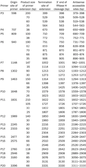
Table 1. Antisense accessible regions of hTERT shown by primer extension analysis
Primer site of Origin primer
Product size of primer extension (bp)
Termination site of extension
3’ end of RNase H cleavage (bp)
Antisense accessible sites (bp)
P3 598 200 399 398 379~398
70 529 528 509~528
60 539 538 519~538
35 564 563 544~563
P4 707 38 670 669 650~669
P5 809 100 710 709 690~709
38 772 771 752~771
P6 940 190 751 750 731~750
82 859 858 839~858
70 871 870 851~870
66 875 874 855~874
35 906 905 896~905
P7 1148 147 1002 1001 982~1001
35 1114 1113 1094~1113
P8 1302 90 1213 1212 1193~1212
30 1273 1272 1253~1272
P9 1463 150 1314 1313 1294~1313
66 1398 1397 1378~1397
38 1426 1425 1406~1425
P10 1648 70 1579 1578 1559~1578
26 1623 1622 1603~1622
P11 1831 210 1622 1621 1602~1621
105 1727 1726 1707~1726
30 1802 1801 1782~1801
25 1807 1806 1787~1806
P12 1989 140 1850 1849 1830~1849
30 1960 1959 1940~1959
P13 2245 30 2216 2215 2196~2215
P14 2333 82 2252 2251 2232~2251
30 2304 2303 2284~2303
P15 2477 30 2448 2447 2428~2447
P16 2575 66 2510 2509 2490~2509
30 2546 2545 2526~2545
P17 2760 118 2643 2642 2623~2642
P18 2946 38 2909 2908 2889~2908
P19 3160 85 3076 3075 3056~3075
30 3131 3130 3111~3130
P20 3396 66 3331 3330 3311~3330
reaction volume was 50 μl and was incubated for 2 hours at 37°C. RNase-free DNase I was added to a concentration of 1 U DNase/1 μg template DNA and then incubated for 15 min at 37°C to remove the DNA templates. 85 µl of diethylpyro-carbonate (DEPC)-treated wa- ter was then added with 15 µl of 3 M sodium acetate. The cRNA was extracted first with an equal volume of 1:1 phenol/ chloroform mixture, and then, twice, with an equal volume of chloroform. The aqueous pha- se was collected and trans-ferred into a new tube. The cRNA was then precipitated, by adding 2 volumes of ethanol and incubating at -20°C for 30 min. After centrifugation, to re- move the supernatant, the pel-let was rinsed with 500 µl of cold 70% ethanol. The cRNA was resuspended in 20 µl of DEPC-treated water and then stored at -70°C after its con-centration was determined by UV spectrophotometry and it was identified by 1% agarose gel electrophoresis.
RNase H cleavage reaction
we used primer premier 5.0 to design a reverse extension primer every 150 to 300 bases from the 5’ to 3’ end of the RNA [19]. There were a total of 20 primers designed and synthesized by Beijing Sunbiotech Co., Ltd (Table 1).
Primer extension reaction
The primer extension reaction involved primer labeling, marker labeling and the primer ex-tension reaction, which were all performed by standard techniques and according to the man-ufacturer’s guidelines (purchased from Beijing Yahui Biological Engineering Company).
Polyacrylamide gel electrophoresis and auto-radiography
The primer extension products were analyzed on a 16×18 cm denaturing polyacrylamide gel
[image:4.612.93.527.74.271.2]RNase H digestion and screening, the best significant stem-loop structures of the region were designated as the best antisense binding sites. We based these best binding sites on the stem-loop structures because we expect these to be easily accessible because the activity of a stem-loop structure is higher than that of other common mRNA structures. Seven of the best antisense accessible sites were screen- ed and named AS-ODN1~AS-ODN7 respectively. A selected region (1468~1487 bp), which had an obvious stem-loop structure according to the computer analysis, but did not have cleav-age sites in the random oligonucleotide library/ RNase H digest method was used as a control antisense accessible site (see Figure 2) and the synthesized complementary control anti-sense oligonucleotide was named AS-ODN0, the sequence was 5’-GAGATGAACTTCTTGGTG-
Figure 1. Seven obvious binding site regions and fragment size obtained by primer extension. Primer extension anal-ysis for the selection of antisense accessible sites of hTERT by random oligonucleotide library/RNase H cleavage method. The seven selected sites are highlighted and labeled with their corresponding serial number. M: φX174/ Hinf I DNA Marker; P1~P20: Primer extension products of P1~P20; the positive control product is an 87 mer.
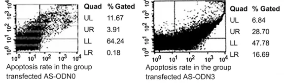
Figure 2. In vitro experiments showed a higher rate of apoptosis of antisense AS-ODN3 in comparison with AS-ODN0 (control) of prostate cancer cells. The right hand section of the flow cytometry graphs represents the apoptotic cell population. AS-ODN3 had higher apoptosis rates than the AS-ODN0 control.
containing 8% acrylamide (19:1 acrylamide:bis), 7 M ur- ea and TBE 1×buffer.
Antisense binding site selec-tion and the design of control antisense oligonucleotides
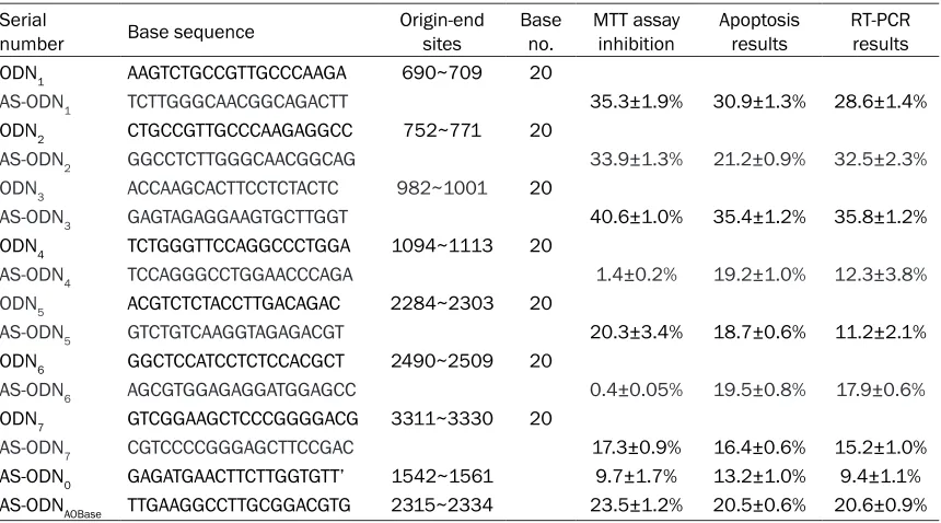
[image:4.612.89.377.346.430.2]TT-3’. Another control was the most efficient hTERT antisense oligonucleotide of the five that were published by AOBase, the sequence was 5’-TTGAAGGCCTTGCGGACGTG-3’ and this was named AS-ODNAOBase. This control sequence was one used previously by Kraemer et al. [3] and was listed as ASt2315 in their study to reflect its location start point of 2315 in the mRNA sequence.
Cell culture and gene transfection
Human prostate cancer cell lines, androgen-independent PC-3 cells [21] and DU145 cells were obtained from the American Type Culture Collection and maintained in RPMI 1640 medi-um for PC-3 cells or DMEM for DU145 cells, with 10% fetal calf serum, 100 U/ml penicillin and 100 μg/ml streptomycin. Cells were cul -tured at 37°C in a humidified 5% CO2 atmo-sphere and passaged once every 3 days. At logarithmic growth phase the PC-3 cells were seeded in culture plates, following the protocol of the liposome Oligofectamine™ Reagent kit (Invitrogen Company, Cat. No. 12252-011) for the transfection of the AS-ODNs (200 nM). To avoid oligonucleotide decomposition in the cells, three bases at both ends of oligonucle-otides were phosphorothioated at the time of synthesis [22]. The transfection efficiency was between 57.7% and 58.1%.
MTT assay for detecting cell growth activity
100 μl PC-3 prostate cancer cells were seeded in each well of 96-well plates at a density of 5×10³/100 μl. The wells were set as the un-transfected control group, control group (AS- ODN0), AOBase control group (AS-ODNAOBase) and AS-ODN1~AS-ODN7 experimental groups, each oligonucleotide was also set at 200, 400 and 600 nmol/L concentrations. Each group had five duplicate wells. 10 μl of 5 mg/ml MTT (pH = 7.4) was added after transfection, for 12, 24 and 36 hours. The cells were continued in culture for 4 hours and the supernatant was removed after centrifuging at 4000 r/min for 10 minutes. 100 μl DMSO was added and left at room temperature for 10 minutes to fully dissolvethecrystals. A microplate reader was used to detect the absorbance (A490, 630 dual-wavelength method). The cell growth inhibition rate (%) = (1-the average A490, 630 value of ex- perimental group/the average A490, 630 value of control group)×100% [23].
Detection of apoptosis
DU145 prostate cancer cells were seeded in 24-well plates at a density of 5×10³/100 μl with each well inoculated with 500 μl. The con -ditions were set as the untransfected control group, control group (AS-ODN0), AOBase con- trol group (AS-ODNAOBase) and AS-ODN1 ~AS-ODN7 experimental groups, each oligonucle-otide was also used at 200, 400 and 600 nmol/L concentrations. Each group used five duplicate wells. Annexin V-FITC and PI double staining flow cytometry was used to detect cell apoptosis after transfection at 12, 24 and 36 hours [24].
Detection of hTERT expression levels
Cultured cells were collected 12-24 h after transfection and Trizol reagent was used to isolate total RNA. This was reverse transcribed into cDNA. PCR primers were synthesized ac- cording to references, P1: 5’-CGGCTTTTGTTC- AGATGCC-3’ (sense), P2: 5’-AGCACACATGCGT- GAAACCT-3’ (antisense). The product length was expected to be 301 bp (30). The PCR in- ternal reference was GAPDH, 5’-GAAGGTGAA- GGTCGGAGTC-3’ (sense), 5’-GAAGATGGTGATG- GGATTTC-3’ (antisense), the product length was expected to be 226 bp. The PCR reaction system was: 1 μl DNA template, 0.5 μl 10 mM dNTPs, 2.5 μl 10×PCR buffer, 1.5 μl 25 mM MgCl2, 1 μl of two 10 mM primers, 1 μl 25 U/μl Taq enzyme, 16.5 μl ddH2O to a total volume of 25 μl. The cycling conditions were a first pre-denaturation at 95°C for 5 min, then 94°C for 40 sec, 60°C for 45 sec and 72°C for 60 sec for 30 cycles and a final extension step at 72°C for 10 min. The products were electrophoresed on 2.5% agarose gels and observed under a UV lamp and photographed. The band density was measured using ImageJ and calculated for each group: gray value of hTERT/GAPDH. hTERT expression inhibition rate (%) = [1-the average (hTERT/GAPDH) value of experimental group/ the average (hTERT/GAPDH) value of untrans-fected control group]×100%.
Morphological observation
con-ventional embedding and slicing, the changes in the cell’s ultrastructure were observed under a transmission electron microscope.
Statistical analysis
SPSS11.0 statistical software (SPSS Inc., Chi- cago, IL, USA) was used for data analysis and the outcomes were expressed as mean ± stan-dard error. Differences were evaluated using two independent samples with Student’s t test, a p value of <0.05 was considered statistical- ly significant.
Results
hTERT mRNA primer extension reaction and autoradiography
As shown in Figure 1, P1 and P2 located in the non-coding region of hTERT had no clear exten-sion product that could be seen after the prim-er extension reaction and autoradiography. There were 26 antisense binding sites select- ed after analysis of the extension products of P3~P20. The size of the fragment obtained from the primer binding with antisense and the start and end sites of the product are shown in Table 1. These are the primer initiation site, extension of the product size, RNase H 3’ end cleavage site and the corresponding antisense binding sites.
RNA structure analysis
Analysis of the secondary structure of hTERT mRNA, showed that among the 26 antisense accessible sites, seven sites had obvious stem-loop structures as forecast by RNA structure software. The 7 antisense accessible sites of the hTERT gene that were considered to be the best because of the high chance of being a stem-loop are represented as ODN1-7 in Table 2, and were located in the 690 bp~709 bp, 752 bp~771 bp, 982 bp~1001 bp, 1094 bp~1114 bp, 2284 bp~2303 bp, 2490 bp~2509 bp, and 3311 bp~3330 bp areas of the hTERT mRNA. The seven complementary antisense oligonu-cleotide sites were AS-ODN1~AS-ODN7 (shown in Table 2).
Results of the MTT assay
[image:6.612.93.523.95.334.2]Cell growth was investigated as a measure of the antisense oligonucleotide’s potential for anti-cancer therapy. After transfection with 200 nmol/L of AS-ODN0, AS-ODNAOBase, and AS-ODN1~AS-ODN7 for 12-36 h, the growth of the PC-3 cell growth slowed down, each to a different extent. Each group exhibited the most significant inhibition after 24 h transfec -tion, and the inhibition rates were 9.7±1.7%, 23.5±1.2%, 35.3±1.9%, 33.9±1.3%, 40.6± 1.0%, 1.4±0.2%, 20.3±3.4%, 0.4±0.05%, and
Table 2. Antisense accessible sites and their complementary oligonucleotide sequences selected by random oligonucleotide library/RNase H cleavage combined with RNA structure analysis
Serial
number Base sequence Origin-end sites Base no. MTT assay inhibition Apoptosis results RT-PCR results ODN1 AAGTCTGCCGTTGCCCAAGA 690~709 20
AS-ODN1 TCTTGGGCAACGGCAGACTT 35.3±1.9% 30.9±1.3% 28.6±1.4%
ODN2 CTGCCGTTGCCCAAGAGGCC 752~771 20
AS-ODN2 GGCCTCTTGGGCAACGGCAG 33.9±1.3% 21.2±0.9% 32.5±2.3%
ODN3 ACCAAGCACTTCCTCTACTC 982~1001 20
AS-ODN3 GAGTAGAGGAAGTGCTTGGT 40.6±1.0% 35.4±1.2% 35.8±1.2%
ODN4 TCTGGGTTCCAGGCCCTGGA 1094~1113 20
AS-ODN4 TCCAGGGCCTGGAACCCAGA 1.4±0.2% 19.2±1.0% 12.3±3.8%
ODN5 ACGTCTCTACCTTGACAGAC 2284~2303 20
AS-ODN5 GTCTGTCAAGGTAGAGACGT 20.3±3.4% 18.7±0.6% 11.2±2.1%
ODN6 GGCTCCATCCTCTCCACGCT 2490~2509 20
AS-ODN6 AGCGTGGAGAGGATGGAGCC 0.4±0.05% 19.5±0.8% 17.9±0.6%
ODN7 GTCGGAAGCTCCCGGGGACG 3311~3330 20
AS-ODN7 CGTCCCCGGGAGCTTCCGAC 17.3±0.9% 16.4±0.6% 15.2±1.0%
17.3±0.9% respectively. The difference was statistically significant between the groups (P<0.05) so different oligonucleotides had dif-ferent efficiencies at targeting cell growth and AS-ODN3 was most effective. The three differ-ent concdiffer-entrations of oligonucleotides had no
[image:7.612.90.370.71.354.2]0.05) so different oligonucleotides had differ-ent efficiencies at inhibition of hTERT and AS-ODN3 was most effective. The effect of differ-ent concdiffer-entrations of oligonucleotides was not significant on the inhibition of hTERT mRNA’s expression (P>0.05).
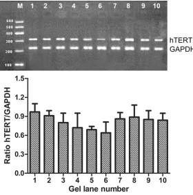
Figure 3. After seven antisense oligonucleotides (AS-ODNn) and control group were transfected into PC-3 cells, hTERT was blocked as detected by RT-PCR electrophoresis. RT-PCR detection of the blocking effects of antisense oligo -nucleotides on hTERT mRNA expression of PC-3 cells: 1. not transfected con-trol. 2. AS-ODN0. 3. AS-ODNAOBase. 4. AS-ODN1. 5. AS-ODN2. 6. AS-ODN3. 7. AS-ODN4. 8. AS-ODN5. 9. AS-ODN6. 10. AS-ODN7. M: PCR marker (100 bp~600 bp); GAPDH as an inner control (226 bp). The experiment was repeated six times and the mean values are presented in the graph.
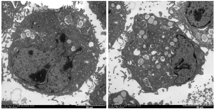
Figure 4. Antisense (AS-ODN3) and the control group were observed 24 hours after transfection of PC-3 cells in cell morphology. A: The cell morphology before antisense oligonucleotide was transfected. B: The cell morphology 24 h after 200 nmol/L AS-ODN3 was transfected.
significant difference in the inhibition of growth (P>0.05).
Detection of apoptosis
After transfection with 200 nmol/L of AS-ODN0, AS- ODNAOBase, and AS-ODN1 ~AS-ODN7 for 12-36 h, the cells in each group showed the most obvious apoptosis after 24 h. The apoptosis rates were 13.2±1.0%, 20.5±0.6%, 30.9±1.3%, 21.2±0.9%, 35.4± 1.2%, 19.2±1.0%, 18.7±0.6%, 19.5±0.8% and 16.4±0.6% respectively. The difference was statistically significant between the groups (P<0.05) so different oligonucleotides had different efficiencies at causing apoptosis and AS- ODN3 was most effective. The three different concentrat- ions of oligonucleotides sh- owed no significant differenc -es in the rate of apoptosis (P>0.05) (Figure 2).
Detection of hTERT expres-sion levels
[image:7.612.90.375.466.577.2]Morphological observation
Under an inverted microscope, the prostate cancer cells in the control group were found to be polygonal in shape. After the cells were transfected with the antisense oligonucleoti- des, the cell volume decreased and the cell morphology was irregular (Figure 4). Most of the cells showed changes typical of apoptosis under electron microscopy after transfection, including condensed nucleus, shrunk nuclear membrane, and marginalized and budded ch- romatin, compared to control cells with large nucleus and dense microvilli (Figure 5).
Discussion
The aim of this study was to use a random oli-gonucleotide library/RNase H cutting method combined with software analysis to explore tar-geted therapy of the hTERT gene in prostate cancer. The results showed that targeting seven optimal antisense accessible sites decreased hTERT mRNA expression levels in the prostate cancer cell lines and significantly inhibited cell growth producing detectable levels of apopto-sis. An alternative method for targeted anti-cancer therapy is RNA interference (interfer-ence RNA, RNAi) which induces sequ(interfer-ence-spe- sequence-spe-cific posttranscriptional gene silencing by use
[image:8.612.87.529.70.293.2]of double-stranded small interfering RNA (SiRNA) is characterized by strong inhibition, high stability, and easier cellular uptake. This method has been used successfully to study gene function and in anti-tumor research [25, 26]. However, SiRNA has some disadvantages; it cannot be used to bind anti-cancer drugs because of its double-stranded structure which has lower specificity than antisense oligodeoxy -nucleotides and has a less efficient transfer into cells, decreased specificity due to energy diffusion in later stage silencing, and poor sta-bility in the body, also, it has not been widely used in mammalian systems [27]. For these re- asons, we used antisense therapy in this study. It can be challenging finding the optimum anti -sense accessible sites in the target gene and there are many different methods for select- ing them, including oligonucleotide arrays [28], reverse transcription with random oligodeoxy-nucleotide libraries [29], mRNA accessible site tagging [30] serial analysis of antisense bind-ing sites [31], and mRNA antisense-accessible sites library using native mRNA [32]. A random oligonucleotide library/RNase H cutting meth-od combined with computer software analysis has been used successfully elsewhere and in this study we referred to the experience of Ho et al. [16] and Lloyd et al. [33], and synthesized
an oligonucleotide library which contained all possible random 20 mer sequences with a nu- mber of methylated bases on both ends. The sites identified for the oligonucleotides hybrid -ized with the hTERT template chain sites, also referred to antisense accessible sites. In this study, 26 antisense accessible sites were scr- eened in the hTERT gene, 7 of which were in significant stem-loop regions suggesting better accessibility when combined with RNA struc-ture computer software analysis, so these were considered to be the best antisense accessible sites. Synthesis of the complementary strands of the best antisense accessible sites were designated as AS-ODN, we transfected the AS- ODN into prostate cancer cells and this sig- nificantly inhibited their growth. The highest inhibition rate was with AS-ODN3 (40.6±1.0%). Apoptosis was highest for the AS-ODN3 group (35.39±1.2%), the hTERT mRNA expression le- vel was also significantly inhibited and by the highest amount with AS-ODN3 (35.8±1.2%). In this study, of the 7 selected binding sites of the antisense oligonucleotides targeted against hTERT, AS-ODN3 was most effective at shut- ting down hTERT expression. Each oligonucle-otide was used at three different concentra-tions 200, 400 and 600 nmol/L but we found that there were no significant differences in the slowing of cell growth, apoptosis rates or ex- pression levels between the different concen-trations used. The fact that these concentra-tions are quite low and that the lowest concen-tration had similar effects to the highest one, suggests that these oligonucleotides are act- ing in a sequence specific manner rather than by general nonspecific inhibition. The most ef-fective time point was apparently 24 hours in terms of cell growth inhibition, apoptosis and hTERT expression levels. We investigated at 12, 24 and 36 hours and so this is slightly surpris-ing as the longest time period might be expect-ed to show greater effects.
The Beijing Institute of Nuclear Medicine has built a database of different antisense oligonu-cleotides that have been identified in publish-ed literature. The database uses an English interface and is free of charge. We used this to inquire into different antisense oligonucleoti- des [34] (http://www.bioit.org.cn/ao/aobase). The database contains five antisense oligonu -cleotides targeted at hTERT mRNA. These five are all located in different areas to the seven
best locations we discovered using the RNase H method in combination with computer soft-ware. We selected the most efficient antisense oligonucleotide of the five as an experimental control. We also selected an obvious stem-loop structure on the basis of the hTERT mRNA sec-ondary structure prediction by the RNA struc-ture software, which does not have cleavage sites in the region of the random oligonucle-otide library/RNase H cutting method as anoth-er control. Ultimately, these results show that these two control AS-ODNs have no significant difference in efficiency, and are lower than the experimental screening of the AS-ODN3. An important point to consider is the time course of these studies. We found effects at very short time periods when compared to previous stud-ies where effects were seen at 15 days [7]. It is probably to be expected that a longer time period would be needed to fully shutdown the telomere’s influence in cancer cells because they are usually very long and take time to degrade [35].
This study has some limitations, we have con-sidered the secondary and tertiary structures that the RNA is likely to adopt, but in vivo the RNA will form a complex with proteins, these may interfere with access to these sequences. Before the efficacy of the different AS-ODNs can be fully understood, they need to be tested in vivo and over longer time periods. We there-fore intend to undertake further studies in cell culture and animals.
for the dual function treatment of tumors by targeted therapy combined with anti-cancer drugs, such as curcumin and theprubicin as a prodrug and trigger for the catalytic release of a drug in a highly sequence specific manner. This approach could potentially be used to se- lectively kill prostate cancer cells. However, this would not be possible with siRNA because of its double stranded nature [37].
Acknowledgements
This research was supported by the National Natural Science Foundation of China (No. 30- 860284), the Education Department Empha- sis Foundation of Guizhou province (Science and Education No. 2007027 in Guizhou) and the Guiyang Science and Agriculture Key Foundation of Guiyang City (No. 3-009).
Disclosure of conflict of interest
None.
Address correspondence to: Dr. Fangmin Chen, De- partment of Urology, Tianjin Third Central Hospital, Tianjin 300171, China; Department of Urology, Affi-liated Hospital of Guizhou Medical University, Gui- yang 550004, China. Tel: +86-13984319766; Fax: +86-21-64085875; E-mail: [email protected]; Jiaqi Shi, Department of Urology, Affiliated Hospital of Guizhou Medical University, Guiyang 550004, China. Tel: 86-13809420456; E-mail: shijiaqigy@ aliyun.com
References
[1] Takakura M, Kyo S, Kanaya T, Tanaka M and Inoue M. Expression of human telomerase subunits and correlation with telomerase ac-tivity in cervical cancer. Cancer Res 1998; 58: 1558-1561.
[2] Krams M, Claviez A, Heidorn K, Krupp G, Par-waresch R, Harms D and Rudolph P. Regula-tion of telomerase activity by alternate splicing of human telomerase reverse transcriptase mRNA in a subset of neuroblastomas. Am J Pathol 2001; 159: 1925-1932.
[3] Kraemer K, Fuessel S, Schmidt U, Kotzsch M, Schwenzer B, Wirth MP and Meye A. Antisense-mediated hTERT inhibition specifically reduces the growth of human bladder cancer cells. Clin Cancer Res 2003; 9: 3794-3800.
[4] Kaighn ME, Narayan KS, Ohnuki Y, Lechner JF and Jones LW. Establishment and character-ization of a human prostatic carcinoma cell line (PC-3). Invest Urol 1979; 17: 16-23.
[5] Stone KR, Mickey DD, Wunderli H, Mickey GH and Paulson DF. Isolation of a human prosta- te carcinoma cell line (DU 145). Int J Cancer 1978; 21: 274-281.
[6] Glybochko PV, Zezerov EG, Glukhov AI, Alyaev YG, Severin SE, Polyakovsky KA, Varshavsky VA, Severin ES and Vinarov AZ. Telomerase as a tumor marker in diagnosis of prostatic in -traepithelial neoplasia and prostate cancer. Prostate 2014; 74: 1043-1051.
[7] Schindler A, Fiedler U, Meye A, Schmidt U, Fussel S, Pilarsky C, Herrmann J and Wirth MP. Human telomerase reverse transcriptase anti-sense treatment downregulates the viability of prostate cancer cells in vitro. Int J Oncol 2001; 19: 25-30.
[8] Yatabe N, Kyo S, Kondo S, Kanaya T, Wang Z, Maida Y, Takakura M, Nakamura M, Tanaka M and Inoue M. 2-5A antisense therapy directed against human telomerase RNA inhibits telom-erase activity and induces apoptosis without telomere impairment in cervical cancer cells. Cancer Gene Ther 2002; 9: 624-630.
[9] Gabler A, Krebs S, Seichter D and Forster M. Fast and accurate determination of sites along the FUT2 in vitro transcript that are accessible to antisense oligonucleotides by application of secondary structure predictions and RNase H in combination with MALDI-TOF mass spec-trometry. Nucleic Acids Res 2003; 31: e79. [10] Sohail M and Southern EM. Selecting optimal
antisense reagents. Adv Drug Deliv Rev 2000; 44: 23-34.
[11] Zheng L, Tong Q, Chen F, Zeng F, Wang L and Dong J. Selection of optimal antisense acces-sible sites of uroplakin II mRNA for bladder uro -thelium. J Huazhong Univ Sci Technolog Med Sci 2009; 29: 344-349.
[12] Scherr M, Rossi JJ, Sczakiel G and Patzel V. RNA accessibility prediction: a theoretical ap-proach is consistent with experimental studies in cell extracts. Nucleic Acids Res 2000; 28: 2455-2461.
[13] Ding Y and Lawrence CE. A statistical sampling algorithm for RNA secondary structure predic-tion. Nucleic Acids Res 2003; 31: 7280-7301. [14] Joli F, Bouchemal N, Laigle A, Hartmann B
and Hantz E. Solution structure of a purine rich hexaloop hairpin belonging to PGY/MDR1 mRNA and targeted by antisense oligonucle-otides. Nucleic Acids Res 2006; 34: 5740-5751.
[15] Yano A, Horiya S, Minami T, Haneda E, Ikeda M and Harada K. Identification of antisense RNA stem-loops that inhibit RNA-protein interac-tions using a bacterial reporter system. Nucleic Acids Res 2010; 38: 3489-3501.
multidrug resistance-1 mRNA are rationally selected by mapping RNA-accessible sites with oligonucleotide libraries. Nucleic Acids Res 1996; 24: 1901-1907.
[17] Ho SP, Bao Y, Lesher T, Malhotra R, Ma LY, Flu-harty SJ and Sakai RR. Mapping of RNA acces -sible sites for antisense experiments with oli-gonucleotide libraries. Nat Biotechnol 1998; 16: 59-63.
[18] Liu CL, Bernstein BE and Schreiber SL. Whole genome amplification by T7-based linear am -plification of DNA (TLAD): III. Sample purifica -tion. CSH Protoc 2008; 2008: pdb prot5004. [19] Calzone FJ, Britten RJ and Davidson EH.
Map-ping of gene transcripts by nuclease protection assays and cDNA primer extension. Methods Enzymol 1987; 152: 611-632.
[20] Seetin MG and Mathews DH. TurboKnot: rapid prediction of conserved RNA secondary struc-tures including pseudoknots. Bioinformatics 2012; 28: 792-798.
[21] Suenaga M, Soda H, Oka M, Yamaguchi A, Nakatomi K, Shiozawa K, Kawabata S, Kasai T, Yamada Y, Kamihira S, Tei C and Kohno S. Histone deacetylase inhibitors suppress telom-erase reverse transcriptase mRNA expression in prostate cancer cells. Int J Cancer 2002; 97: 621-625.
[22] Stessl M, Marchetti-Deschmann M, Winkler J, Lachmann B, Allmaier G and Noe CR. A pro-teomic study reveals unspecific apoptosis in -duction and re-duction of glycolytic enzymes by the phosphorothioate antisense oligonucle-otide oblimersen in human melanoma cells. J Proteomics 2009; 72: 1019-1030.
[23] Sargent JM. The use of the MTT assay to study drug resistance in fresh tumour samples. Re-cent Results Cancer Res 2003; 161: 13-25. [24] Mulik RS, Monkkonen J, Juvonen RO, Mahadik
KR and Paradkar AR. Transferrin mediated solid lipid nanoparticles containing curcumin: enhanced in vitro anticancer activity by induc-tion of apoptosis. Int J Pharm 2010; 398: 190-203.
[25] Motavaf M and Alavian SM. RNA interference: a promising approach for the treatment of viral hepatitis. Hepat Mon 2012; 12: 7-8.
[26] Chen G, Kronenberger P, Teugels E, Umelo IA and De Greve J. Targeting the epidermal growth factor receptor in non-small cell lung cancer cells: the effect of combining RNA interference with tyrosine kinase inhibitors or cetuximab. BMC Med 2012; 10: 28.
[27] Ashihara E. [RNA interference for cancer thera-pies]. Gan To Kagaku Ryoho 2010; 37: 2033-2041.
[28] Sun Y, Duan M, Lin R, Wang D, Li C, Bo X and Wang S. A novel integrated strategy (full length gene targeting) for mRNA accessible site tagging combined with microarray hybrid-ization/RNase H cleavage to screen effective antisense oligonucleotides. Mol Vis 2006; 12: 1364-1371.
[29] Allawi HT, Dong F, Ip HS, Neri BP and Lyamichev VI. Mapping of RNA accessible sites by exten-sion of random oligonucleotide libraries with reverse transcriptase. RNA 2001; 7: 314-327. [30] Zhang HY, Mao J, Zhou D, Xu Y, Thonberg H,
Liang Z and Wahlestedt C. mRNA accessible site tagging (MAST): a novel high throughput method for selecting effective antisense oligo-nucleotides. Nucleic Acids Res 2003; 31: e72. [31] Fang H, Yue X, Li X and Taylor JS. Identifica-tion and characterizaIdentifica-tion of high affinity anti -sense PNAs for the human unr (upstream of N-ras) mRNA which is uniquely overexpressed in MCF-7 breast cancer cells. Nucleic Acids Res 2005; 33: 6700-6711.
[32] Fang H, Shen Y and Taylor JS. Native mRNA antisense-accessible sites library for the selec-tion of antisense oligonucleotides, PNAs, and siRNAs. RNA 2010; 16: 1429-1435.
[33] Lloyd BH, Giles RV, Spiller DG, Grzybowski J, Tidd DM and Sibson DR. Determination of opti-mal sites of antisense oligonucleotide cleav-age within TNFalpha mRNA. Nucleic Acids Res 2001; 29: 3664-3673.
[34] Bo X, Lou S, Sun D, Yang J and Wang S. AOBase: a database for antisense oligonucleotides se-lection and design. Nucleic Acids Res 2006; 34: D664-667.
[35] Corey DR. Telomerase inhibition, oligonucle-otides, and clinical trials. Oncogene 2002; 21: 631-637.
[36] Vickers TA, Koo S, Bennett CF, Crooke ST, Dean NM and Baker BF. Efficient reduction of target RNAs by small interfering RNA and RNase H-dependent antisense agents. A comparative analysis. J Biol Chem 2003; 278: 7108-7118. [37] Ma Z and Taylor JS. Nucleic acid triggered cata