Audible Language Tasks
F. Zerrin Yetkin, Thomas A. Hammeke, Sara J. Swanson, George L. Morris, Wade M. Mueller, Timothy L. McAuliffe, and Victor M. Haughton
PURPOSE: To compare word generation tasks performed silently and aloud as paradigms for
functional MR. METHODS: Images were obtained at 1.5 T, with echoplanar acquisition in nine subjects performing word generation aloud or silently. Functional images created from the echo-planar images by means of cross-correlation techniques were superimposed on anatomic refer-ence images. The location of activation from the two tasks was tabulated; the number of activated pixels in each region from the two tasks was compared. RESULTS: Both silent and aloud word generation produced activation in the inferior frontal lobes, sensorimotor cortex regions, supple-mentary motor areas, and anterior cingulate gyri, predominantly in the dominant hemisphere. Significantly more activated pixels and fewer artifacts were detected with silent word generation than with word generation aloud. CONCLUSION: Word generation silently or aloud produce activation in the brain. Greater activation can be detected in the left frontal lobe with silent word generation, although the subject’s performance of the task cannot be monitored independently during silent word generation.
Index terms: Speech; Magnetic resonance, functional; Brain, magnetic resonance
AJNR Am J Neuroradiol16:1087–1092, May 1995
Functional magnetic resonance (MR) imag-ing may be applied to the mappimag-ing of function-ally eloquent regions of the brain for research or for surgical planning. The “activation” second-ary to motor, sensory and cognitive tasks (1– 13) is detected in functional MR as the result of changes in regional cerebral blood flow and de-oxyhemoglobin that accompany neuronal ac-tivity. In susceptibility-weighted functional MR acquisitions, the major contrast mechanism is the reduction in deoxyhemoglobin that occurs when blood flow increases in excess of that needed to meet the increased oxygen needs of functioning neurons (6, 10). To apply functional MR to cerebral mapping in patients, techniques that minimize acquisition time, motion artifacts,
and random variation are needed. Head move-ment is an obvious cause of artifact that must be minimized (14). A less obvious cause of artifact is the movement of the tissues in the neck and face during swallowing or vocalization, which also degrade image quality (Haughton VM, Yetkin FZ, Cox RW, ‘‘Motion-Related Artifacts in Functional MR Imaging,’’ presented at the Ra-diological Society of North America 80th Scien-tific Assembly and Annual Meeting, Chicago, Ill, November 27–December 2, 1994). Functional imaging studies based on tasks that require no movement of the face or neck are likely to pro-duce images with less artifact than tasks requir-ing patient movement. In this study we com-pared the functional MR activation patterns secondary to language tasks performed silently and performed audibly.
Methods
A “bird cage” type head coil designed to optimize signal detection from the cerebrum (Wong EC, Boskamp E, Hyde JS, ‘‘A Volume Optimized Quadrature Elliptical End-cap Birdcage Brain Coil,’’ presented at the 11th Annual Meeting of the Society of Magnetic Resonance in Medicine, Received July 27, 1994; accepted after revision December 27.
From the Departments of Radiology (F.Z.Y., V.M.H.), Neurology (T.A.H., S.J.S., G.L.M.), Neurosurgery (W.M.M.), and Biostatistics (T.L.M.), The Medical College of Wisconsin, Milwaukee.
Address reprint requests to Victor M. Haughton, MD, Department of Radiology, Medical College of Wisconsin, 8700 W Wisconsin Ave, Milwau-kee, WI 53226.
AJNR 16:1087–1092, May 1995 0195-6108/95/1605–1087
qAmerican Society of Neuroradiology
Berlin, 1992) and a blipped echoplanar acquisition (Wong EC, Bandettini PA, Hyde JS, ‘‘Echoplanar Imaging of the Human Brain Acquired with a Three Axis Local Gradient Coil,” presented at the 11th Annual Meeting of the Society of Magnetic Resonance in Medicine, Berlin, 1992) were used with a commercial 1.5-T imager. Sub-jects between 20 and 40 years of age who had no symp-toms of neurologic disease were recruited. Inclusion crite-ria included: English speaking; healthy; no contrain-dication to MR; and no previous head trauma, craniotomy, or neurologic disease. Handedness was determined by means of the Edinburgh Handedness Inventory (15). After signing a consent form, the subjects were instructed and rehearsed in the word generation task, asked to remain as still as possible in the scanner, and, having inserted ear plugs, were positioned on the gantry. Anatomic and func-tional MR images were obtained in each subject. A series of locater images was obtained in the axial plane. On the basis of these images, a series of 13 parasagittal planes, 1 cm in thickness, encompassing the entire brain was se-lected for obtaining the anatomic reference images. Typ-ically these anatomic reference images were obtained with spin-echo acquisitions, 800/25/2 (repetition time/echo time/excitations), 24 cm field of view, 1283256 matrix, and 10-mm section thickness. A series of functional im-ages was then obtained in the same planes. The functional images were created from 140 gradient-echo echoplanar images in the selected plane at 2-second intervals while the subject alternately rested for 20 seconds and per-formed the specified language task for 20 seconds. Tech-nical parameters for the images included 2000/40/1; ma-trix, 64392; field of view, 20; section thickness, 10 mm; acquisition time, 40 milliseconds. The series of images was screened for head movement by viewing the images in cine mode. In the acquisitions without visible head motion, the time course of the signal intensity in each pixel was compared with a synthesized square reference wave func-tion with a period of 40 seconds. By means of a cross-correlation program (16) with a threshold set at a correla-tion coefficient of 0.7, pixels temporally correlated change in signal intensity were displayed as activated pixels. The activated pixels were overlayed on the anatomic reference images. Activated pixels that did not correlate with brain tissue or intracranial vessels were considered artifacts probably caused by motion.
Two tasks were performed by each subject. For the silent word generationtask, the subject was cued to think
of as many words as possible starting with a letter of the alphabet specified by the investigator. The subject contin-ued the word generation silently until ccontin-ued by the investi-gator to stop after 20 seconds. The subjects were in-structed to avoid moving and thinking of words as much as possible during the 20-second intervals (“rest”) between tasks. The audible word generation task was identical except that the subject said words aloud for 20 seconds when cued by the investigator. Different letters were sup-plied by the investigator for each task period. Each func-tional acquisition consisted of subjects performing the word generation task three times, interspersed with four intervals of rest.
The order in which the tasks were performed was audi-ble first and then silent in each subject. The activation was measured by counting the number of activated pixels in specific brain regions in the functional images. Brain re-gions were identified by conventional methods used for parcellation in functional imaging studies (17, 18). The Sylvian fissure and its anterior and posterior ascending rami and the anatomically related gyri were identified. The anterior ascending and horizontal rami were used to iden-tify the inferior frontal gyrus. Activation adjacent to the central sulcus was classified as occurring in the sensori-motor cortex. On the midline images, the anterior cingu-late gyrus, marginal sulcus, paracentral lobule, and the supplementary motor areas were identified by criteria pre-viously used (17, 18). Activated pixels in the superior temporal, middle temporal, inferior temporal gyri, and pa-rietal gyri also were counted. The excess of activated pix-els in left hemisphere regions compared to the right in each brain region was calculated as (L2R)/(L1R), where L and R are the number of activated pixels in the region in the left and right hemispheres, respectively.
Results
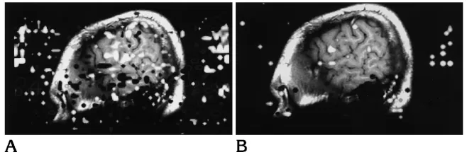
[image:2.612.228.557.101.212.2]Eight right-handed subjects and one left-handed subject were studied. Functional im-ages without observable head motion were ob-tained in each subject. Regions of activation lying outside the brain, attributable to artifact, were more frequently identified in the images of the aloud condition than in the silent condition (Fig 1). Activation in the word generation tasks performed silently and audibly was distributed Fig 1. Functional images in parasagittal
in a similar pattern (Figs 1–3). The inferior fron-tal gyrus and/or adjacent regions of the middle and precentral frontal gyrus were regularly ac-tivated by the two tasks, as were the sensorim-otor cortex, the anterior cingulate gyrus, and the supplementary motor area. Activation also was seen in the thalami and parietal, temporal, and occipital lobes in some subjects during silent or aloud word generation.
The number of pixels demonstrating activa-tion in the left hemisphere by silent word gen-eration is summarized in Fig 4. In the left hemi-sphere, both the inferior frontal gyrus and the adjacent middle frontal and precentral gyrus were activated by silent word generation in all cases. The average number of pixels activated in the inferior frontal and adjacent frontal gyri
was 12 and 16, respectively. On average, in the sensorimotor cortex 6 pixels and in the anterior cingulate gyrus 8 pixels were activated by word generation silently. In the supplementary motor area and in the temporal gyri, on average 5 or fewer activated pixels were identified.
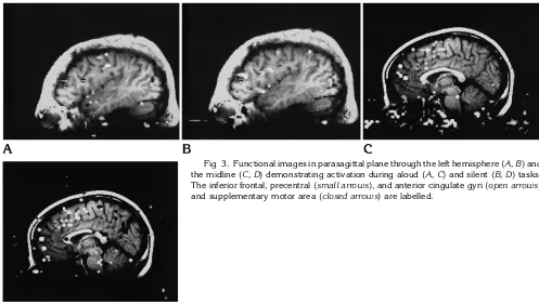
[image:3.612.57.382.101.215.2]Activation in the left hemisphere by word generation aloud is summarized in Figure 5. In the left hemisphere, on average 7 pixels were activated in the inferior frontal gyrus and 14 in the adjacent middle, frontal, and precentral gyri by word generation aloud. In the sensorimotor cortex, anterior cingulate gyrus, supplementary motor area, and temporal gyri 2 to 7 activated pixels were seen on average from word gener-ation aloud (Fig 5). On average, more pixels were activated in the silent word generation task Fig 2. Functional images in parasagittal plane in another subject during aloud (A) and silent word generation (B).
[image:3.612.55.552.453.735.2]than in the word generation task aloud. The difference in activation from silent and aloud word generation in the seven right-handed sub-jects is shown in Figure 6. On average, for right handers, 7 more pixels were activated in the left frontal lobe (inferior frontal and adjacent pre-central and middle frontal gyri) by word gener-ation silently than by word genergener-ation aloud. The difference was statistically significant (P 5
.05, Student’s t test for a paired sample). For the sensorimotor cortex, on average fewer pix-els were activated in right handers by the word generation task aloud than by word generation silently (difference 51.25, not significant). For the anterior cingulate region, 2.75 more pixels were on average activated by the silent com-pared to the aloud word generation (P 5 .09). For the supplementary motor area and the
tem-poral gyri, the difference in activation between word generation silently and aloud was small and insignificant.
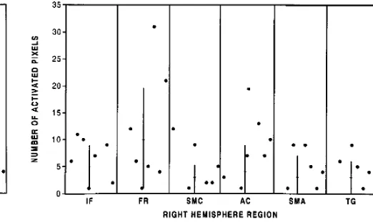
Activation in the right hemisphere secondary to silent word generation is tabulated in Figure 7. In the silent word generation task, on average 5 activated pixels were identified in the inferior right frontal lobe and 10 in the adjacent right middle frontal and precentral gyri. In one case in which activation was identified in the left frontal lobe, no activation was identified in the right frontal region. On average fewer than 4 acti-vated pixels were identified in the other regions that were tabulated.
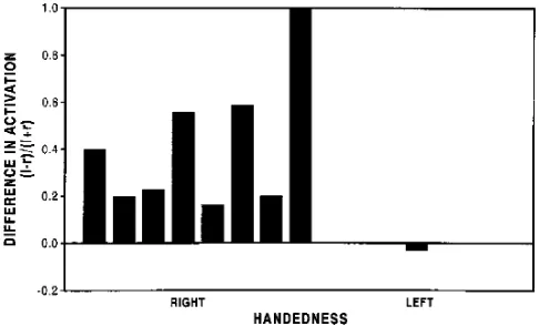
[image:4.612.60.305.102.255.2]The activation in the right and left frontal lobes from silent word generation is compared in Figure 8. In all right-handed subjects, more activation was seen in the left than in the right Fig 4. Activation in nine subjects in each left hemisphere
[image:4.612.187.542.103.258.2]re-gion performing word generation silently.Key to abbreviations:IF indicates inferior frontal gyrus; FR, adjacent frontal gyri; SMC, sensorimotor cortex;AC, anterior cingulate gyrus;SMA, supple-mentary motor area; andTG, temporal gyri.
Fig 5. Activation in each left hemisphere region in each sub-ject performing word generation aloud. (See Figure 4 legend for key to abbreviations.)
[image:4.612.296.557.324.480.2]Fig 6. Difference in number of activated pixels in the left hemisphere of right-handed subjects performing word generation silently and aloud. (See Figure 4 for key to abbreviations.)
[image:4.612.59.305.324.478.2]frontal lobe. In one subject, the ratio of (L2R)/ (L1R) was 1 because no right frontal lobe activation was detected. In right handers, activation in the left frontal lobe exceeded that in the right frontal lobe by 37%. In the one left-handed subject, activation on the right frontal lobe exceeded that on the left (Fig 8). In six subjects, the (L2R)/(L1R) ratio was similar in the silent and aloud word generation tasks. In the one left-handed subject, a slight excess of activation in the left frontal lobe (ratio 5 0.1) was seen with word generation aloud. On another subject the ratio dropped from 1 to 0.27, and in one other it increased from 0.2 to 0.5.
Discussion
This functional MR study showed activation in the inferior frontal lobe, the sensorimotor cortex region, the anterior cingulate gyrus, and the supplementary motor area in subjects perform-ing word generation silently or aloud. Similar results have been reported previously (19 –22), although silent and aloud word generation have not been compared (3, 4, 12). Functional MR imaging with word generation showed more ac-tivation in the left hemisphere than in the right hemisphere in right-handed subjects. It also showed greater right hemisphere activation in a left-handed subject.
The study showed more activation in the dominant frontal lobe in subjects performing the word generation task silently than aloud. The greater number of activated pixels in si-lent word generation might be attributable to physiologic differences in the two tasks, or to technical differences, such as decreased
noise during the task, minimizing movement of the head and facial structures. The in-creased activation in the sensorimotor cortex secondary to tasks performed audibly may reflect actual movement of the lips, tongue, and facial muscles. Activation in sensorimo-tor cortex, secondary to silently performed tasks, observed in functional MR imaging and positron emission tomography (19), is not yet explained. Differences in activation between subjects may represent differences in the way individuals performed the tasks. The physio-logic significance of regions activated sec-ondary to these tasks is beyond the scope of this study.
Motion of the head or of objects in the mag-netic field during the acquisition of functional MR data degrades the functional information (14). Head motion was monitored in this study, and any acquisition with head motion was ex-cluded. Movement of facial structures was not monitored, and subtle mouth or jaw movement during silent tasks cannot be excluded. Sub-voxel head movement also is not excluded. The increased number of artifactual activations in the audible tasks compared with silent tasks is consistent with the increased facial movements needed to perform the tasks.
The precision and accuracy of functional MR have not been studied systematically. Anatomic location of pixels adjacent to sulci (eg, near the inferior frontal gyrus or central sulcus) may be inexact. Therefore, in some tabulations we combined the activation in the inferior frontal and adjacent gyri and the activation in precen-tral and postcenprecen-tral gyri as sensorimotor cortex. Because of susceptibility artifacts near the base of the skull, detection of activation in the inferior temporal gyrus may be less reliably detected than activation elsewhere. Practice effects (ie, decreased activation in the repetitions) were not observed in this study.
[image:5.612.57.300.102.249.2]This study shows that for the purposes of mapping language functions with functional MR, the word generation task may be per-formed silently or aloud. An advantage of si-lent word generation is that more activation and fewer artifacts result from motion of the head, jaw, tongue, or throat. A disadvantage of the silent version of the language task is the inability to monitor the patient’s performance of the task.
References
1. Frahm J, Bruhn H, Merboldt K, et al. Dynamic MR imaging of human brain oxygenation during rest and photic stimulation.J Magn Reson Imaging1992;2:501–505
2. Kwong KK, Belliveau JW, Chesler DA, et al. Dynamic magnetic resonance imaging of human brain activity during primary sen-sory stimulation.Proc Natl Acad Sci1991;89:5675–5679 3. Binder JR, Rao SM, Hammeke TA, et al. Functional magnetic
resonance imaging of human auditory cortex.Ann Neurol1994; 35:662– 672
4. Rueckert L, Appollonio I, Grafman J, et al. MRI functional activa-tion of left frontal cortex during covert word producactiva-tion.J Neuro-imaging1994;4:67–70
5. Cao Y, Towle VL, Levin DN, Balter J. Functional mapping of human cortical activation by conventional MRI at 1.5T.J Magn Reson Imaging1993;3:869 – 875
6. Bandettini PA, Wong EC, Hincks RS, et al. Time course EPI of human brain function during task activation.Magn Res Med1992; 25:390 –397
7. Blamire AM, Ogawa S, Ugurbil K, et al. Dynamic mapping of the human visual cortex by high speed magnetic resonance imaging. Proc Nat Acad Sci1992;89:11069 –11073
8. Menon RS, Ogawa S, Kim S-G, et al. Functional brain mapping using magnetic resonance imaging: signal changes accompany-ing visual stimulation.Invest Radiol1992;27:S47–S53
9. Belliveau JW, Kwong KK, Kennedy DN, et al. Magnetic resonance image mapping of brain function: human visual cortex.Invest Radiol1992;27:S59 –S65
10. Ogawa S, Tank DW, Menon R, et al. Intrinsic signal changes accompanying sensory stimulation: functional brain mapping with magnetic resonance imaging.Proc Nat Acad Sci1992;89: 5951–5999
11. Jack CR, Thompson R, Butts RK, et al. Sensory motor cortex: presurgical mapping with functional MR imaging and invasive cortical mapping.Radiology1994;190:85–92
12. Hinke R, Hu X, Stillman AE, et al. Functional magnetic resonance imaging of Broca’s area during internal speech. Neuro Report 1993;4:675– 678
13. Morris GL, Mueller WM, Yetkin FZ, et al. Functional magnetic resonance imaging in partial epilepsy.Epilepsia1994;35:1194 – 1198
14. Hajnal J, Myers R, Oatridge A, Schwieso JE, Young IR, Bydder GM. Epilepsia: Artifacts due to stimulus correlated motionin func-tional imaging in brain.Magn Reson Med1994;31:283–291 15. Oldfield RC. The assessment and analysis of handedness: the
Edinburgh inventory.Neuropsychologia1971;9:97–113 16. Bandettini PA, Jesmanowicz A, Wong EC, et al. Processing
strat-egies for time-course data sets in function MRI of the human brain. Magn Reson Med1993;30:161–173
17. Sobel DF, Gallen CC, Schwartz BJ, et al. Locating the central sulcus: comparison of MR anatomic and magnetoencephalo-graphic functional methods.AJNR Am J Neuroradiol1993;14: 915–925
18. Rademacher J, Galaburda AM, Kennedy DN, Filipek PA, Caviness VS. Human cerebral cortex: localization, parcellation, and mor-phometry with magnetic resonance imaging.J Cognitive Neurosci 1992;4:352–358
19. Friston KJ, Frith CD, Liddle PF, Frackowiak RSJ. Investigating a network model of word generation with positron emission tomog-raphy.Proc R Soc Lond [Biol]1991;244:101–106
20. Wise R, Chollet F, Hadar U, Friston K, Hoffner E, Frackowiak R. Distribution of cortical neural networks involved in word compre-hension and word retrieval.Brain1991;114:1803–1817 21. Haxby JV, Grady CL, Ungerleider LG, Horwitz B. Mapping the
functional neuroanatomy of the intact human brain with brain work imaging.Neuropsychology1991;29:539 –555