Original Research Article
Maternal risk factors and its association with low birth weight:
a study from rural area of Maharashtra
Dnyaneshwar N. Digole
1, A. S. Nagaonkar
2, Anant A. Takalkar
1*
INTRODUCTION
Low birth weight is single most important factor determining the survival chances of the child. A high percentage of low birth weight points to deficient health status of pregnant women, inadequate prenatal care and the need for improved care of the newborn.1
Reduction of low birth weight incidence was one of the major goals of the ‘World Fit for Children’ plan adopted
by the United Nations General Assembly in 2002. It also forms an important contribution to the millennium development goal (MDG) for the reduction of child mortality.2
Krammer has identified 43 potential factors for low birth weight.3 Not all the factors should be present in a given area. The factors vary from one area to another, depending upon geographic, socioeconomic and cultural factors. Thus it is necessary to identify factors prevailing ABSTRACT
Background: Low birth weight is single most important factor determining the survival chances of the child. A high percentage of low birth weight points to deficient health status of pregnant women, inadequate prenatal care and the need for improved care of the newborn. Present hospital based study was undertaken to identify the maternal risk factors associated with low birth weight babies.
Methods: The present hospital based descriptive study was conducted at Swami Ramanand Teerth Rural Govt. Medical College and Hospital. The data collection was done during 1st March 2012 to 28th February 2013. All deliveries occurring on alternate days were included in the study which comes to, 1154 deliveries. Data was analysed by using SPSS 16.0 version.
Results: The percentage of low birth weight babies was more in primipara mothers (25.53%). The percentage of low birth weight babies was high (34.56%) when pregnancy interval was 1 year. Percentage of low birth weight was maximum among mothers with no antenatal visits (42.93%). Percentage of low birth weight babies was more (38.75%) in mothers who had not consumed iron and folic acid tablets. Percentage of low birth weight babies was higher (35.62%) among mothers who had antenatal history of radiological exposure. Percentage of low birth weight babies was more (30.27%) in mothers who received inadequate afternoon rest.
Conclusions: In our study low birth weight was commonly observed in primipara mothers, pregnancy interval less than a year, mothers with no antenatal visits, in mothers who had not consumed iron and folic acid tablets and with history of radiological exposure.
Keywords: Maternal risk factors, Low birth weight, Rural area Department of Community Medicine, 1
MIMSR Medical College, 2Government Medical College, Latur, Maharashtra, India
Received: 25 May 2019
Accepted: 11 June 2019
*Correspondence:
Dr. Anant A. Takalkar,
E-mail: [email protected]
Copyright: © the author(s), publisher and licensee Medip Academy. This is an open-access article distributed under the terms of the Creative Commons Attribution Non-Commercial License, which permits unrestricted non-commercial use, distribution, and reproduction in any medium, provided the original work is properly cited.
in a particular area responsible for low birth weight, so as to plan the strategy to tackle this important problem.
Present hospital based study was undertaken to identify the maternal risk factors associated with low birth weight babies.
METHODS
The present hospital based Descriptive study was conducted at Swami Ramanand Teerth Rural Govt. Medical College and Hospital. The data collection was done during 1st March 2012 to 28th February 2013. The study subjects comprised of postnatal mothers having full term normal delivery with single ton live born baby delivered during the study period. Multiple births, preterm births, still births and those PNC cases who did not give consent for study were excluded. The sample size was calculated by using the formula n=4pq/L2.4 So, based on national incidence of low birth weight 28% sample size at 95% confidence level with 10% allowable error will be 1029.1 Present study being hospital based, 50% of the deliveries occurring during the study period were included. Therefore, all deliveries occurring on alternate days were included in the study which comes to, 1154 deliveries.
By international agreement, low birth weight has been defined as a birth weight less than 2.5 kg (up to and including 2499 grams), the measurement being taken preferably within the first hour of life, before significant weight loss has occurred.1
Approval form the Institutional Ethical Committee (IEC) was taken before the study. Investigator himself visited PNC ward on alternate days during study period, by interviewing and examining mothers, data was collected in predesigned pretested proforma. Also available record of the mothers including antenatal record was scrutinized.
Collected data was entered in MS Excel sheet. Statistical analysis was done by using percentages and Chi square test. SPSS 16.0 version software was used for calculation of Chi square test and p value.
RESULTS
Out of 1154 mothers, maximum i.e. 995 mothers were Primipara followed by 125 with 2nd para, 22 with 3rd para and 12 were having parity 4.
The percentage of low birth weight babies was more in Primipara mothers (25.53%). Percentage of low birth weight babies decreased with increase in parity up to 3 and again it was more for 4th parity (25.00%).
Significant association was found between parity and birth weight of baby (p<0.05).
Table 1: Distribution of newborns according to birth weight and parity.
Parity
Normal birth weight
Low birth
weight Total
N (%) N (%) N (%) 1 741 (74.47) 254 (25.53) 995 (100)
2 106 (84.80) 19 (15.20) 125 (100)
3 19 (86.36) 3 (13.64) 22 (100)
4 9 (75.00) 3 (25.00) 12 (100)
Total 875 (75.82) 279 (24.18) 1154 (100)
Figures in parenthesis indicate row percentage; X2=7.823, Df=3, p<0.05.
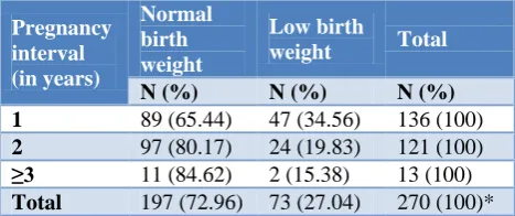
Table 2: Distribution of newborns according to birth weight and pregnancy interval.
Pregnancy interval (in years)
Normal birth weight
Low birth
weight Total
N (%) N (%) N (%) 1 89 (65.44) 47 (34.56) 136 (100)
2 97 (80.17) 24 (19.83) 121 (100)
≥3 11 (84.62) 2 (15.38) 13 (100)
Total 197 (72.96) 73 (27.04) 270 (100)*
*These mothers were having multiple pregnancies (884 were Primipara without history of abortion excluded form analysis). Figures in parenthesis indicate row percentage. X2=7.977, Df=2, p<0.05.
Out of 270 mothers maximum number of mothers i.e. 136 were having pregnancy interval of up to 1 year followed by 121 mothers having pregnancy interval of 2 years and 13 mothers having pregnancy interval of 3 and more years.
The percentage of low birth weight babies was high (34.56%) when pregnancy interval was 1 year. The percentage decreased to 19.83% and 15.38% when pregnancy interval was increased to 2 and 3 or more.
Significant association was found between pregnancy interval and birth weight of baby (p<0.05).
Table 3: Distribution of newborns according to birth weight and number of ANC visits by mother.
No. of ANC visits by mother
Normal birth weight
Low birth
weight Total
N (%) N (%) N (%) No visit 121 (57.07) 91 (42.93) 212 (100)
<3 visit 233 (68.13) 109 (31.87) 342 (100)
≥3 visit 521 (86.83) 79 (13.17) 600 (100)
Total 875 (75.82) 279 (24.18) 1154 (100)
Out of 1154 mothers, 212 did not give ANC visits whereas 342 had inadequate i.e. less than 3 visits and remaining 600 mothers had adequate antenatal visits i.e. more than or equal to 3.
Percentage of low birth weight was maximum among mothers with no antenatal visits (42.93%) and percentage of low birth weight decreased to 13.17% when visits were adequate i.e. more than or equal to 3.
Highly significant association was found between number of antenatal care visits and birth weight of baby (p<0.001).
Table 4: Distribution of newborns according to birth weight and antenatal consumption of iron and folic
acid tablets.
Antenatal consumption of iron and folic acid tablets
Normal birth weight
Low birth
weight Total
N (%) N (%) N (%) Nil 98 (61.25) 62 (38.75) 160 (100)
<100 281 (73.18) 103 (26.82) 384 (100)
100 496 (81.31) 114 (18.69) 610 (100)
Total 875 (75.82) 279 (24.18) 1154 (100)
Figures in parenthesis indicate row percentage, X2=30.03, Df=2, p<0.001.
Out of 1154 mothers, 160 mothers did not consumed any iron and folic acid tablets during ANC period whereas 384 mothers had inadequate consumption i.e. less than 100 iron and folic acid tablets and 610 mothers consumed adequate i.e. 100 iron and folic acid tablets.
Percentage of low birth weight babies was more (38.75%) in mothers who had not consumed iron and folic acid tablets and was 26.82% in mothers who consumed less than 100 iron and folic acid tablets. Percentage of low birth weight babies was very less (18.69%) in those who consumed 100 iron and folic acid tablets.
Highly significant association was observed between iron and folic acid tablets consumed by mother and birth weight of baby (p<0.001).
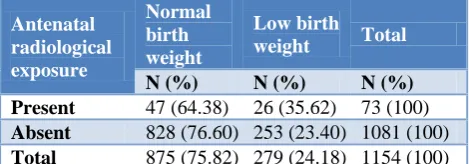
Table 5: Distribution of new borns according to birth weight and antenatal radiological exposure.
Antenatal radiological exposure
Normal birth weight
Low birth
weight Total
N (%) N (%) N (%) Present 47 (64.38) 26 (35.62) 73 (100)
Absent 828 (76.60) 253 (23.40) 1081 (100)
Total 875 (75.82) 279 (24.18) 1154 (100)
Figures in parenthesis indicate row percentage, X2=5.563, Df
Out of 1154 mothers, only 73 mothers had antenatal history of radiological exposure.
Percentage of low birth weight babies was higher (35.62%) among mothers who had antenatal history of radiological exposure than who did not have any antenatal history of radiological exposure (23.40%).
Significant association was observed between antenatal history of radiological exposure and birth weight of baby (p<0.05).
Table 6: Distribution of newborns according to birth weight and afternoon rest by mother.
Afternoon rest by mother (hrs)
Normal birth weight
Low birth
weight Total
N (%) N (%) N (%) <2 546 (69.73) 237 (30.27) 783 (100)
≥2 329 (88.68) 42 (11.32) 371 (100)
Total 875 (75.82) 279 (24.18) 1154 (100)
Figures in parenthesis indicate row percentage, X2=49.3, Df=1, p<0.001.
Out of 1154 mothers, maximum i.e. 783 mothers did not received adequate afternoon rest and 371 mothers had adequate afternoon rest.
Percentage of low birth weight babies was more (30.27%) in mothers who received inadequate afternoon rest as compared to percentage of low birth weight babies (11.32%) in mothers who received an adequate afternoon rest.
Highly significant association was observed between rest taken by mother in afternoon and birth weight of baby (p<0.001).
DISCUSSION
Out of 1154 mothers, Maximum i.e. 995 mothers were Primipara followed by 125 with 2nd para, 22 with 3rd para and 12 were having parity 4.
The percentage of low birth weight babies was more in Primipara mothers (25.53%). This may be because of early marriages and subsequently early first pregnancy, poor awareness of ANC services in primiparous mothers. Poor nutritional profile of primiparous mothers as most of them belong to teenage groups also contribute to poor pregnancy outcome like low birth weight babies.
Percentage of low birth weight babies decreased with increase in parity up to 3 and again it was more for 4th parity (25.00%). Significant association was found between parity and birth weight of baby (p<0.05).
et al, Brotane et al, Anand and Garg, Negi et al and Agarwal et al.5-12
However present study findings are not in confirmation with, Chaudhary et al who reported that parity did not have any stastistically significant association with birth weight.13 This variation may be because of sample size and geographic area.
Out of 270 mothers maximum number of mothers i.e. 136 were having pregnancy interval of up to 1 year followed by 121 mothers having pregnancy interval of 2 years and 13 mothers having pregnancy interval of 3 and more years.
The percentage of low birth weight babies was high (34.56%) when pregnancy interval was 1 year. This is because of more chance of poor pregnancy outcome as abortion, more stress on mother for care of baby, increased nutritional demands which are poorly met due to short pregnancy interval. The percentage decreased to 19.83% and 15.38% when pregnancy interval was increased to 2 and 3 or more. Significant association was found between pregnancy interval and birth weight of baby (p<0.05).
The present study findings are in confirmation with, Hirve and Ganatra, Negi et al, Deswal et al, and Metgud et al.5,11,14,15
Out of 1154 mothers, 212 did not give ANC visits whereas 342 had inadequate i.e. less than 3 visits and remaining 600 mothers had adequate antenatal visits i.e. more than or equal to 3.
Percentage of low birth weight was maximum among mothers with no antenatal visits (42.93%) and percentage of low birth weight decreased to 13.17% when visits were adequate i.e. more than or equal to 3.
Adequate number of antenatal visits would prevent low birth weight babies by decreasing anaemia, toxemia, by ensuring early treatment of other morbid conditions and by making availability of promotive, preventive and curative services to mother. Highly significant association was found between number of antenatal care visits and birth weight of baby (p<0.001).
The present study findings are in confirmation with Gawande et al, Anand and Garg, Negi et al, Metgud et al, Kogan et al, Joshi et al, Aghamolaei et al, Khatun and Rahman.6,10,11,14,16-19
However present study findings are not in confirmation with Krammer and Bortane et al where they found no significant association of birth weight with ANC visits by mother.3,9 Variations in the observation conducted by Kramer may be because of the fact that meta-analysis included studies conducted in developed countries where perhaps women had less need for ANC care.
Out of 1154 mothers, 160 mothers did not consumed any iron and folic acid tablets during ANC period whereas 384 mothers had inadequate consumption i.e. less than 100 iron and folic acid tablets and 610 mothers consumed adequate i.e. 100 iron and folic acid tablets.
Mothers with no ANC visits may have taken the Iron and folic acid tablets from private practitioners or other sources.
Percentage of low birth weight babies was more (38.75%) in mothers who had not consumed iron and folic acid tablets and was 26.82% in mothers who consumed less than 100 iron and folic acid tablets. Percentage of low birth weight babies was very less (18.69%) in those who consumed 100 iron and folic acid tablets.
Adequate iron consumption during ANC is associated with improved hemoglobin concentration in mother which improves the pregnancy outcome. Highly significant association was observed between iron and folic acid tablets consumed by mother and birth weight of baby (p<0.001).
The present study findings are in confirmation with Bortane et al, Metgud et al and Palma et al.9,15,20 Whereas the present study findings are not in confirmation with Grover et al who reported no significant association between birth weight and iron and folic acid consumption.21 The author explained this fact in the background of non compliance to iron and folic acid consumption.
Out of 1154 mothers, only 73 mothers had antenatal history of radiological exposure. Percentage of low birth weight babies was higher (35.62%) among mothers who had antenatal history of radiological exposure than who did not have any antenatal history of radiological exposure (23.40%).
Antenatal radiological exposure affects organogenesis resulting in IUGR which resulting in poor birth weight. Significant association was observed between antenatal history of radiological exposure and birth weight of baby (p<0.05).
However, these findings are not in confirmation with, Mortazavi et al who reported that there were no statistical significant differences between the mean weight of newborns whose mothers had been exposed to some common sources of ionizing and non-ionizing radiations and those of non-exposed mothers.22 This may be due to the fact that study included all types of radiation including mobile phone, cordless phone and cathode ray tube.
Percentage of low birth weight babies was more (30.27%) in mothers who received inadequate afternoon rest as compared to percentage of low birth weight babies (11.32%) in mothers who received an adequate afternoon rest. The adequate afternoon rest by mother is necessary for optimal growth of baby, restore health of mother and better pregnancy outcome. Highly significant association was observed between rest taken by mother in afternoon and birth weight of baby (p<0.001). The present study findings are in confirmation with, Chaudhary et al (2013).13
CONCLUSION
In our study low birth weight was commonly observed in Primipara mothers, pregnancy interval less than a year, mothers with no antenatal visits, in mothers who had not consumed iron and folic acid tablets and with history of radiological exposure.
Funding: No funding sources Conflict of interest: None declared
Ethical approval: The study was approved by the Institutional Ethics Committee
REFERENCES
1. Park K. Park’s text book of Preventive and Social Medicine. Chapter 10, 22nd ed. Jabalpur: M/s Banarsidas Bhanot; 2013: 495-592.
2. Low birth weight country, Regional and Global estimates. United Nation’s Children’s Fund, New York: United Nation’s Children’s Fund and World Health Organization; 2004.
3. Kramer MS. Determinant of low birth weight: Methodological assessment and met analysis. Bulletin of WHO. 1987;65(5):663-737.
4. Dixit J.V. Principles and Practice of Biostatistics. Chapter 5, 4th ed. Jabalpur: M/s Banarasiadas Bhanot Publishers; 2009: 65.
5. Hirve SS, Ganatra BR. Determinants of Low Birth Weight. Community based prospective cohort study, Indian J Pediatr. 1994;31:1221-5.
6. Gavande UH, Pimpalgaonkar MS, Bentharia SH. Biosocial determinants of birth weight in rural urban Nagpur. Indian J Community Med. 1994;21(3):64-7. 7. Joshi SM, Pai NP. Effect of maternal biosocial determinants on the birth weight in a slum area greater Mumbai. Indian J Community Med. 2000;25(2):121-3.
8. Chhabra P, Sharma AK, Grover VL, Aggarwal OP. Prevalence of low birth weight and its determinants in an urban resettlement area of Delhi. Asia Pac J Public Health. 2004;16(2):95-8.
9. Bortane A, Gupta S, Datta S, Mehendale A, Garg B. Determinants of low birth weight in rural Wardha. Indian J Matern Child Heal. 2012;14(2):1-9.
10. Anand K, Garg BS. A study of factors affecting low birth weight. Indian J Community Med. 2000;25(2):57-62.
11. Negi K, Kandpal S, Kukreti M. Epidemiological factors affecting low birth weight. JK Sci. 2006;8(1):31-4.
12. Agarwal G, Sartaj A, Goel K, Kumar V, Goel P, Garg M. Maternal Risk Factors Associated with Low Birth Weight Neonates in a Tertiary Care Hospital, Northern India. J Community Med Health Educ. 2012;02(09):9-12.
13. Choudhary AK, Tiwari SC, Dwivedi R. Factors associated with low birth weight among newborns in an urban slum community in Bhopal. Indian J Public Health. 2013;57(1):20-3.
14. Deswal B, Singh J, Kumar D. A study of risk factors for low birth weight. Indian J Community Med. 1999;24(3):127-31.
15. Metgud CS, Naik VA, Mallapur MD. Factors affecting birth weight of a newborn – a community based study in rural Karnataka, India. PLoS One. 2012;7(7):1-4.
16. Kogan MD. Social causes of low birth weight. J R Soc Med. 1995;88(11):611-5.
17. Joshi HS, Subba SH, Dabral SB, Dwiwedi S, Kumar D, Singh S. Risk Factors associated with low birth weight in newborns. Indian J Community Med. 2005;30(4):142-3.
18. Aghamolaei T, Eftekhar H, Zare S. Risk factors associated with intrauterine growth retardation (IUGR) in Bandar Abbas. J Med Sci. 2007;7(4):665-9.
19. Khatun S, Rahman M. Socio-economic determinants of low birth weight in Bangladesh: A multivariate approach. Bangladesh Med Res Counc Bull. 2009;34(3):81-6.
20. Palma S, Perez-Iglesias R, Prieto D, Pardo r, Llorca J, Delgado-Rodriuez M. Iron but not folic acid supplementation reduces the risk of low birth weight in pregnant women without anaemia: a case-control study. J Epidemiol Community Health. 2008;62(2):120-4.
21. Grover V, Agarwal OP, Gupta A, Kumar P, Tiwari RS. Effect of daily and alternate day iron and folic acid supplementation to pregnant females on the weight of newborn. Indian journal of community medicine 1998;23(4):165-8.
22. Mortazavi SMJ, Shirazi KR, Mortazavi G. The study of the effects of ionizing and non-ionizing radiations on birth weight of newborns to exposed mothers. J Nat SciBiol Med. 2013;4(1):213-7.