Development of Compton X-Ray Spectrometer
for Fast Ignition Experiment
∗
)
Sadaoki KOJIMA, Yasunobu ARIKAWA, Takahito IKENOUCHI, Takahiro NAGAI, Yuki ABE,
Shouhei SAKATA, Hiroaki INOUE, Takuya NAMIMOTO, Shinsuke FUJIOKA, Mitsuo NAKAI,
Hiroyuki SHIRAGA, GEKKO-XII&LFEX Team, Tetsuo OZAKI
1), Makoto R. ASAKAWA
2),
Ryukou KATO
3)and Hiroshi AZECHI
Institute of Laser Engineering, Osaka University, 2-6 Yamada-oka, Suita 565–0871, Japan
1)National Institute for Fusion Science, 322-6 Oroshi-cho, Toki 509-5292, Japan 2)Faculty of Engineering Science, Kansai University, 3-3 Yamate, Suita 564-8680, Japan
3)Institute of Scientific and Industrial Research, Osaka University, 2-6 Yamada-oka, Suita 565-0871, Japan
(Received 22 July 2013/Accepted 20 February 2014)
In the context of Fast Ignition, the fast electron temperature is a key parameter to determine the laser-to-fast electron energy conversion efficiency. Bremsstrahlung X-ray (γ-ray) emission represents an attractive alternative to measure this fundamental parameter. In this study, a single-shot high-energyγ-ray spectrometer with sensitivity ranging from 0.5 MeV to 7 MeV was developed, allowing to estimate theγ-ray spectrum in the fast ignition.
c
2014 The Japan Society of Plasma Science and Nuclear Fusion Research
Keywords: fast ignition, inertial confinement fusion, fast electron, x-ray spectrometer, filter stack spectrometer DOI: 10.1585/pfr.9.4405109
1. Inroduction
Fast Ignition (FI) is an alternative approach to Iner-tial Confinement Fusion, consisting in the separation of the compression and heating stages. In FI, the heating of the compressed Deuterium-Tritium (DT) fuel is pro-duced by a ultra-high intensity laser-generated fast electron beam depositing its energy in the high density plasma by Coulomb interaction. The coupling efficiency is strongly dependent on the fast electron energy distribution, there-fore representing a crucial parameter to be diagnosed [1]. From simulations, in order to ignite a compressed DT pel-let having density of 300 g/cm3, 18 kJ of fast electron
en-ergy has to be deposited within a diameter of 40µm in the compressed fuel. Fast electrons in the 1-3 MeV energy range are responsible for the energy deposition in such a small volume, allowing for the creation of a lateral hot spot. Thus high conversion efficiency into 1-3 MeV energy range fast electrons is fundamental for the achievement of Fast Ignition [2–4]. However, it is difficult to measure ini-tial fast electron temperature from the outside of plasma because of the strong sheath potential around the target. The bremsstrahlung X-ray is an attractive alternative diag-nostic for this aim. In previous works, many kind ofγ-ray detectors with different sensitivities have been developed. Various techniques have been adopted to measure the fast electron energy spectrum, such as vacuum electron
spec-author’s e-mail: [email protected]
∗)This article is based on the presentation at the Conference on Laser and
Accelerator Neutron Source and Applications (LANSA ’13).
troscopy, nuclear activation, Bremsstrahlung, buried fluo-rescent foils, proton emission, and coherent transition radi-ation [5–9]. Filter stack technique uses differential filtering to discriminate theγ-ray spectrum [10, 11]. The spectrom-eter consists of thirteen filters of increasing Z, five 100µm thick filters Al, Ti, Fe, Cu and Mo, followed by 150µm Ag, 500µm Sn and Ta filters, and one 1.58 mm Au filter. Finally four Pb filters with thickness varying from 1 mm to 4 mm for differential filtering are positioned in the stack. Nuclear activation technique uses photo nuclear activation to discriminate theγ-ray spectrum. In this method,γ-ray spectrum is measured from the activation ratio of a pseudo-alloy consisting of gold (197Au), tantalum (181Ta), niobium
(93Nb), and chromium (50Cr,52Cr, 53Cr, 54Cr) with mass
fraction being 43.25%, 24.25%, 16.25%, 16.25%, respec-tively [12]. However, observable energy range of these two detectors are respectively up to 0.5 keV and above 7 MeV, respectively. Thus, these materials are unsuitable to mea-sureγ-ray s in the range 1-3 MeV which represents the en-ergy range of our interest. In this study, a single-shot high-energyγ-ray spectrometer, with sensitivity from 0.5 keV to 7 MeV, was developed, allowing for the first time to esti-mate of the fast electron-generated bremmstrahlung X-ray spectrum.
2. Diagnostic Design
2.1
Measurement of X-ray energy
Photons with energy around 1 MeV mainly interact with matter by Compton scattering, the energy of incident
c
2014 The Japan Society of Plasma
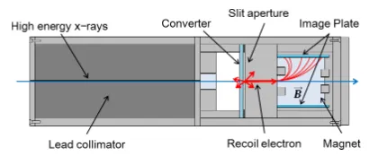
Fig. 1 The structure of developed detector. The structure of the Compton X-ray spectrometer consists of a lead collima-tor, converter and an electron spectrometer.
X-rays can be measured by measuring the energy and the angle of recoil electrons. The structure of the Compton X-ray spectrometer is shown in Fig. 1. The high energy X-rays emitted from the target are initially collimated by means of a lead collimator. Then, the collimated X-rays recoil electrons via Compton scattering in the converter. The energy of the electrons crossing the aluminum slit set in front of the converter is measured by means of an elec-tron spectrometer (ESM) and the data recorded on Image Plate (IP).
2.2
Converter
The material composing the converter is chosen in order to have high conversion efficiency from X-rays to recoiled electrons and low energy absorption in the con-verter. The material is selected by estimating these two parameters from X-ray-matter interaction cross sections, electron stopping power and Coulomb scattering.
Cross sections of each factor can be approximated as
σcompton∝NZ(hν)−1 (1)
σphoto∝NZ5(hν)−
7
2 (2)
σpair∝NZ2(hν−1.02) (3)
−dE
dx ∝NZ (4)
σscatter∝
Z2
(hν)2 (5)
whereNis the number of the atom per cm3,Zis the atomic number andhνis the incident photon energy.
Cross sections for Compton scattering and stopping power depend linearly on the atomic number. On the other hand, Cross sections for photoelectric absorption, pair cre-ation and Coulomb scattering have a non-linear depen-dence on Z.
Figure 2 shows Monte Carlo simulation results of Coulomb scattering in the converter simulated by Casino ver.2.42 [13]. The red lines and the blue lines show respec-tively the trajectories of back-scattered and forward scat-tered electrons. In high-Z material, efficiency of the back scattering increase due to strong Coulomb potential around the nuclear.
Figure 3 shows the cross sections for photelectric ef-fect, Compton scattering and pair production in low and
Fig. 2 Trajectory of electrons in the converter simulated by Casino ver.2.42. (a) Gold foil (high Z) and (b) Silica plate (low Z). In high-Z material, efficiency of the back scat-tering shown by red lines increase due to strong Coulomb potential around the nuclear.
Fig. 3 Cross section for the three interactions. (a) Gold (high Z) and (b) Silica (low Z). Low Z material must be chosen in order to maximize the Compton scattering produced electron.
high Z material, respectively SiO2and Au. Being the cross
section for photelectric effect proportional to Z to the fifth power, low Z material must be chosen in order to maximize the Compton scattering produced electron.
Following the discussion above, a thick, low Z con-verter material is the optimal choice. Due to alignment requirements, a transparent material was needed. A BK glass provides at the same time the characteristics of trans-parency and low Z material.
2.3
Background reduction
A lead box and two magnets were designed to shield respectively X-rays and charged particle produced back-ground on the IP detector. A second IP is positioned in front of the recoil electron IP detector in order to measure positron signal in the unlikely case of pair production in the converter.
3. Calibration Experiments by Using
60Co
Cobalt60 decays in two possible ways, emitting two
γ rays which energy is respectively 1.1732 MeV and 1.3325 MeV. The new detector was tested by diffracting
γ-rays emitted by60Co. As converter, a thin 50µm Au foil
was chosen, in order to guarantee low recoil electron en-ergy loss in the converter. Figure 4 represents the recoil electron spectrum from60Co. Experimental data are
de-Fig. 4 Recoil electron signal from60Co. Experimental data are
represented by red dots while the Black line represents the Monte Carlo simulation. The energy resolution is obtained via Gaussian fitting of the two peaks, and is estimated to be 10.4% for 1.1732 MeV and 11.2% for 1.3325 MeV respectively.
tector energy resolution. In Fig. 4, the experimental result is represented for60Co gamma-ray detection. The energy
resolution is high enough to allow detecting two distinct peaks corresponding to the twoγphoton energy. The en-ergy resolution is obtained via Gaussian fitting of the two peaks, and is estimated to be 10.4% for 1.1732 MeV and 11.2% for 1.3325 MeV respectively.
4. Fast Ignition Experiment
4.1
Experimental setup
The experiment was performed at GEKKO laser fa-cility, Institute of Laser Engineering, Osaka University. GEKKO laser delivers up to 12 beams of∼300 J energy arranged in spherical geometry. Coupled to GEKKO laser LFEX, a ultra-high intensity beam capable of deliver∼5 kJ of laser light onto a∼50µm focal spot in 1-10 ps.
Typical γ-rays were generated by using LFEX&GEKKO laser. The target was constituted by a cone-in-shell, adopting an hemispherical shell config-uration, as shown in Fig. 5. The shell was mounted on a cubic Ta block of 1 mm lateral size. The distance from the cone tip to the Ta block was 50µm. Three frequency doubled (527 nm) GEKKO beams, delivering 30 J each of laser energy, were focused onto the hemispheric CD shell producing a low Z and low density (ne∼1.4×1022cm−3)
plasma medium between the cone and the Ta block. Sub-sequently, 568 J, 1054 nm and 1 ps long LFEX pulse were focused at the cone tip to produce a fast electron beam, propagating through the low density CD plasma into the Ta block. Part of the fast electron energy is converted in
γ-rays into the Ta block, which emission is measured by means of the Compton scattering spectrometer (Compton) and a High Energy X-ray Spectrometer (HEXS), the latest set at 21◦relatively to the LFEX laser axis.
The HEXS diagnostic is based on the widely used multiple filter stack detector technique. Experimental
re-Fig. 5 Experimental setup. The target was constituted by a cone-in-shell, adopting an hemispherical shell configura-tion. The shell was mounted on a cubic Ta block.
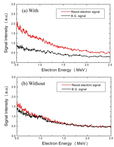
Fig. 6 Signals recorded by the detector. (a) with converter, (b) without converter.
sults for the Compton spectrometer are reported in Figs. 6, 7. The Compton spectrometer has been operated with and without converter foil during the experiment and results are represented in Fig. 6. The recoil electron signal is represented in red while the background signal in black. From experimental data it appears clear that the back-ground electron signal collected without converter layer is much smaller than the recoil electron signal in presence of converter.
4.2
Analysis method
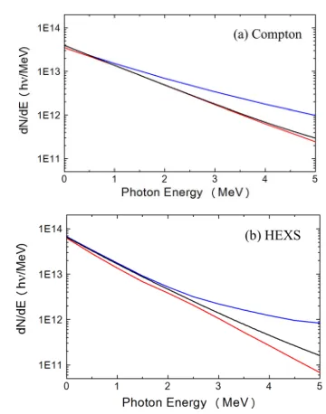
experimen-Fig. 7 (a)γ-ray spectrum measured by Compton spectrometer. (b) Filter stack detector. The black solid line show the best fit distribution, red and blue lines represent respec-tively the lower and the upper limit of our solution.
tal data with the Monte Carlo simulation.
f(E)=R1exp
−E
T1
+R2exp
−E
T2
+R3exp
−E
T3
(6) in the simulation T1, T2, T3 varied from 0.1 MeV to
10 MeV andR1andR2respectively varied from 1 to 10000
and 1 to 1000 whileR3is considered to be 1. Figures 7 (a)
and (b) show the X-ray spectrum obtained by the filter stack detector (HEXS) and the Compton spectrometer, re-spectively. In Fig. 7, the black solid line show the best fit distribution, red and blue lines represent respectively the lower and the upper limit of our solution, imposed by the fitting error and detector response error.
We successfully measuredγ-ray spectrum in fast igni-tion related experiment and data from the two diagnostics are in good agreement.
5. Conclusions
In this study, a single-shot, high-energy Compton X-ray spectrometer with the sensitivity up to 7 MeV has been developed. From experimental data, the energy res-olution of the detector is estimated to be the 10.4% for 1.1732 MeVγ-rays, and the 11.2% for 1.3325 MeVγ-rays.
γ-rays in the energy range from 0.5 to 2.5 MeV have been measured. These correspond to fast electrons which pene-tration range is optimum for compressed DT plasma heat-ing in a Fast Ignition scenario. By comparison with the HEXS diagnostic, the Compton scattering diagnostic al-lows for the determination of theγ-ray spectrum and the determination of the absolute photon number per unit
en-ergy, allowing, at the same time, for a more accurate deter-mination of theγ-ray energy spectrum compared to HEXS. Indeed for a specific data acquisition with the HEXS, there are multiple γ-ray spectra that can reproduce the same data. The Compton spectrometer data instead are univo-cally determined. Moreover the energy spectrum recorded by the Compton spectrometer is much larger than that one recorded by the HEXS, allowing to detectγ-rays having energies up to 7 MeV.
Acknowledgment
The authors gratefully acknowledge the support of the GEKKO XII operation group, the LFEX develop-ment and operation group, the target fabrication group, and the plasma diagnostics operation group of the Insti-tute of Laser Engineering, Osaka University. T. Yamamoto and S. Suemine are acknowledged for the technical sup-port. This work was partly supported by the Japan So-ciety for the Promotion of Science under the contracts of Grant-in-Aid for Scientific Research (A) No.24244095 and (B) No.23360413, Grant-in-Aid for Young Scientists (A) No.24686103, Grant-in-Aid for Challenging Exploratory Research No.25630419, and the auspices of Japanese Min-istry of Education, Culture, Sports, Science and Technol-ogy (MEXT) project on “Promotion of relativistic nuclear physics with ultra-intense laser.”
[1] R. Kodama, H. Shiraga, K. Shigemori, Y. Toyama, S. Fujioka, H. Azechi, H. Fujita, H. Habara, T. Hall, Y. Izawa, T. Jitsuno, Y. Kitagawa, K.M. Krushelnick, K.L. Lancaster, K. Mima, K. Nagai, M. Nakai, H. Nishimura, T. Norimatsu, P.A. Norreys, S. Sakabe, K.A. Tanaka, A. Youssef, M. Zepf and T. Yamanaka, Nature418(6901), 933 (2002).
[2] S. Atzeni, A. Schiavi and C. Bellei, Phys. Plasmas14(5), 052702 (2007).
[3] R. Kodama, P.A. Norreys, K. Mima, A.E. Dangor, R.G. Evans, H. Fujita, Y. Kitagawa, K. Krushelnick, T. Miyakoshi, N. Miyanaga, T. Norimatsu, S.J. Rose, T. Shozaki, K. Shigemori, A. Sunahara, M. Tampo, K.A. Tanaka, Y. Toyama, T. Yamanaka and M. Zepf, Nature
412(6849), 798 (2001).
[4] M. Tabak, J. Hammer, M.E. Glinsky, W.L. Kruer, S.C. Wilks, J. Woodworth, E.M. Campbell, M.D. Perry and R.J. Mason, Phys. Plasmas1(5), 1626 (1994).
[5] K.A. Tanaka, R. Kodama, H. Fujita, M. Heya, N. Izumi, Y. Kato, Y. Kitagawa, K. Mima, N. Miyanaga, T. Norimatsu, A. Pukhov, A. Sunahara, K. Takahashi, M. Allen, H. Habara, T. Iwatani, T. Matusita, T. Miyakosi, M. Mori, H. Setoguchi, T. Sonomoto, M. Tanpo, S. Tohyama, H. Azuma, T. Kawasaki, T. Komeno, O. Maekawa, S. Matsuo, T. Shozaki, K. Suzuki, H. Yoshida and T. Yamanaka, Phys. Plasmas7(5), 2014 (2000).
[6] T. Iwawaki, H. Habara, T. Tanimoto, N. Nakanii, K. Shimada, T. Yabuuchi, K. Kondo and K.A. Tanaka, Rev. Sci. Instrum.81, 10E535 (2010).
Singh, R.A. Snavely, M.A. Stoyer, S.C. Wilks and K. Yasuike, Phys. Plasmas7, 2076 (2000).
[8] K. Yasuike, M.H. Key, S.P. Hatchett, R.A. Snavely and K.B. Wharton, Rev. Sci. Instrum.72(1), 1236 (2001). [9] W. Theobald, K. Akli, R. Clarke, J.A. Delettrez, R.R.
Freeman, S. Glenzer, J. Green, G. Gregori, R. Heathcote, N. Izumi, J.A. King, J.A. Koch, J. Kuba, K. Lancaster, A.J. MacKinnon, M. Key, C. Mileham, J. Myatt, D. Neely, P.A. Norreys, H.-S. Park, J. Pasley, P. Patel, S.P. Regan, H. Sawada, R. Shepherd, R. Snavely, R.B. Stephens, C. Stoeckl, M. Storm, B. Zhang and T. C. Sangster, Phys. Plas-mas13(4), 043102 (2006).
[10] B. Westover, C.D. Chen, P.K. Patel, M.H. Key, H. McLean,
R. Stephens and F.N. Beg, Phys. Plasmas 18, 063101
(2011).
[11] C.D. Chen, P.K. Patel, D.S. Hey, A.J. Mackinnon, M.H. Key, K.U. Akli, T. Bartal, F.N. Beg, S. Chawla, H. Chen, R.R. Freeman, D.P. Higginson, A. Link, T.Y. Ma, A.G. MacPhee, R.B. Stephens, L.D. Van Woerkom and B. Westover, Phys. Plasmas16, 082705 (2009).
[12] M.M. Günther, K. Sonnabend, E. Brambrink, K. Vogt, V. Bangnoud, K. Harres and M. Roth, Phys. Plasmas18, 083102 (2011).
[13] D. Drouin, A. Réal Couture, D. Joly, X. Tastet, V. Aimez and R. Gauvin, Scanning29, Issue3, 92 (2007).