One month of cocaine abstinence potentiates rapid dopamine
signaling in the nucleus accumbens core
Courtney M. Cameron1, R. Mark Wightman2, and Regina M. Carelli1
1Department of Psychology and Neuroscience, The University of North Carolina, Chapel Hill, NC
2Department of Chemistry, The University of North Carolina, Chapel Hill, NC
Abstract
Cocaine addiction is a chronic relapsing disorder that is difficult to treat in part because addicts relapse even after extended periods of abstinence. Given the importance of the mesolimbic dopamine (DA) system in drug addiction, we sought to characterize cocaine abstinence induced changes in rapid DA signaling in the nucleus accumbens (NAc). Here, rats were trained to self-administer cocaine for 14 consecutive days, then divided into two groups. Day 1 rats (D1; n = 7) underwent 24 hours of abstinence; Day 30 rats (D30; n = 7) underwent one month of abstinence. After abstinence, all rats underwent a single extinction session. Immediately after, rats were deeply anesthetized and fast scan cyclic voltammetry (FSCV) was used to measure DA release and uptake dynamics in the NAc core before and following a single cocaine injection. We show that one month of cocaine abstinence potentiates the peak concentration of electrically evoked DA in the NAc core following an acute injection of cocaine. This potentiation is not related to alterations in DA uptake parameters, which are unchanged following abstinence, but may reflect alterations in release. These results further support the abundance of literature showing that cocaine abstinence induces neuroplasticity in brain areas implicated in drug reward and relapse. The present findings also demonstrate critical differences between abstinence-induced neuroadaptations in DA signaling and those caused by drug exposure itself.
Keywords
electrochemistry; voltammetry; behavior; abstinence; addiction; relapse; fast-scan cyclic voltammetry; self-administration; cocaine; dopamine; rat
1. Introduction
Cocaine addiction is characterized by cycles of excessive drug use, intermittent periods of abstinence, and resumption of drug taking, or relapse (Gawin, 1991; Koob and Volkow, 2010). The high propensity of addicts to relapse even after extended periods of abstinence is
Corresponding author: Regina M. Carelli, PhD, Professor, Department of Psychology and Neuroscience, University of North Carolina, Chapel Hill, CB#3270 Davie Hall, Chapel Hill, NC 27599, Phone: 919-962-8775, Fax: 919-962-2537, [email protected].
Publisher's Disclaimer: This is a PDF file of an unedited manuscript that has been accepted for publication. As a service to our
customers we are providing this early version of the manuscript. The manuscript will undergo copyediting, typesetting, and review of
HHS Public Access
Author manuscript
Neuropharmacology
. Author manuscript; available in PMC 2017 December 01.Published in final edited form as:
Neuropharmacology. 2016 December ; 111: 223–230. doi:10.1016/j.neuropharm.2016.09.006.
A
uthor Man
uscr
ipt
A
uthor Man
uscr
ipt
A
uthor Man
uscr
ipt
A
uthor Man
uscr
often attributed to a progressive increase in cocaine craving during drug-free periods (Gawin and Kleber, 1986). In support, research using rodent models has consistently shown that drug-seeking behavior increases with longer periods of abstinence from cocaine self-administration, a phenomenon termed ‘incubation of craving’ (Grimm et al., 2001; Li et al., 2015; Lu et al., 2004b; Pickens et al., 2011; Tran-Nguyen et al., 1998). Drug-seeking continues to increase until 3 months of abstinence before returning to baseline levels by approximately 6 months (Lu et al. 2004b). Importantly, a similar phenomenon has also been confirmed in human drug addicts (Bedi et al., 2011; Li et al., 2015). Therefore, a better understanding of abstinence-induced alterations in brain and behavior is critical for the development of effective relapse-prevention therapies.
Many studies have demonstrated that abstinence-induced changes in motivated behaviors are correlated with neuroadaptations in brain areas important for reward processing, including the nucleus accumbens (NAc) and associated regions (Conrad et al., 2008; Loweth et al., 2014; Lu et al., 2003; Pickens et al., 2011; Robinson et al., 2001). Our work using in vivo electrophysiology has shown that the percentage of NAc core neurons that encode goal-directed behaviors for cocaine or cocaine-associated cues is dramatically increased following 30 days (compared to 1 day) of cocaine abstinence (Cameron and Carelli, 2012; Hollander and Carelli, 2007, 2005). Similar findings were observed in the medial prefrontal (prelimbic) cortex, a primary afferent to the NAc core (West et al., 2014). While these findings demonstrate a neural correlate in the NAc core of increased cocaine-seeking following abstinence, less is known about changes in rapid dopamine (DA) signaling dynamics under similar situations.
Previous work using fast-scan cyclic voltammetry (FSCV) in anaesthetized rats has shown that even a short-term withdrawal paradigm (7 consecutive days of experimenter-delivered cocaine injections followed by 1 day of withdrawal) can cause a potentiation of DA signaling in the NAc in response to a subsequent cocaine challenge (Addy et al., 2010). Given the well-established cellular (Ferrario et al., 2005; Hollander and Carelli, 2007, 2005; Lee et al., 2013; Loweth et al., 2014; Robinson et al., 2001), molecular (Conrad et al., 2008; Kalivas et al., 2003; Lu et al., 2003; Pierce and Wolf, 2013; Toda et al., 2002; Xi et al., 2003) and behavioral (Grimm et al., 2001; Lu et al., 2004b; Pickens et al., 2011; Tran-Nguyen et al., 1998) changes associated with prolonged drug abstinence (30 days), it is possible that unique changes in DA signaling may also be observed following long term cocaine abstinence, and as a result of self-administered versus experimenter-administered drug.
Here, we used FSCV to examine rapid DA release and uptake dynamics in the NAc core of rats that had undergone 14 days of cocaine self-administration followed by 1 or 30 days of cocaine abstinence. To confirm incubation of craving after abstinence all rats underwent a single extinction session (lever pressing had no programmed consequence) immediately prior to electrochemical recordings. Rats were then deeply anesthetized and DA was measured in the NAc core in response to electrical stimulation of the ventral tegmental area (VTA) at baseline and following a single cocaine injection. Using this approach, we reveal distinct alterations in DA signaling as a result of prolonged cocaine abstinence.
A
uthor Man
uscr
ipt
A
uthor Man
uscr
ipt
A
uthor Man
uscr
ipt
A
uthor Man
uscr
2. Methods and Materials
2.1. Subjects
Male Sprague-Dawley rats (Harlan Sprague Dawley, Indianapolis, IN, USA; n = 14) aged 90-120 days and weighing 260–350 g were used. Rats were individually housed with a 12/12 h light/dark cycle and body weights were maintained at no < 85% of pre-experimental levels by food restriction (10–15 g of Purina laboratory chow each day). Water was available ad libitum. This regimen was in place for the duration of the experiment, except during the post-operative recovery period when food was given ad libitum. A total of 30 rats began the study. Animals were excluded for catheter failure, anesthetic complications, or failure to elicit electrically-evoked DA. A total of 14 rats completed all self-administration and anesthetized voltammetry procedures. All efforts were made to minimize animal suffering, to reduce the number of animals used, and to utilize alternatives to in vivo techniques. Animal procedures were conducted in accordance with the National Institutes of Health Guidelines for the Care and Use of Laboratory Animals, and were approved by the University of North Carolina at Chapel Hill Institutional Animal Care and Use Committee (IACUC).
2.2. Behavioral Training
All training was conducted in custom-made experimental chambers that consisted of a 43 × 43 × 53 cm Plexiglass chamber housed within a commercial sound-attenuated cubicle (Med Associates Inc., St Albans, VT, USA). One side of the chamber was equipped with two retractable levers (Coulbourn Instruments, Allentown, Pennsylvania) 17 cm apart,
corresponding cue lights positioned 6 cm above each lever, and a reward receptacle located equidistantly between the levers.
Rats were surgically implanted with an intravenous catheter using established procedures, described previously (Carelli and Deadwyler, 1994). Following recovery from surgery, rats were initially trained to self-administer food (45 mg pellet; FR1) to establish lever pressing behavior. Following acquisition of lever pressing (at least 50 presses in 30 min), rats were trained to self-administer cocaine under an FR1 schedule of reinforcement in daily 2 hour sessions. The start of the self-administration session was signaled by the onset of the cue light positioned above the active lever and extension of the lever into the chamber. Each response on the lever resulted in 3 simultaneous events: (1) an intravenous cocaine delivery (0.33 mg/infusion, approximately 1 mg/kg/infusion, 6 s) via a computer-controlled syringe pump, (2) termination of the cue light and onset of a compound tone (65 dB, 2900 Hz) and houselight cue (20 s), and (3) retraction of the lever (20 s).
Rats were then divided into two groups (D1, n = 7; D30, n = 7). During abstinence rats remained in their home cages without access to cocaine. Following 1 or 30 days of
abstinence, rats underwent a single extinction session that took place in the same behavioral chamber as cocaine self-administration sessions. During extinction, the cue light above the cocaine-associated lever was illuminated and the lever was extended into the chamber; however, lever depression had no programmed consequences. The extinction session ended when an animal made no lever press response for 30 min. Immediately following the
A
uthor Man
uscr
ipt
A
uthor Man
uscr
ipt
A
uthor Man
uscr
ipt
A
uthor Man
uscr
extinction session, rats were deeply anaesthetized with urethane (1.2 g/kg urethane, i.p., Sigma-Aldrich, St. Louis, MO) and prepared for voltammetric recording in the NAc core as previously described (Addy et al., 2010).
2.3. Electrochemistry
Changes in DA concentration during electrical stimulation of the VTA before and following a single cocaine injection were assessed using FSCV, described previously (Addy et al., 2010; Day et al., 2007; Roitman et al., 2004). Briefly, for each animal, a fresh carbon-fiber microelectrode was initially inserted 6 mm into the brain above the NAc core (+1.3 mm anterior, −1.3mm lateral from bregma), and a triangular waveform (−0.4 to +1.3 V, 400 V/ sec) was applied at a frequency of 60 Hz for 20 min. Waveform application was then changed to 10 Hz, and the electrode was lowered into the NAc core (ranging from −6.5 to −7.5 mm ventral to brain surface). Microelectrode depth was optimized based on the presence of a characteristic cyclic voltammogram for DA. Stimulating electrode placement was optimized along the dorsal-ventral axis by the presence of a slight whisker twitch in response to stimulation of the VTA (−5.2 mm posterior, −1.0 mm lateral from bregma, −8.2 to 8.5 mm ventral to brain surface). Electrical stimulation was applied to the VTA and consisted of 24 biphasic pulses (300 μA), 2 ms each phase, applied at frequencies of 10, 20, 40, and 60 Hz with train lengths of 2.4, 1.2, 0.6, and 0.4 s, respectively. These stimulation parameters were repeated 5 times each in the predrug state. Rats then received an acute injection of cocaine (20 mg/kg, i.p.). 60-Hz stimulations of the VTA were performed every 5 min until the cocaine effect on rapid DA reached an asymptote. The asymptote was reached at a mean ± sem of 15.71 ± 1.7 min and 20.00 ± 1.1 min in D1 and D30 animals,
respectively. This difference did not quite meet statistical significance (t12=2.12, p=0.055). VTA stimulations were then repeated at 10, 20, 40, and 60 Hz to evaluate the effect of cocaine on DA signaling dynamics at multiple stimulation frequencies.
2.4. Data Analysis
DA signals from FSCV were identified as previously described (Roitman et al., 2004). For analyte identification, current during a voltammetric scan was plotted against applied potential to yield a cyclic voltammogram (the chemical signature of the analyte). A background signal from 1 voltammetric scan (100 ms time bin) before stimulation was subtracted from the remainder of the scans. After the experiment, changes in current were converted to changes in DA concentration by post calibration, using established methods (Addy et al., 2010; Roitman et al., 2004). Briefly, the carbon-fiber microelectrode was removed from the brain and placed into a flow-injection analysis system in which known DA concentrations, ranging from 100 nM to 3 μM, were presented at the electrode surface. The calibration curve was generated using 12 different electrodes and gave a linear response across dopamine concentrations with an r2 value of 0.95 (data not shown). The equation generated from the linear response was used to determine electrode response of 27.03nA/ μM.
First, we examined [DA]max, defined as peak evoked concentration of DA observed in the NAc core after stimulation of the VTA. For all D1 and D30 animals, [DA]max was compared with a three-way mixed design ANOVA (stimulation frequency × drug condition ×
A
uthor Man
uscr
ipt
A
uthor Man
uscr
ipt
A
uthor Man
uscr
ipt
A
uthor Man
uscr
abstinence condition) followed by Tukey HSD post hoc tests of significant effects. Next, we examined the DA uptake by calculating clearance half-life (t1/2) (Yorgason et al., 2011). t1/2 was calculated in Clampfit (Molecular Devices, LLC, USA) as the decay time between 100% and 50% evoked concentration for each animal. The percent of pre-drug baseline was calculated by dividing [DA]max (or t1/2) achieved post-drug injection by [DA]max (or t1/2) achieved pre-drug injection. Percent of pre-drug baseline was then collapsed across stimulation parameters and analyzed with an unpaired t-test.
All lever press events were recorded during performance of cocaine self-administration and the extinction session. A two-way mixed design ANOVA (self-administration day × abstinence condition) was performed on the number of lever presses to compare cocaine self-administration history between D1 and D30 animals. The average number of cocaine-reinforced lever presses as well as the number of extinction presses was compared between D1 and D30 animals with unpaired t-tests.
All statistics were performed with commercially available software (Statistica, Tulsa, Oklahoma; GraphPad Software, La Jolla, CA).
2.5. Histology
After completion of each experiment, electrode placement was verified as previously described (Day et al., 2007). To mark the placement of electrode tips, a 209 μA current was passed for 10s through a stainless steel electrode lowered to the location of the carbon-fiber microelectrode. Brains were removed and post fixed in 10% formalin. After post fixing and freezing, 50 μm coronal brain sections were taken on a cryostat and mounted throughout the rostral–caudal extent of the NAc. The specific position of individual electrodes was assessed by visual examination of successive coronal sections for electrolytic lesions.
3. Results
3.1. Behavioral responding during cocaine self-administration and extinction
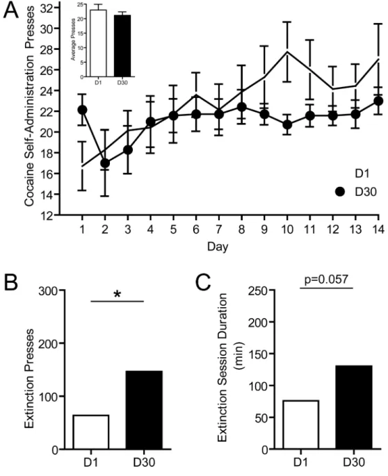
Behavioral data was obtained from rats (n=7) that underwent 1 day (D1 group) or 30 days (n=7; D30 group) of abstinence following self-administration training. Importantly, there were no differences in cocaine self-administration in animals destined for the D1 or D30 groups. A two-way mixed design ANOVA with a within-subjects factor of day of cocaine self-administration training and a between-subjects factor of group conducted on the number of cocaine-reinforced lever presses revealed no main effect of group (F(1,12) = 1.29, p > 0.05), no main effect of day of self-administration (F(9,108) = 1.336, p > 0.05) and no group × day interaction (F(9,108) = 1.375, p > 0.05; Fig. 1A). This statistic was performed on data from the last 10 days of cocaine self-administration when all animals had achieved stable responding. Further, there were no differences in the average number of daily presses between groups over the entire 14 days of cocaine self-administration (t12 = 0.7812; p > 0.05; Fig. 1A, inset).
Similar to previous studies (Grimm et al., 2001; Hollander and Carelli, 2007, 2005), extinction responding revealed an ‘incubation’ effect in D30 animals compared to D1 animals. That is, D30 animals lever pressed significantly more times during extinction than
A
uthor Man
uscr
ipt
A
uthor Man
uscr
ipt
A
uthor Man
uscr
ipt
A
uthor Man
uscr
D1 animals (t12 = 2.700; p < 0.05; Fig. 1B). Furthermore, there was a trend for D30 animals to take longer to completely extinguish responding compared to D1 animals (t12=2.11, p=0.057; Fig. 1C).
3.2. [DA]max is potentiated following an acute cocaine injection, and this potentiation is
greater following 30 days of abstinence
To evaluate the effects of cocaine abstinence on rapid dopamine dynamics, we first
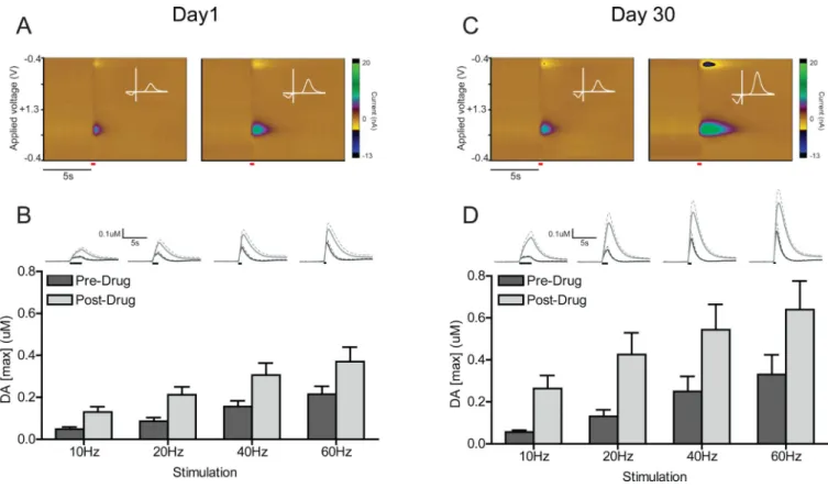
examined [DA]max, the peak electrically-evoked concentration of DA, in rats that underwent 1 day versus 30 days of abstinence. Baseline FSCV recordings were first performed before experimenter-delivered drug administration, then following an injection of cocaine (20 mg/kg, i.p.). In each condition (pre- and post-drug), we measured DA responses at 4 different electrical stimulation parameters (10, 20, 40, and 60Hz). Fig. 2 shows average pre- and post-drug background-subtracted colorplots and cyclic voltammograms for the 60Hz stimulation parameter in representative D1 (Fig. 2A) and D30 (Fig. 2C) animals. [DA]max was compared with a three-way mixed design ANOVA (stimulation frequency as within-subjects factor × drug condition as within-within-subjects factor × abstinence condition as between-subjects factor; Fig. 2B and D). This analysis revealed no main effect of abstinence
condition (F(1,12) = 2.75, p > 0.05), but a main effect of drug condition (F(1,12) = 38.34, p < 0.0001), and a main effect of stimulation frequency (F(3,36) = 32.51, p < 0.0001). There was no abstinence × drug × stimulation interaction (F(3,36) = 0.66, p > 0.05); however, there was a significant abstinence × drug interaction (F(1,12) = 5.13, p < 0.05). Tukey HSD post-hoc tests showed that, collapsed across all stimulation parameters, the post-drug increase in [DA]max was significantly different (i.e., higher) compared to pre-drug in D30 animals (Tukey HSD, p < 0.05). This was not the case in D1 animals.
3.3. Potentiated [DA]max following 30 days of cocaine abstinence is not related to changes
in clearance half-life
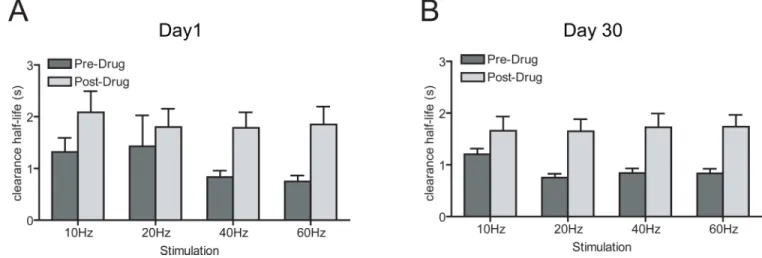
The potentiation of electrically-evoked [DA]max after cocaine challenge in D30 animals suggested a possible increase in DA release in response to an injection of cocaine following a month of abstinence; however, it was also possible that uptake of DA was affected by the abstinence period. To determine the contribution of uptake following an injection of cocaine, we examined clearance half-life with a three-way mixed design ANOVA (stimulation frequency as within-subjects factor × drug condition as within-subjects factor × abstinence condition as between-subjects factor; Fig. 3A and B), and confirmed that the data met the assumption of homogeneity of variance with Hartly F-max, Cochran C, Bartlett Chi-square (p > 0.05). The ANOVA revealed no main effect of abstinence condition (F(1,11) = 0.18, p > 0.05), but a main effect of drug condition (F(1,11) = 23.42, p < 0.001), and a main effect of stimulation frequency (F(3,33) = 18.11, p < 0.0001). There was no abstinence × drug × stimulation interaction (F(3,36) = 0.66, p > 0.05). Unlike [DA]max, there was no abstinence × drug interaction (F(1,12) = 0.19, p > 0.05). Thus, cocaine increased clearance half-life to the same extent in both D1 and D30 animals.These data show that the potentiated
electrically-evoked DA response following a cocaine challenge in D30 animals is not due to changes in clearance half-life and is therefore unlikely to be a result of abstinence-induced changes in DA reuptake.
A
uthor Man
uscr
ipt
A
uthor Man
uscr
ipt
A
uthor Man
uscr
ipt
A
uthor Man
uscr
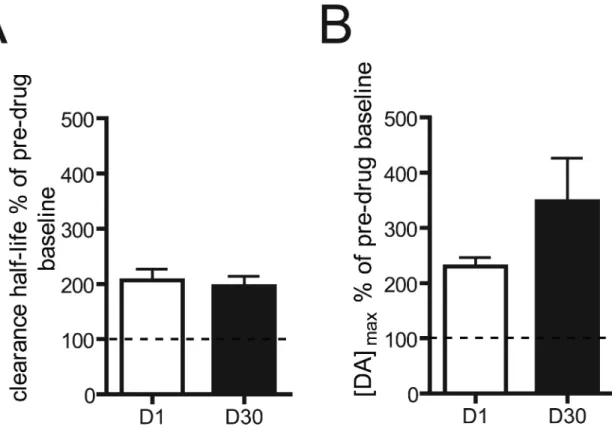
We further explored the potential contributions of release and uptake following an injection of cocaine by calculating percent of pre-drug baseline for both [DA]max and clearance half-life. In this analysis, any percent higher than 100% would indicate an increase in [DA]max or clearance half-life in the post-drug compared to pre-drug condition. Pre-drug baselines were collapsed across all stimulation frequencies for [DA]max and clearance half-life and
compared between D1 and D30 animals. There was no difference in clearance half-life percent of pre-drug baseline between D1 and D30 animals (t12 = 0.40; p > 0.05; Fig. 4A), indicating that the two abstinence groups did not differ in this measure after cocaine. We also calculated clearance rate (the slope from 60% to 20% evoked concentration for each animal) to rule out other effects of altered DAT function. Similar to clearance half-life, a cocaine challenge increased clearance rates in both groups. The mean ± sem percent of pre-drug baseline was 166.0 ± 32.63 and 165.4 ± 26.52 in D1 and D30 animals, respectively. As with clearance half-life, there were no differences between D1 and D30 animals in this measure (t9=0.015, p>0.05), suggesting that the observed potentiation in [DA]max in D30 animals following a cocaine challenge is not related to changes in this uptake parameter. [DA]max percent of pre-drug baseline was not significantly different between D1 and D30 animals (t12 = 1.49; p > 0.05; Fig. 4B). Thus, we cannot make a definitive claim about the contributions of release and uptake with the data normalized to pre-drug baseline. However, given that the non-normalized data showed that length of abstinence had no effect on the uptake inhibiting properties of cocaine as measured by clearance half-life and (Fig. 3), it is possible that the observed abstinence-induced changes in electrically-evoked DA after a cocaine challenge may be driven by changes in release.
3.4 Relationship between extinction responding and [DA]max
No significant correlation was observed between individual animals’ extinction responding (lever pressing) and [DA]max percent of pre-drug baseline in D1 (F(1,5) = 0.4363, p > 0.05, r2 = 0.0803) or D30 (F(1,5) = 1.673, p > 0.05, r2 = 0.2507) rats.
3.5. Histology
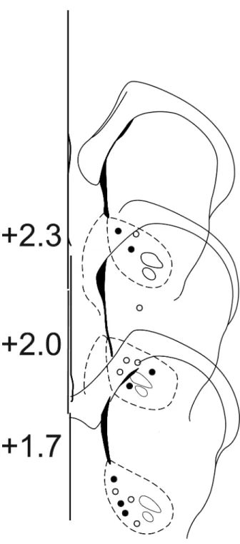
Histological reconstruction of carbon fiber electrode placements confirmed the location of recording sites in the NAc core. Placements ranged from +2.5 to +1.7 mm from bregma. Only data from electrode placements within the borders of the NAc core, as depicted in the atlas of Paxinos & Watson (1997), were included in the analysis (Fig. 5).
4. Discussion
Numerous studies have reported that cocaine abstinence following repeated drug exposure results in neuroadaptations in brain areas important for reward processing, including the nucleus accumbens (NAc) and associated regions (Conrad et al., 2008; Loweth et al., 2014; Lu et al., 2003; Pickens et al., 2011; Robinson et al., 2001). Here, we examined if changes occur in rapid DA signaling dynamics in the NAc core of rats that had undergone 14 days of cocaine self-administration followed by either 1 or 30 days of cocaine abstinence. At baseline (before cocaine challenge) there were no differences in peak evoked DA between abstinence groups. Administration of cocaine increased the peak concentration of
electrically-evoked DA in the NAc core in both D1 and D30 rats; however, this increase was
A
uthor Man
uscr
ipt
A
uthor Man
uscr
ipt
A
uthor Man
uscr
ipt
A
uthor Man
uscr
potentiated in rats that underwent 30 days of abstinence. Examination of release and uptake parameters revealed that the potentiated [DA]max observed in D30 animals (compared to D1 rats) was not related to a potentiation of uptake inhibition as measured by clearance half-life and clearance rate, but may be related to facilitation of release. These findings indicate that simply removing drug access for an extended period of time following cocaine self-administration has long lasting effects on discrete features of rapid dopamine signaling in the NAc.
Several mechanisms could account for the abstinence-induced increases in [DA]max
observed in the present study. One possibility is that there is an increase in vesicular release. Another possibility is that neuroadaptations occur in the VTA during extended cocaine abstinence that can potentiate presynaptic DA release. For example, self-administration of cocaine produces a persistent potentiation of VTA excitatory synapses that remain after 3 months of abstinence (Chen et al., 2008). Likewise, a study by Bocklisch and colleagues also suggests that post-synaptic alterations in NAc neurons can alter DA release from the VTA (Bocklisch et al., 2013). Exposure to cocaine evokes synaptic potentiation in a subset of NAc medium spiny neurons (MSNs) that project to GABA neurons of the VTA, leading to greater disinhibition of VTA neurons and thus increased DA release in the NAc. While the primary pharmacological action of cocaine is inhibition of the dopamine transporter, cocaine can also directly enhance DA release by mobilizing a reserve pool of dopamine-containing synaptic vesicles (Venton, 2006). As such, it is possible that prolonged abstinence enhances this effect of cocaine in a yet to be determined manner. Ultimately, we cannot ascribe any single factor to the increase in electrically-evoked DA concentration we observed in the present study. These changes could be due to increased number of vesicles available for release, larger dopamine content in the vesicles, lower modulation of release by
autoreceptors, an increase in dopamine neuron excitability, or an alteration in afferent inputs to dopamine neurons, among other possibilities.
While some studies report that a history of cocaine self-administration reduces electrically-evoked DA concentration (Calipari et al., 2014; Ferris et al., 2015; Mateo et al., 2005; Siciliano et al., 2015), the experimental designs differed markedly from our own. For example, Calipari and colleagues (2014) assessed presynaptic DA signaling parameters in the NAc core to determine the effects of cocaine self-administration on DA release and uptake in the absence of drug. They reported significant reductions in DA release elicited by single pulse electrical stimulations after cocaine self-administration. However, differences between studies included the use of slice versus in vivo electrochemistry, drug presence during time of testing, type of self-administration paradigm (e.g., 2 hour versus 6 hour drug access), and whether drug was available in a continuous or intermittent schedule during self-administration sessions. These findings underscore the complexity of factors influencing dopamine release/uptake dynamics in animals with a history of cocaine exposure.
An important finding of our study was that the potentiated electrically-evoked [DA]max following cocaine challenge observed in D30 animals was not related to a potentiation of clearance half-life, a measure of uptake inhibition. This is in contrast to other studies that reported a potentiation in uptake inhibition by cocaine following a history of drug exposure (Addy et al., 2010; Calipari et al., 2013). While various aspects of the experimental designs
A
uthor Man
uscr
ipt
A
uthor Man
uscr
ipt
A
uthor Man
uscr
ipt
A
uthor Man
uscr
differed across studies as noted above, another critical difference between these studies and the present work is our inclusion of a prolonged (30 day) abstinence period. Another study that examined DA release/uptake parameters following cocaine self-administration and extended abstinence (3 weeks) found no differences between cocaine- and saline-treated rats on multiple measures of DA reuptake in the NAc (Ramamoorthy et al., 2010). Recently however, others demonstrated that a brief abstinence period (7 days) augmented cocaine's ability to increase DA release as well as inhibit uptake in vitro (Calipari et al., 2015). Thus, it appears that prolonged abstinence (at least 1 month) produces fundamentally different changes in DA signaling than cocaine self-administration alone, increasing cocaine-evoked DA concentration without affecting uptake inhibition.
The precise influence of the potentiated evoked DA response observed in this study on behavioral responding remains to be elucidated. We observed no significant correlation between individual animals’ extinction responding (lever pressing) and [DA]max percent of pre-drug baseline for D1 or D30 rats. However, on average rats in the D30 group showed increased lever press responding during extinction compared to the D1 group (consistent with ‘incubation of craving,’) as well as a potentiation of electrically-evoked [DA]max after cocaine challenge. As such, it is possible that the observed changes in DA signaling following abstinence may drive the observed increase in lever pressing in D30 compared to D1 rats. While classic studies have demonstrated the importance of mesolimbic DA in cocaine reinforcement (de Wit and Wise, 1977; Roberts et al, 1977; Woolverton 1986), those studies examined only maintenance of cocaine self-administration and did not include a period of abstinence, which could fundamentally change the role of DA in cocaine
reinforcement. Furthermore, this interpretation is complicated by the finding that a priming infusion of cocaine, which increases NAc DA levels, abolishes the behavioral incubation effect rather than enhancing cocaine-seeking (Lu et al., 2004a).
Given DAs role as a neuromodulator, it is likely that it has complex interactions with other neurotransmitter systems that may be more responsible for driving the behavioral incubation effect, especially glutamate (Conrad et al., 2008; Kalivas and Volkow, 2005). Prior work by Wolf and colleagues has shown that glutamatergic signaling is altered during incubation by insertion of calcium-permeable AMPA receptors into NAc MSNs, leading to an increase in the responsiveness of MSNs to glutamate (Conrad et al., 2008; Purgianto et al., 2013), and the insertion of calcium-permeable AMPA receptors is causally linked to the behavioral manifestation of incubation (Conrad et al., 2008).
Considering these findings, another possible interpretation is that the abstinence-induced changes in DA signaling observed in the present study may in fact be compensatory, mitigating the effects of other neuroadaptations, primarily glutamatergic, that increase extinction responding following abstinence. Previous work from our lab has shown that there is increased encoding of cocaine-associated stimuli and cocaine-seeking behavior following cocaine abstinence in both the NAc core and prelimbic cortex (Hollander and Carelli, 2007, 2005; West et al., 2014). Thus, a possible role of DA following abstinence may be modulation of glutamate signaling from the prefrontal cortex (PFC) to the NAc (Kalivas, 2009; Kalivas et al., 2005). DA dampens the effect of glutamatergic input from the PFC, and this inhibitory action appears to be mediated through D2 receptors (Brady and
A
uthor Man
uscr
ipt
A
uthor Man
uscr
ipt
A
uthor Man
uscr
ipt
A
uthor Man
uscr
O'Donnell, 2004; Goto and Grace, 2005). Following abstinence, these two neurotransmitters may thus exert opposing actions on behavior, with glutamate potentiating drug-seeking and DA blunting this effect (Doyle et al., 2014; Ramôa et al., 2014). Future experiments could investigate the link between glutamate and abstinence-induced alterations in dopamine signaling, for example, by employing voltammetry and targeted inotophoretic delivery of agonists and antagonists (Owesson-White et al. 2016).
4.1. Conclusions
Overall, the results of this study support the abundance of literature showing that cocaine abstinence induces neuroplasticity in brain areas implicated in drug reward and relapse. The finding that increases in extracellular DA are not related to an increase in uptake inhibition also demonstrates a critical difference between abstinence-induced neuroadaptations in DA signaling and those caused by drug exposure itself. Finally, these results provide insight into the neurobiological processes that control addictive behavior, specifically the impact of altered DA signaling on cocaine-seeking behavior following abstinence, and can inform the development of improved relapse prevention therapies.
Acknowledgements
This research was supported by DA029978 to CMC and DA017318 and DA014339 to RMC. The authors thank Dr. Michael Saddoris for help with statistical analysis, Dr. Elyse Dankoski and Fei Fei Wang for assistance in running experiments, Elina Thomas for help with data analysis, and Dr. Travis Moschak for comments on an earlier version of the manuscript.
References
Addy NA, Daberkow DP, Ford JN, Garris PA, Wightman RM. Sensitization of rapid dopamine signaling in the nucleus accumbens core and shell after repeated cocaine in rats. J. Neurophysiol. 2010; 104:922–31. [PubMed: 20554845]
Bedi G, Preston KL, Epstein DH, Heishman SJ, Marrone GF, Shaham Y, De Wit H. Incubation of cue-induced cigarette craving during abstinence in human smokers. Biol. Psychiatry. 2011; 69:708–711. [PubMed: 20817135]
Bocklisch C, Pascoli V, Wong JCY, House DRC, Yvon C, de Roo M, Tan KR, Lüscher C. Cocaine disinhibits dopamine neurons by potentiation of GABA transmission in the ventral tegmental area. Science. 2013; 341:1521–5. [PubMed: 24072923]
Brady AM, O'Donnell P. Dopaminergic modulation of prefrontal cortical input to nucleus accumbens neurons in vivo. J. Neurosci. 2004; 24:1040–9. [PubMed: 14762122]
Calipari ES, Ferris MJ, Jones SR. Extended access of cocaine self-administration results in tolerance to the dopamine-elevating and locomotor-stimulating effects of cocaine. J. Neurochem. 2014;
128:224–32. [PubMed: 24102293]
Calipari ES, Ferris MJ, Zimmer BA, Roberts DCS, Jones SR. Temporal pattern of cocaine intake determines tolerance vs sensitization of cocaine effects at the dopamine transporter.
Neuropsychopharmacology. 2013; 38:2385–92. [PubMed: 23719505]
Calipari ES, Siciliano CA, Zimmer BA, Jones SR. Brief intermittent cocaine self-administration and abstinence sensitizes cocaine effects on the dopamine transporter and increases drug seeking. Neuropsychopharmacology. 2015; 40:728–35. [PubMed: 25212486]
Cameron CM, Carelli RM. Cocaine abstinence alters nucleus accumbens firing dynamics during goal-directed behaviors for cocaine and sucrose. Eur. J. Neurosci. 2012; 35:940–951. [PubMed: 22356698]
A
uthor Man
uscr
ipt
A
uthor Man
uscr
ipt
A
uthor Man
uscr
ipt
A
uthor Man
uscr
Carelli RM, Deadwyler SA. A comparison of nucleus accumbens neuronal firing patterns during cocaine self-administration and water reinforcement in rats. J. Neurosci. 1994; 14:7735–46. [PubMed: 7996208]
Chen BT, Bowers MS, Martin M, Hopf FW, Guillory AM, Carelli RM, Chou JK, Bonci A. Cocaine but not natural reward self-administration nor passive cocaine infusion produces persistent LTP in the VTA. Neuron. 2008; 59:288–97. [PubMed: 18667156]
Conrad KL, Tseng KY, Uejima JL, Reimers JM, Heng L-J, Shaham Y, Marinelli M, Wolf ME. Formation of accumbens GluR2-lacking AMPA receptors mediates incubation of cocaine craving. Nature. 2008; 454:118–21. [PubMed: 18500330]
Day JJ, Roitman MF, Wightman RM, Carelli RM. Associative learning mediates dynamic shifts in dopamine signaling in the nucleus accumbens. Nat. Neurosci. 2007; 10:1020–8. [PubMed: 17603481]
Doyle SE, Ramôa C, Garber G, Newman J, Toor Z, Lynch WJ. A shift in the role of glutamatergic signaling in the nucleus accumbens core with the development of an addicted phenotype. Biol. Psychiatry. 2014; 76:810–5. [PubMed: 24629536]
Ferrario CR, Gorny G, Crombag HS, Li Y, Kolb B, Robinson TE. Neural and behavioral plasticity associated with the transition from controlled to escalated cocaine use. Biol. Psychiatry. 2005; 58:751–9. [PubMed: 16098484]
Ferris MJ, Calipari ES, Rose JH, Siciliano CA, Sun H, Chen R, Jones SR. A Single Amphetamine Infusion Reverses Deficits in Dopamine Nerve-Terminal Function Caused by a History of Cocaine Self-Administration. Neuropsychopharmacology. 2015; 40:1826–36. [PubMed: 25689882] Gawin FH. Cocaine addiction: psychology and neurophysiology. Science. 1991; 251:1580–6.
[PubMed: 2011738]
Gawin FH, Kleber HD. Abstinence symptomatology and psychiatric diagnosis in cocaine abusers. Clinical observations. Arch. Gen. Psychiatry. 1986; 43:107–13. [PubMed: 3947206]
Goto Y, Grace AA. Dopaminergic modulation of limbic and cortical drive of nucleus accumbens in goal-directed behavior. Nat. Neurosci. 2005; 8:805–12. [PubMed: 15908948]
Grimm JW, Hope BT, Wise RA, Shaham Y. Neuroadaptation. Incubation of cocaine craving after withdrawal. Nature. 2001; 412:141–2. [PubMed: 11449260]
Hollander JA, Carelli RM. Cocaine-associated stimuli increase cocaine seeking and activate accumbens core neurons after abstinence. J. Neurosci. 2007; 27:3535–9. [PubMed: 17392469] Hollander JA, Carelli RM. Abstinence from cocaine self-administration heightens neural encoding of
goal-directed behaviors in the accumbens. Neuropsychopharmacology. 2005; 30:1464–74. [PubMed: 15856078]
Kalivas PW. The glutamate homeostasis hypothesis of addiction. Nat. Rev. Neurosci. 2009; 10:561–72. [PubMed: 19571793]
Kalivas PW, McFarland K, Bowers S, Szumlinski K, Xi Z-X, Baker D. Glutamate transmission and addiction to cocaine. Ann. N. Y. Acad. Sci. 2003; 1003:169–75. [PubMed: 14684444]
Kalivas PW, Volkow N, Seamans J. Unmanageable motivation in addiction: a pathology in prefrontal-accumbens glutamate transmission. Neuron. 2005; 45:647–50. [PubMed: 15748840]
Kalivas PW, Volkow ND. The neural basis of addiction: a pathology of motivation and choice. Am. J. Psychiatry. 2005; 162:1403–13. [PubMed: 16055761]
Koob GF, Volkow ND. Neurocircuitry of addiction. Neuropsychopharmacology. 2010; 35:217–238. [PubMed: 19710631]
Lee BR, Ma Y-Y, Huang YH, Wang X, Otaka M, Ishikawa M, Neumann PA, Graziane NM, Brown TE, Suska A, Guo C, Lobo MK, Sesack SR, Wolf ME, Nestler EJ, Shaham Y, Schlüter OM, Dong Y. Maturation of silent synapses in amygdala-accumbens projection contributes to incubation of cocaine craving. Nat. Neurosci. 2013; 16:1644–51. [PubMed: 24077564]
Li X, Caprioli D, Marchant NJ. Recent updates on incubation of drug craving: A mini-review. Addict. Biol. 2015
Loweth JA, Tseng KY, Wolf ME. Adaptations in AMPA receptor transmission in the nucleus accumbens contributing to incubation of cocaine craving. Neuropharmacology. 2014
A
uthor Man
uscr
ipt
A
uthor Man
uscr
ipt
A
uthor Man
uscr
ipt
A
uthor Man
uscr
Lu L, Grimm JW, Dempsey J, Shaham Y. Cocaine seeking over extended withdrawal periods in rats: different time courses of responding induced by cocaine cues versus cocaine priming over the first 6 months. Psychopharmacology (Berl). 2004a; 176:101–8. [PubMed: 15071719]
Lu L, Grimm JW, Hope BT, Shaham Y. Incubation of cocaine craving after withdrawal: a review of preclinical data. Neuropharmacology. 2004b; 47(Suppl 1):214–26. [PubMed: 15464139] Lu L, Grimm JW, Shaham Y, Hope BT. Molecular neuroadaptations in the accumbens and ventral
tegmental area during the first 90 days of forced abstinence from cocaine self-administration in rats. J. Neurochem. 2003; 85:1604–13. [PubMed: 12787079]
Mateo Y, Lack CM, Morgan D, Roberts DCS, Jones SR. Reduced dopamine terminal function and insensitivity to cocaine following cocaine binge self-administration and deprivation.
Neuropsychopharmacology. 2005; 30:1455–63. [PubMed: 15702135]
Owesson-White C, Belle AM, Herr NR, Peele JL, Gowrishankar P, Carelli RM, Wightman RM. Cue-evoked dopamine release rapidl modulates D2 neurons in the nucleus accumbens during motivated behavior. J. Neurosci. 2016; 36(22):6011–6021. [PubMed: 27251622]
Pickens CL, Airavaara M, Theberge F, Fanous S, Hope BT, Shaham Y. Neurobiology of the incubation of drug craving. Trends Neurosci. 2011; 34:411–20. [PubMed: 21764143]
Pierce RC, Wolf ME. Psychostimulant-Induced Neuroadaptations in Nucleus Accumbens AMPA Receptor Transmission. Cold Spring Harb. Perspect. Med. 2013; 3
Purgianto A, Scheyer AF, Loweth J. a, Ford K. a, Tseng KY, Wolf ME. Different adaptations in AMPA receptor transmission in the nucleus accumbens after short vs long access cocaine
self-administration regimens. Neuropsychopharmacology. 2013; 38:1789–97. [PubMed: 23546386] Ramamoorthy S, Samuvel DJ, Balasubramaniam A, See RE, Jayanthi LD. Altered dopamine
transporter function and phosphorylation following chronic cocaine self-administration and extinction in rats. Biochem. Biophys. Res. Commun. 2010; 391:1517–21. [PubMed: 20035724] Ramôa CP, Doyle SE, Lycas MD, Chernau AK, Lynch WJ. Diminished role of dopamine D1-receptor
signaling with the development of an addicted phenotype in rats. Biol. Psychiatry. 2014; 76:8–14. [PubMed: 24199666]
Robinson TE, Gorny G, Mitton E, Kolb B. Cocaine self-administration alters the morphology of dendrites and dendritic spines in the nucleus accumbens and neocortex. Synapse. 2001; 39:257– 66. [PubMed: 11169774]
Roitman MF, Stuber GD, Phillips PEM, Wightman RM, Carelli RM. Dopamine operates as a subsecond modulator of food seeking. J. Neurosci. 2004; 24:1265–71. [PubMed: 14960596] Siciliano CA, Ferris MJ, Jones SR. Cocaine self-administration disrupts mesolimbic dopamine circuit
function and attenuates dopaminergic responsiveness to cocaine. Eur. J. Neurosci. 2015 Toda S, McGinty JF, Kalivas PW. Repeated cocaine administration alters the expression of genes in
corticolimbic circuitry after a 3-week withdrawal: a DNA macroarray study. J. Neurochem. 2002; 82:1290–9. [PubMed: 12358776]
Tran-Nguyen LT, Fuchs RA, Coffey GP, Baker DA, O'Dell LE, Neisewander JL. Time-dependent changes in cocaine-seeking behavior and extracellular dopamine levels in the amygdala during cocaine withdrawal. Neuropsychopharmacology. 1998; 19:48–59. [PubMed: 9608576]
Venton BJ. Cocaine Increases Dopamine Release by Mobilization of a Synapsin-Dependent Reserve Pool. J. Neurosci. 2006; 26:3206–3209. [PubMed: 16554471]
West EA, Saddoris MP, Kerfoot EC, Carelli RM. Prelimbic and infralimbic cortical regions differentially encode cocaine-associated stimuli and cocaine-seeking before and following abstinence. Eur. J. Neurosci. 2014; 39:1891–902. [PubMed: 24690012]
Xi Z-X, Ramamoorthy S, Shen H, Lake R, Samuvel DJ, Kalivas PW. GABA transmission in the nucleus accumbens is altered after withdrawal from repeated cocaine. J. Neurosci. 2003; 23:3498– 505. [PubMed: 12716959]
Yorgason JT, España RA, Jones SR. Demon voltammetry and analysis software: analysis of cocaine-induced alterations in dopamine signaling using multiple kinetic measures. J. Neurosci. Methods. 2011; 202:158–64. [PubMed: 21392532]
A
uthor Man
uscr
ipt
A
uthor Man
uscr
ipt
A
uthor Man
uscr
ipt
A
uthor Man
uscr
Highlights
• FSCV was used to measure NAc core DA after 1 day or 1 month cocaine abstinence
• Rats showed an incubation of craving after 1 month cocaine abstinence
• Distinct changes in DA signaling occur following prolonged cocaine abstinence
• 1 month of cocaine abstinence potentiated cocaine-evoked peak [DA]
• Changes in cocaine-evoked DA appear driven by changes in release facilitation
A
uthor Man
uscr
ipt
A
uthor Man
uscr
ipt
A
uthor Man
uscr
ipt
A
uthor Man
uscr
Figure 1. Behavioral responding during cocaine self-administration and extinction
A. Mean ± sem presses during 14 days of cocaine self-administration training for animals destined for D1 (open circles) or D30 (closed circles) group. Inset: comparison of mean ± sem daily presses for animals destined for D1 (open bar) or D30 (closed bar) group. B. Comparison of mean ± sem presses during extinction for animals that underwent 1 day of abstinence (open bar) versus animals that underwent 30 days of abstinence (closed bar), *p < 0.05. Circles represent data points from individual animals. C. Comparison of mean ± sem extinction session duration for animals that underwent 1 day of abstinence versus animals that underwent 30 days of abstinence.
A
uthor Man
uscr
ipt
A
uthor Man
uscr
ipt
A
uthor Man
uscr
ipt
A
uthor Man
uscr
Figure 2. Electrically-evoked DA release in the NAc core
A. Color representations of cyclic voltammetric data collected for one D1 animal in response to electrical stimulation (60Hz, 24 pulses) of the VTA before (left) and after (right) injection of cocaine (data represents the average of 5 trials; stimulation represented by red line). The y-axis is applied voltage, the x-axis is time, and current measured at the carbon fiber electrode is plotted in false color. Inset: cyclic voltammagram taken at peak evoked
concentration of DA ([DA]max; white line). B. Average concentration traces and [DA]max at 10, 20, 40, and 60 Hz stimulation frequencies in animals that underwent 1 day of abstinence. Lines represent average [DA] traces + sem in all D1 animals before (dark gray line) and after (light gray line) injection of cocaine (black bars represent time of VTA stimulation). Dark gray bars denote [DA]max before cocaine injection; light gray bars denote [DA]max following cocaine injection. C. Color representations of cyclic voltammetric data collected for one D30 animal in response to electrical stimulation (60Hz, 24 pulses) of the VTA before (left) and after (right) injection of cocaine (data represents the average of 5 trials; stimulation represented by red line). Conventions as in A. D. Average concentration traces and [DA]max at 10, 20, 40, and 60Hz stimulation frequencies in animals that underwent 30 days of abstinence. Conventions as in B. Error bars represent sem in all panels.
A
uthor Man
uscr
ipt
A
uthor Man
uscr
ipt
A
uthor Man
uscr
ipt
A
uthor Man
uscr
Figure 3. DA reuptake is not affected by length of abstinence
A. Clearance half-life at 10, 20, 40, and 60Hz stimulation frequencies in animals that underwent 1 day of abstinence. B. Clearance half-life in animals that underwent 30 days of abstinence. Dark gray bars denote [DA]max before cocaine injection; light gray bars denote [DA]max following cocaine injection. Error bars represent sem in all panels.
A
uthor Man
uscr
ipt
A
uthor Man
uscr
ipt
A
uthor Man
uscr
ipt
A
uthor Man
uscr
Figure 4. Influence of DA release and uptake parameters
A. Clearance half-life percent of pre-drug baseline. 100% of pre-drug baseline (indicated by dotted line) represents no change in [DA]max in the post-drug (cocaine on board) compared to pre-drug (no cocaine) condition. Percentages higher than 100% represent an increase in clearance half-life in the post-drug compared to pre-drug condition. Comparison of D1 (open bars) and D30 (closed bars) collapsed across all stimulation frequencies. B. [DA]max percent of pre-drug baseline. Comparison of D1 (open bars) and D30 (closed bars) collapsed across all stimulation frequencies.
A
uthor Man
uscr
ipt
A
uthor Man
uscr
ipt
A
uthor Man
uscr
ipt
A
uthor Man
uscr
Figure 5. Histology
Schematic representation of electrode tip placements in the NAc core. Numbers to the left of coronal sections indicate distance anterior to bregma (Paxinos & Watson, 2007). Open circles indicate recording locations from D1 animals; closed circles represent recording locations from D30 animals.