INFLUENCE OF DIFFERENT DENTIN DRYING METHODS ON ADHESIVE BEHAVIOR
OF COMPOSITE RESTORATIONS
Helena Reis de Souza1, Stéfanie Thieme Perotto1, Maria Carolina Guilherme Erhardt2, Fabio Garcia Lima3, Ana Maria Spohr4, Celso Afonso Klein Júnior5, Fabio Herrmann Coelho de Souza2
1 Federal University of Rio Grande do Sul, Porto Alegre, RS, Brazil
2 Department of Operative Dentistry, Federal University of Rio Grande do Sul, Porto Alegre, RS, Brazil 3 Department of Operative Dentistry, Federal University of Pelotas, Pelotas, RS, Brazil 4 Department of Operative Dentistry, Catholic PontiQical University, Porto Alegre, RS, Brazil
5 Department of Operative Dentistry, Lutheran University of Brazil, Canoas, RS, Brazil
CORRESPONDING AUTHOR: [email protected]
ABSTRACT
Aim:The aim of this study was to evaluate, in vitro, the influence of four different dentin drying methods (air drying for 10s, absorbent paper, endodontic suction cannula and air drying for 10s with subsequent re-wetting with distilled water), after etching with phosphoric acid gel 37%, on bond strength and microleakage of composite restorations.
Material and Methods: Twenty sound bovine incisors were selected and sectioned transversely. The buccal surfaces were frayed until exposure of dentin, etched and washed with distilled water. Each specimen was subjected to one of the four different drying methods. A 3-step total-etch adhesive system (Adper Scotchbond™ Multi-Purpose) was used on 10 teeth and on the other 10, a 2-step total-etch adhesive system (Adper Single BondT™ 2) was used. Composite resin restorations in cylindrical shapes were made and, after thermocycling, the teeth were subjected to the microshear bond strength test. Cervical areas of the teeth were prepared and restored with the same techniques describe above. Cervical restorations were cut and subjected to microleakage analysis in the cervical margin. Data obtained were tabulated and statistically analyzed, using ANOVA, Chi-square and Kruskal-Wallis tests, and the significance level was set at 5%.
Results:The results showed no statistically significant differences among the groups tested.
Conclusions: It was concluded that all drying methods tested represent feasible alternatives to clinical application for total etch dentin-bonding agents.
KEYWORDS: dentin-bonding agents, dentin moisture, adhesive system
INTRODUCTION
Over the last few years, restorative dentistry had significant changes, mainly on restorative materials
and adhesive systems. Before this evolution, one of the main aims of restorative cosmetic dentistry is obtain materials able to adhere properly to
dental tissues, reduce microleakage and improve the bond strength1.
surfaces to increase the restorative materials adhesion to the tooth. Previous phosphoric acid etching changes the enamel structure, increases the surface area and the adhesion becomes more favorable to restorative materials, further seal properly pits and fissures, and prevent marginal failures. On the other hand, dentin tissue is more complex in the case of adhesive systems, when compared to enamel, due to its heterogeneous morphology and composition formed by an organic matrix, collagen fibers, hydroxyapatite and water3. Besides, dentin has tubular nature and the smear layer, which means a residue layer deposited on the dentin surface during the cavity preparation4. However, with development of new materials able to remove the smear layer, and of bifunctional primers, restorative materials adhesion to dentin surface became more favorable5.
A d h e s i v e s y s t e m s w h i c h perform total smear layer removal recommend acid etching of enamel and dentin simultaneously6, following the application of primer and adhesive in distinct bottles or combined in a same one. After dentin etching, the acid component is removed through water washing. It is very important because determines the tissue humidity for posterior application of primer and adhesive1,4, and induces the formation of an appropriate hybrid layer and consequent restorative material adhesion to the tooth structure7. The overdrying would change the collagen fibers and preclude the appropriate penetration of primer and adhesive6. The water kept inside the intertubular space of dentin tissue is responsible by maintain the collagen matrix and preserve the structure necessary to the adhesive penetrate in the tubular and inter tubular dentin1,8. In contrast, moisture excess
would influence negatively the adhesive performance, which would be diluted and would not polymerize properly. Tay et al.9 affirmed that water excess dilute the primer and its contents in more than one stage, what results in blistering. Besides, they considered that dentin humidity is indispensable for effective adhesion, but water excess can result in a very spoiled adhesion, characterized as overwet.
Because of the absence of a defined protocol, mainly regarding to the humidity after acid etching, for restorative materials appropriate adhesion to the tooth structure, many studies were performed comparing drying methods. Pereira et al.1 evaluated the bond strength by microshear of two adhesive systems applied to dentin surfaces with different humidity degrees, and they obtained higher bond strength for an adhesive system used after dry dentin with wet cotton balls (23.2 MPa) and air drying for 5 seconds (21.3 MPa). For other adhesive system used, the best result was found when the drying method was air drying for 30 seconds (19.5 MPa). For the two adhesive systems tested, the worst result regarding to the bond strength was related to overwet dentin (2.7 MPa and 2.4 MPa). Mitchem and Gronas10 had tested shear strength in composite restorations in dentin and also found values significantly low when the dentin was kept humid.
However, Spazzin et al.11 demonstrated better performance for bond strength when the dentin remained humid (drying by cotton) compared to the overdrying (air). Magne et al.12 showed there was no difference between dry dentin with air or suction cannula regarding to the bond strength by microtensile using a three-step adhesive system. Drying dentin with absorbing paper after acid etching is also an interesting alternative, according to
Jayaprakash et al.13, presenting better bond strength when compared to other drying methods.
Thus, several ways to remove water excess after acid etching rinsing are employed; but few studies compare these different methods in an effective way, and the results are not satisfactorily conclusive. The result of restorative procedure with composite in dentin is closely linked to the appropriate carry out all stages of surface preparation, presenting great technical sensitivity because each step is important for the success and the durability of the restoration. Therefore, the aim of this study was to evaluate in vitro the influence of different drying methods for dentin after phosphoric acid etching on the bond strength and marginal microleakage in composite restorations.
MATERIAL AND METHODS
Inferior bovine incisors were used in this study. This research was evaluated and approved by the Commission Research of the Faculty of Dentistry of UFRGS (project number 63649).
For this study, 20 healthy bovine incisors were selected, using an n of 10 units by group, according to the sample calculation previously performed. The sample size was calculated from a standard deviation of 8.1 MPa associated to a confidence interval of 95% and estimated margin of error of 5%, achieving an n of 10. The restoration was considered sample unit and performed four restorations in each tooth section.
drying for 10s, absorbent paper, endodontic suction cannula and air drying for 10s with subsequent re-wetting with distilled water by microbrush (KG BrushTM, KG Sorensen).
Two types of adhesive system were used for evaluation, a three-step etch-and-rinse (Adper ScotchbondTM Multi-Purpose Plus, 3M ESPE), and a two-step etch-and-rinse (Adper Single BondTM 2,
3M ESPE).
The groups was described in the table 1.
Table 1. Distribution of the groups tested.
Each tooth selected, after disinfection in formalin 2% during 15 days, was transversally sectioned with Diamond disc under water cooling; divided in two parts, one of them composed by the incisal third and the middle third of the crown (crown); the other part is composed by the cervical third of the crown and root (root). In the first (crown), vestibular surface had the enamel wore out with Diamond bur 3098 (KG Sorensen) until dentin exposition, in which four cylinders of low viscosity composite were confectioned for each tooth, using a silicone pipe of 2mm diameter. After preparation, the surfaces were clean and dry, and etched with phosphoric acid 37% during 15s, and washed in plenty distilled water (according to manufacturer’s instructions). Then the dentin was dry; this procedure occurred in a different way for each group.
Group 1 was subjected to air drying with oil-free compressor during 10s, with the tip of the syringe at 10 cm from dentin surface. In the Group 2, dentin drying was performed with circular sections of 6mm diameter of absorbent paper (filter paper
MelittaTM), applied with clinic tweezers, touching the dentin during 10s. Group 3 had drying carried out with endodontic suction cannula (Indusbello), 1mm from dentin surface during 5s. Group 4 was dried with air drying, oil-free compressor during 10s, with the tip of the syringe at 2cm from dentin surface, with subsequent re-wetting with distilled water by m i c r o b r u s h ( K G B r u s hT M, K G Sorensen) impregnated with distilled water. A three-step adhesive system (Adper ScotchbondTM Multi-Purpose) was used in the Groups 1-4, according to manufacturer’s instructions. The primer was applied actively to the conditioned surface, with an applicator type microbrush (KG BrushTM, KG Sorensen), dried with air during 5s for solvent evaporation. Then, the adhesive was applied on the enamel and dentin surfaces treated and lightcured with curing unit LED (Smart LightTM, Dentisply), with intensity measured by a radiometer, over 600 mW/cm2 during 10s.
In the Groups 5-8 were used the two-steps adhesive system (Adper Single BondTM 2), according to manufacturer’s instructions. Two
consecutive layers of adhesive system were applied actively with applicator microbrush, during 15s, and right after, air drying by 5s from 5cm distance for solvent evaporation, and lightcured by the same LED device.
A f t e r t h e a p p r o p r i a t e treatment of surfaces, restorations were confectioned using a cylindrical matrix in transparent plastic with low viscosity composite resin (Fill Magic FlowTM, Vigodent), color A3, lightcured during 20s with the same curing unit LED. After restored, teeth were stored in water by 24 hours and then passed by thermocycler with 500 cycles of 30s each (from 5° to 55°C). After thermocycling, crown surfaces were included in metal cylinders with selfcured acrylic resin (JETTM, Classic) with the buccal surface up, allowing carry out the microshear bond strength test.
On the other part of tooth (root), cavity preparation were performed in 3mm x 3mm dimension, with cervical finishing in dentin (class V) through diamond bur 3098 in high speed with constant water cooling. Each tooth (root) received 04 preparations (1 by surface - buccal,
Drying method Adhesive system
Adper Scotchbond™ Multi-Purpose Adper Single Bond™ 2
Air-drying Group 1 Group 5
Absorbent paper Group 2 Group 6
Suction cannula Group 3 Group 7
lingual, mesial, and distal). Cavities were conditioned with phosphoric acid and divided into the same previous groups according to the drying methods of dentin and adhesive systems. Then, cavities were restored with composite resin Filtek Supreme (3M ESPE), color A3E by incremental technique (increments of 2mm), singly lightcured by the same LED described before. The finishing/polishing was performed immediately through Enhance system (Dentsply).
After restored, teeth were stored in water during 24 hours and then thermocycled with 500 cycles of 30 seconds each (from 5° and 55°C). After thermocycling, colorless nail varnish was applied externally on the restoration margins. From that, teeth were put in dye solution, staying 24 hours in rhodamine B. Then, a section of teeth was obtained with Diamond disc under constant water cooling, first separating restorations in two parts in a cut in occlusal-cervical direction (divided on the center of the restoration), to analyze the marginal microleakage.
Bond strength was evaluated by microshear in a Universal test machine (EMIC) that measured a value in Newton (N). This force value was divided by the area of composite resin cylinder (3.14 mm2), resulting in a mega pascal (MPa). The composite resin c y l i n d e r s w e r e d i s p o s e d perpendicularly along the machine shaft, which worked by traction with travel speed of 0.5 mm/min, using an orthodontic wire 0.25 mm diameter. After carry out the test, dentin surfaces were analyzed in stereomicroscope with magnifying 30 times (Wild Heerbrugg, M5-26293, Switzerland) to evaluate the area of fracture. Type of fracture was classified as adhesive
fracture (occurred in the union line between the specimen and the adhesive system), cohesive fracture (restorative material fracture), dentin fracture (displacement of specimen and dentine portion) and mixed fracture (more than one type of fracture together).
T h e i n t e r f a c e d e n t i n -restoration was evaluated regarding the marginal microleakage (after dying with rhodamine B) in stereoscope magnifying 30 times by blind examiner regarding to the aims of the study. Microleakage was categorized in scores from 0 to 3; 0 when there is no leakage, 1 when the leakage achieves the external half of dentin, 2 when the leakage achieves the internal half dentin toward the axial wall, and 3 when the coloring appears toward the pulp chamber.
Data obtained were subjected to statistical analysis with significance level at 5%. Bond strength was analyzed by ANOVA test. To analyze types of fracture we used the CHI-SQUARE test. Scores related to marginal microleakage were analyzed by Kruskal-Wallis test.
RESULTS
D a t a o b t a i n e d f r o m microshear bond strength test were statistically analyzed through analysis of variance (ANOVA), and it is showed in table 2.
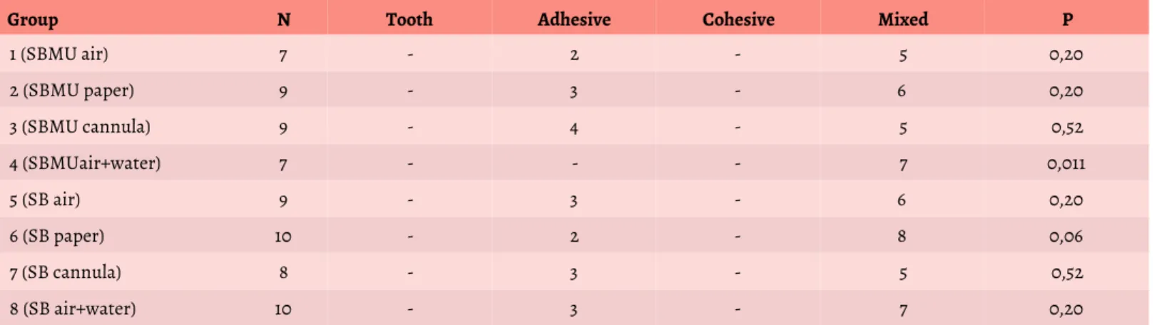
Data regarding to the fracture patterns found after microshear test were analyzed through Chi-square test and it is showed in table 3.
The analysis of groups showed group 4 (Adper ScotchbondTM Multi-Purpose, air drying and posterior rewetting with distilled water) presented significant difference ( p = 0 . 0 1 1 ) , s h o w i n g f a i l u r e s
concentrated in mixed pattern.
Microleakage test data are exposed in table 4, and they were analyzed statistically through Kruskal-Wallis test.
Regarding to marginal microleakage test, there was no difference among drying techniques for each adhesive tested. There was no difference among adhesives when used the same drying techniques. There was significant difference only between the group 1 (SBMU AIR) with group 7 (SB Cannula); group 1 presented lower microleakage level.
DISCUSSION
Results presented in this study showed the bond strength of composite restorations in dentin, evaluated through microshear bond strength test. There was no statistically difference among groups tested, no matter the drying methods used. Similarly, marginal microleakage analysis did not show differences among drying techniques. Thereby, according to these parameters, we can assume that the use of any of the techniques mentioned can be employed with satisfactory performance for both total-etch adhesives used.
adhesive system1,8.
Table 2. Data regarding to microshear bond strength test (MPa).
Adhesive system Adper Scotch BondTM Multi-Purpose, 3M ESPE - 1 (SBMU air) = air drying, 2 (SBMU paper) = absorbent paper drying, 3 (SBMU cannula) = suction cannula. 4 (SBMU air + water) = air drying followed by rewetting. Adhesive system Adper Single BondTM 2, 3M ESPE - 5 (SB air) = air drying, 6 (SB paper) = absorbent paper drying, 7 (SB cannula) = suction cannula and 8 (SB air + water) = air drying followed by rewetting.
Table 3. Fracture patterns in specimens after microshear bond strength test.
Table 4. Data analysis about marginal microleakage.
This work used the in vitro method with bovine incisor teeth selection14,15, maintained in formalin 2% during 15 days for disinfection16. Teeth were transversally sectioned and the buccal surface of crown was worn until dentin exposition. The surfaces
were treated according to the manufacturer’s instructions for application of adhesive systems, except on drying after acid etching, and these methods were tested in this study. After the restorations confectioned in cilyndrical shape with low viscosity
composite resin17,18,19, teeth remained stored in distilled water during 3 weeks, and they were posteriorly submitted to termocycling in order to simulate different temperature changes which occur inside the oral environment and the hydrolysis of
Group Group Mean Standard deviation p
1 (SBMU air) 7 4.35 3.98 0,541
2 (SBMU paper) 9 4.94 3.34
-3 (SBMU cannula) 9 7.28 3.93
-4 (SBMU air + water) 7 6.42 2.20
-5 (SB air) 9 4.87 2.53
-6 (SB paper) 10 5.49 3.87
-7 (SB cannula) 8 4.34 3.53
-8 (SB air + water) 10 4.22 3.65
-Group N Tooth Adhesive Cohesive Mixed P
1 (SBMU air) 7 - 2 - 5 0,20
2 (SBMU paper) 9 - 3 - 6 0,20
3 (SBMU cannula) 9 - 4 - 5 0,52
4 (SBMUair+water) 7 - - - 7 0,011
5 (SB air) 9 - 3 - 6 0,20
6 (SB paper) 10 - 2 - 8 0,06
7 (SB cannula) 8 - 3 - 5 0,52
8 (SB air+water) 10 - 3 - 7 0,20
Group N Microleakage P Dunn's
1 (SBMU air) 10 5/3/1/1 0,002 A
2 (SBMU paper) 10 2/5/2/1 - AB
3 (SBMU cannula) 10 2/2/3/3 - AB
4 (SBMU air + water) 10 3/5/1/1 - AB
5 (SB air) 10 2/4/2/2 - AB
6 (SB paper) 10 2/4/2/2 - AB
7 (SB cannula) 10 1/3/4/2 - B
hybrid layer, what reproduces the bond line aging11,17,20,21.
Microshear test was used to evaluate the bond strength of the restorations in this study, as well as Pereira et al.1, Kanca22, Shimada et al.23 and Dellazzana et al.17. However, different studies tested the bond strength by other tests, and the microtensile bond strength is one of the most found nowadays12,14,24. The preparation of cavities and shape of restorations to test drying methods after surfaces conditioning varied, and cylindrical restorations perpendicular to the buccal surface were used1,22,25, like in this study, or even restorations class II, involving mesial, occlusal and distal surfaces12.
In a study performed by Pereira et al.1, only 3 groups of the 14 tested obtained results of bond strength lower than 10 MPa, other varied from 12 and 24 MPa, as in the study carried out by Kanca22 in which the results were high for all the groups (from 11 to 32 MPa), except by one group, which was lower than 10 MPa. However, this study obtained lower results than those found, from 4 to 8 MPa. These results can be due to methodological differences. Among the factors which can have interfered on the adhesion is the use of bovine teeth, because according to other studies, they can be used in dentistry researches26,27, but caution is necessary, as they can change results in adhesion tests when performed in dentin with random depth and wear, compared to researches in human teeth27,28. Besides, variables in execution of microshear test can interfere in the application of necessary strength to rupture the restoration29. According to Van Noort et al.30, significant changes on specimens can occur due to the
distribution of tensile test during the load application. Geometry, sample sizes, dimension of bonded surface area and the type of composite used determine variables which can make t h e a d h e s i o n v a l u e s v a r y substantially31,32. It is important detach that the load should be applied as juxtaposed as possible to the attached interface plan, in order to avoid influence on the way of application of loads in the test performed29. The microshear test favors the adhesive fractures, because concentrates tensile, mainly with use of low viscosity composite resins, what represents low modulus of elasticity; then the maximum tensile values of the sample which resisted in the moment of fracture becomes less representative.
Different drying methods tested in the literature, after dentin etching, showed statistically significant differences for bond strength tests1,11,13,22, what contraposes this study. According to Kanca22, elevated values of bond strength by microshear were found in the group subjected to air drying at 10cm during 1s. Pereira et al.1 also found differences, and the best results belonged to the group in which drying was performed with wet cotton during 10s. Despite important methodological differences, this study did not show statistically differences among the methods tested, like Magne et al.12, because even using another test to evaluate the bond strength, they did not find discrepancies among the groups (p = 0.54), what indicates that all techniques can be used as alternative for clinical application.
In this work, we observed the fracture pattern of groups tested, determining predominance of fracture pattern type mixed (associated with more than one type of fracture).
Adhesive fractures were also observed, but in lower prevalence. There was not significantly difference on the fracture patterns among different groups, e x c e p t t h e g r o u p 4 ( A d p e r ScotchbondTM Multi-purpose, air drying during 10s from 2cm distance with posterior rewetting with distilled water), in which occurred only mixed fractures (p=0.011). It represents a more stable bond between the adhesive system and dentin, indicating not only adhesive fracture, but also fracture patterns type cohesive and/or dentin.
W i t h i n m e t h o d o l o g i c a l limitations, we highlight that this study b r i n g s i m p o r t a n t s c i e n t i f i c contributions because proposes different clinical alternatives to dry dentin after acid etching, such as: air drying, absorbent paper, suction cannula or complete dry with posterior rewetting. We suggest new studies to e v a l u a t e t h e p e r f o r m a n c e o f restorations after the drying methods tested, involving the analysis of other variables, storage times, as well as longitudinal clinical studies.
CONCLUSIONS
REFERENCES
1.Pereira GD, Paulillo LA, De Goes MF, et al.
How wet should dentin be? Comparison of
methods to remove excess water during moist bonding. J Adhes Dent 2001; 3:257-64.
2. Buonocore M. A simple method of increasing the adhesion of acrylic filling materials to enamel surface. J Dent Res 1955;34:849-53.
3. Marshall GW, Marshall SJ, Kinney JH, et al. The dentin substrate: structure and properties related to bonding. J Dent 1997;25:441-58.
4. Norling BK. Adesão. In: Anusavice KJ. Phillips Materiais dentários. 11. ed. Rio de Janeiro: Elsevier; 2005.
5. Hayakawa T, Kikutake K, Nemoto K.
I n f l u e n c e o f s e l f - e t c h i n g
primer treatment on the adhesion of resin
composite to polished dentin and enamel. Dent Mater 1998;14:99-105.
6. Fusayama T, Nakamura M, Kurosaki N, et al. Non-pressure adhesion of a new restorative resin. J Dent Res 1979;58:1364-70.
7. Nakabayashi N, Kojima K, Masuhara E. The promotion of adhesion by the infiltration of monomers into tooth s u b s t r a t e s . J B i o m e d M a t e r R e s 1982;16:265-73.
8. Pashley DH, Carvalho RM. Dentine permeability and dentine adhesion. J Dent 1997;25:355-72.
9. Tay F, Gwinnett A, Wei SH. The overwet p h e n o m e n o n : a n o p t i c a l , micromorphological study of surface moisture in the acid-conditioned, resin-dentin interface. Am J Dent 1996;9:43-8.
10. Mitchem JC, Gronas DG. Adhesion to dentin with and without smear layers
under varying degrees of wetness. J
Prosthet Dent 1991;66:619-22.
11. Spazzin AO, Carlini Júnior B, Moraes RR, et al. Adesão à dentina úmida e seca:
resistência de união à microtração e infiltração marginal. Rev Odont UNESP 2008;37:91-6.
12. Magne P, Mahallati R, Bazos P, et al. Direct dentin bonding technique sensitivity when using air/suction drying steps. J Esthet Restor Dent 2008;20:130-8.
13. Jayaprakash T, Srinivasan M, Indira R. Evaluation of the effect of surface moisture on dentinal tensile bond strength to dentine adhesive: an in vitro study. J Conserv Dent 2010;13:116-8.
14. Nakaoki Y, Nikaido T, Burrow MF, et al. Effect of residual water on dentin bond strength and hybridization of a one-bottle adhesive system. Oper Dent 2002;27:563-8.
15. Furuse AY, Cunha LF, Moresca R, et al. Enamel wetness effects on bond strength using different adhesive systems. Oper Dent 2011;36:274-80.
16. Arrais C, Giannini M. Morfologia e espessura da difusão da resina através da matriz de dentina desmineralizada com ou sem condicionamento. Pesqui Odontol Bras 2002;16:115-20.
17. Dellazzana F, Coelho-De-Souza FH; Klein-Júnior CA. Avaliação da resistência de união de restaurações de resina composta com diferentes sistemas adesivos, em dois tempos de armazenamento. Rev Fac Odontol Porto Alegre 2008;12:36-40.
18. Francescantonio MD, Aguiar TR, Araújo CTP, et al. Avaliação da resistência de união ao esmalte e à dentina de diferentes sistemas adesivos com carga. Rev Odont UNESP 2008;37:171-6.
19. Aguiar T, Francescantonio MD, Ambrosano G, et al. Avaliação da resistência de união de novos sistemas adesivos ao esmalte e dentina. Rev Bras Odontol 2008;65:177-80.
20. Coelho-de-Souza FH, Camacho GB, Demarco FF, et al. Fracture resistance and gap formation of MOD restorations:
influence of restorative technique, bevel preparation and water storage. Oper Dent 2008;33:37-43.
21. Kato G, Nakabayashi N. The durability of adhesion to phosphoric acid etched, wet d e n t i n s u b s t r a t e s . D e n t M a t e r 1998;14:347-52.
22. Kanca J. Wet bonding: effect of drying time and distance. Am J Dent 1996;9:273-6.
23. Shimada Y, Iwamoto N, Kawashima M, et al. Shear bond strenght of current adhesive systems to enamel, dentin and dentin-enamel junction region. Oper Dent 2003;28:585-90.
24. Foscaldo T, Dos Santos GB, Miragaya
LM, et al. effect of hema phosphate as an
alternative to phosphoric acid for dentin treatment prior to hybridization with
etchand- rinse adhesive systems. J Adhes
Dent 2016; Sep 23.
25. Perdigão J, Swift E, Cloe BC. Effects of etchants, surface moisture, and resin composite on dentin bond strenghts. Am J Dent 1993;6:61-4.
26. Campos M, Campos C, Vitral R. O uso de dentes bovinos como substitutos de dentes humanos em pesquisas odontológicas: uma revisão da literatura. Pesqui Bras Odontopediatria Clín Integr 2008;8:127-32.
27. Matos IC, Sab TBB, Juliboni NC, et al. Utilização de dentes bovinos como possível substituto aos dentes humanos nos testes in vitro: revisão de literatura. UFES Rev Odontol 2008;10:58-63.
28. Nakamichi I, Iwaku M, Fusayama T. Bovine teeth as possible substitutes in the adhesion test. J Dent Res 1983;62:1076-81.
30. Van Noort R, Noroozi S, Howard IC, et a l . A c r i t i q u e o f b o n d s t r e n g t h measurements. J Dent 1989;17:61-7.
31. Swift E, Perdigão J, Heymann HO. Bonding to enamel and dentin: a brief history and state of the art. Quintessence Int 1995;26:95-110.