Effects of in vitro Exposure of Human Respiratory Epithelial Cells to
Formaldehyde
by
Cassandra Ruth O’Lenick
A thesis submitted to the faculty of the University of North Carolina at Chapel Hill in partial fulfillment of the requirements for the degree of Master of Science in the Department of
Environmental Sciences and Engineering (School of Public Health).
Chapel Hill 2008
Approved by:
Advisor: Harvey Jeffries
Professor Ilona Jaspers
ii © 2008
iii
Abstract
Cassandra Ruth O’Lenick
Effects of in vitro Exposure of Human Respiratory Epithelial Cells to Formaldehyde
(under direction of Dr. Harvey E. Jeffries)
Formaldehyde is the most abundant carbonyl in air. As a by-product of combustion
and other natural processes, it is ubiquitous in the environment. Typically, the formaldehyde
concentration levels in ambient air are produced by the photochemical oxidation of volatile
organic compounds (VOCs), including the major biogenics such as isoprene and methane.
Thus, formaldehyde is an important component of atmospheric pollution. Also, it is
commonly used in the medical and health fields, in the manufacturing of building materials,
and is present in substantial quantities in occupational and home environments. People are
likely exposed to low levels of formaldehyde on a daily basis, it has therefore been suggested
that formaldehyde exposure may be linked to increased hospital admissions and increased
prevalence of inflammatory diseases. Other carbonyl compounds similarly produced and
measured in the atmosphere have been shown to produce measurable cytotoxic and
inflammatory responses. However, it is largely unknown how exposure to formaldehyde
effects specific inflammatory mediator production. To evaluate the potential adverse heath
effects elicited by formaldehyde exposure cultured human respiratory epithelial cells (A549)
iv
was measured by the production of pro-inflammatory mediators such as interleukin-8 and
Interleukin-6. Cytotoxicity or cellular death was measured by the release of lactate
dehydrogenase (LDH). Using permeation tube or large chamber air mixtures, formaldehyde
was administered in increasing doses to investigate a dose response of concentrations that
have been observed in ambient and occupational environments. Formaldehyde concentrations
were measured continuously using a permeable membrane sampler, and detected with
fluorometry using an automated Hantzsch derivative reaction (Dasgupta technique). The
concentrations in the 3-hr exposures were approximately 55 ppb, 140 ppb, 220 ppb, 390 ppb,
and 980 ppb. At the 55 to 220 ppb formaldehyde exposure level, there were virtually no
differences between exposed cells and control cells for all endpoints. Cells exposed to 390
and 980 ppb showed no increases in LDH or IL-6, however a concentration dependent
decrease was observed in IL-8 expression for both chamber and permeation tube exposures.
This data suggests that gaseous formaldehyde does not affect common markers of
inflammation or cause cellular death in lung epithelial cells. However the observed decrease
in IL-8 expression implies that there may be a mechanism, unknown to us, by which
v
To my parents, and to Sheri, Randy, and family, my accomplishments are merely a reflection of the unconditional love, support, and guidance you have given me over the years. You have given me the courage to follow my dreams and the fortitude to accept no limitations. To my
vi
Acknowledgements
I would like to thank my adviser, Professor Harvey E. Jeffries, not only did he
provide the opportunity for me to study at UNC but he also, without hesitation, let me pursue an interest in Polish. Language courses are not typically allowed for MS students, however Dr. Jeffries diversion from protocol enabled me to receive a Fulbright grant to Poland. In addition Dr. Jeffries provided me with guidance and encouragement during all phases of my research. I also want to thank Dr. Ilona Jaspers for giving her time, resources, and advice for this project. She financially supported a large portion of my analysis by allowing me to use her equipment, analytical materials, lab space and staff. In addition, Dr. Jaspers provided much needed emotional support and guidance.
I would like to give special recognition and a very special thank you to Dr. Kenneth G. Sexton, who was at all times by my side whenever I needed anything. He provided me support every step of the way from conception of my research idea, to helping me practice my masters defense. Dr. Sexton unselfishly gave his time; he spent endless hours helping me develop an exposure system and he was present for all of my experiments no matter what time of day or night. He encouraged me to work hard, enjoy life, and pursue my dreams even if they weren’t ideal for the research group. Without Dr. Sexton, this project would have never been possible and my experiences at UNC would not have been as great. In addition, I would like to thank Dr. Sexton for his thoughtfulness and generosity. He went out of his way to remember our birthdays, coordinate group social functions, and always bought the
research group and myself food provisions so that we could be productive everyday.
Although Ken was a member of my committee, a technical advisor, and my professor, above all I consider him my friend.
I would especially like to thank the members of Dr. Jeffries research group. From them I received advice, training, encouragement, and compassion that gave me the tools to complete my project. However it was your company and friendship that made the long days and sleepless nights worthwhile.
vii
TABLE OF CONTENTS
1 Background ...1
1.1 Formaldehyde...1
1.2 Significance of Further Formaldehyde Research - Cancer ...2
1.3 Significance of Further Formaldehyde Research – Sensory Irritation ...2
1.4 Cellular Inflammation End-points...3
1.5 Study Objective ...3
2 Materials and Methods ...4
2.1 Lung Cell Characteristics...4
2.2 Formaldehyde-Air Mixture Generation and Exposure ...4
2.2.1 In vitro Exposure System ...5
2.2.2 Permeation Tube System...5
2.2.3 “One-Atmosphere” Outdoor Environmental Chamber...7
2.3 Quantification of Formaldehyde in Air Mixtures...9
2.4 Characterization of Formaldehyde Uptake ...9
2.5 Health Effects Analysis ...10
2.6 Cell Cytotoxicity ...11
2.6.1 ELISA - IL-8 and IL-6 expression...11
2.6.2 Multiplex Antibody Array...12
2.7 Statistical Analysis ...12
3 Results ...13
3.1 Permeation Tube Exposure Results...13
viii
4 Discussion and Conclusions ...16
4.1 Limitations?...18
4.2 Future Research...18
4.3 Final Conclusions ...19
5 Appendices...20
5.1 W.H.O. Formaldehyde Concentrations...20
5.2 Permeation tube generation rate – Weight-loss Histories...20
5.3 Results for Permeation Tube Exposures of 55 ppb to 220 ppb...23
5.3.1 IL-8 Results for Concentrations 55 ppb, 140 ppb, and 220 ppb...23
5.3.2 LDH Results for Concentrations 55 ppb, 140 ppb, and 220 ppb...23
5.4 Characterization of Formaldehyde Loss ...24
ix
TABLE OF FIGURES
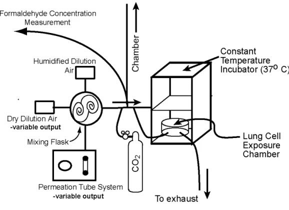
Figure 1: Schematic of Permeation Tube System...6
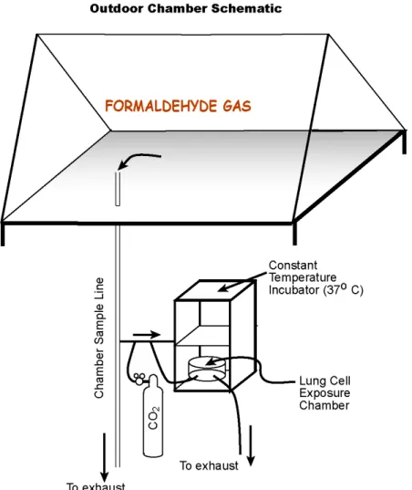
Figure 2: Schematic of Outdoor Smog Chamber...8
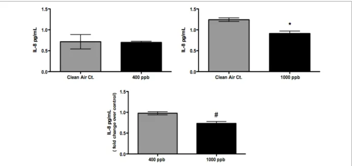
Figure 3: IL-8 responses from cell exposures using the Permeation Tubes...14
Figure 4: IL-6 responses from cell exposures using the Permeation Tubes...14
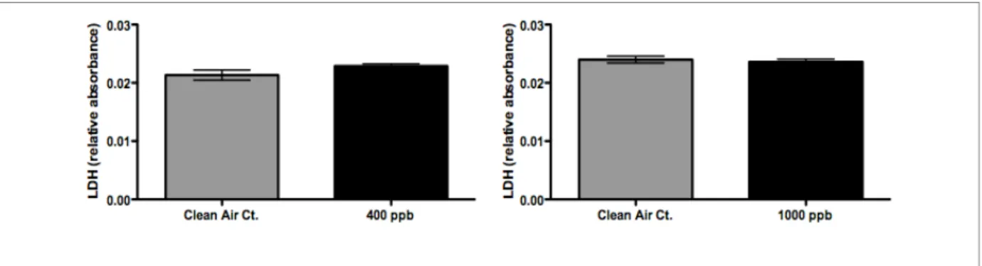
Figure 5: Figure 3: LDH release from cell exposures using the Permeation Tube...15
Figure 6: IL-8 and LDH responses from cell exposures using the outdoor chamber... ...15
Figure 7: Weight-loss history for permeation tube “89”...21
Figure 8: Weight-loss history for permeation tube "88" ...21
Figure 9: Weight-loss history for permeation tube "85". ...22
Figure 10: Weight-loss history for “two ppm” permeation tube ...22
Figure 11: IL-8 results for HCHO exposures of 55 ppb, 140 ppb, and 220 ppb ...23
Figure 12: LDH results for HCHO exposures of 55 ppb, 140 ppb, and 220 ppb ...24
Figure 13: Characterization of Formaldehyde loss...24
Figure 14: Results from RANTES detection assay...25
1 Background
Over the past two decades an unsettling trend in epidemiologic data indicates that the prevalence of asthma and other allergic airway diseases is increasing at alarming rates (Beasley et al). Although there is evidence that air pollution may be the leading cause, the scientific community has not been able to establish an obvious chemical or pollutant as primarily responsible. It has therefore been suggested that formaldehyde, a chemical compound that is widely distributed in the environment and one that can affect a large portion of the population, may be a cause of the increasing prevalence of chronic respiratory diseases (Li Guang-Young, 2007).
1.1 Formaldehyde
Formaldehyde (HCHO) is an organic chemical containing only one terminal carbonyl
group. It is the simplest aldehyde and the most abundant carbonyl in air. It is a chemical of enormous economic impact; estimates from 1992 indicate that over 12 million tons of formaldehyde are produced worldwide. In the U.S. on a daily basis approximately 1.5 million workers are exposed to formaldehyde, and about 1 million in the European Union (Minisymposium 1: Occup. Environ. Med. 2004). As a by-product of incomplete
combustion there are numerous and diverse sources of formaldehyde. It is produced by automobiles, trucks, and industry, and is released into the environment by natural processes. (EPA air toxics website).
Typical industrial uses for formaldehyde include resins, molding compounds,
photographic film, decorative laminates, plywood, and tissue preservative. Not only is HCHO
produced in industry but it is also emitted naturally. Natural sources of formaldehyde include forest fires, animal wastes, plant volatiles, microbial processes in biological systems, and it is even produced within our own bodies as a by-product of our metabolism (EPA air toxics website). Although various sources emit HCHO, combustion processes account for most of
the formaldehyde that enters into the environment. A primary source of HCHO in ambient air
is its direct emission from the incomplete combustion of diesel and gasoline-powered motor vehicles. Formaldehyde is also produced in the atmosphere by the photochemical oxidation of hydrocarbons (VOCs). This secondary source is responsible for the majority of the ambient HCHO. Thus, formaldehyde is an important component of atmospheric pollution
2
Although formaldehyde is ubiquitous in ambient air, the concentrations are relatively low because it is photolyzed and rapidly attacked by the hydroxyl radical. Of special
consideration and concern is the HCHO present in home and occupational environments.
Indoor air is a significant reservoir of HCHO and in some cases the concentrations can be
considerable. The highest concentrations found in indoor air are generally associated with occupational environments and industries that produce HCHO or HCHO-containing products.
Also home environments, especially mobile homes, tend to have higher concentrations of
HCHO than ambient air (W.H.O. see appendix 5.1, Table 1). Most of this HCHO is from the
use of products containing plastics, wood paneling, textiles, carpet, household cleaners, wood stoves, gas burners, and tobacco smoke (TOx probe). Even though different HCHO sources
will lead to various exposure concentrations, when evaluating safety and attempting to minimize exposures, perhaps what is most important to consider is the scientific community’s current stance that, “based on the review of chamber, community, and workplace studies of human exposure, it is not possible to identify a specific no observed adverse effect level or lowest observed adverse effect level for formaldehyde” (Bender et al).
1.2 Significance of Further Formaldehyde Research - Cancer
For decades researchers have investigated formaldehyde and its potential health effects. It is among the most extensively studied industrial chemicals and there is abundant information from chronic animal inhalation studies and research regarding the carcinogenic potential of formaldehyde. The EPA considers formaldehyde to be a probable human carcinogen and has ranked it in EPA's group B1. Animal studies have reported an increased incidence of nasal squamous cell carcinomas by inhalation exposure (EPA air toxics
website). Based on animal inhalation studies, investigators believe that HCHO induced nasal
cancer is caused by HCHO’s ability to form DNA–protein cross-links in the nasal respiratory
mucosa of rats and monkeys. Although genetic effects of HCHO in humans is not well
known, in vitro human cell studies reveal that HCHO induces DNA damage through DNA–
protein cross-links, DNA single-strand breaks, chromosomal aberrations, sister chromatid exchanges, and genetic mutations (TOx Probe).
1.3 Significance of Further Formaldehyde Research – Sensory Irritation
Due to HCHO’s various sources and ubiquity, people are likely exposed to low levels
3
skin rash, nausea, headaches, and difficulty breathing. Although scientific literature is inundated with epidemiologic, community, workplace, and controlled exposure studies, these are mainly observations in human studies concerning eye, nose, and throat irritation (Bender et al.). To date, endpoints other than cancer and sensory irritation are not well characterized; therefore the effects of inhaled formaldehyde on specific inflammatory mediator production are largely unknown. However, one study by Kim et al (2002) reported formaldehyde as a modulating factor of inflammation. This study showed that HCHO has the ability to cause the
release of pro-inflammatory cytokines such as IL-1 and IL-8 from dermal keratinocytes, as well as IL-4 and IL-6 from mast cells that lead to clinical deterioration of atopic eczema.
1.4 Cellular Inflammation End-points
There is limited data to evaluate HCHO’s non-cancerous health effects. Cellular
inflammation, an important non-cancerous health effect, is a typical response to injury from inhaled pollutants and tends to be characterized by specific mediator production. Many diseases with an inflammatory component are associated with the production of cytokines. In the pathogenesis of respiratory diseases involving inflammation typically irritation and mucus production are preceded by an increase in cytokine release from airway epithelium. Cytokines, such as IL-6 and IL-8, can function to stimulate immune responses to trauma by initiating and amplifying the inflammatory cascade. Cytokine production signals the
migration of macrophages and neutrophils to the site of injury and may induce wound repair (Hooper et al.). In addition to being beneficial, cytokines can also havea deleterious effect on the host. Acute and chronic inflammations, as well as thrombosis are among the pathologic conditions thathave a significant cytokine component (Hooper et al.). In vitro cellular
exposure methods have been developed to allow for sensitive assays to evaluate mediators of inflammation. In this paper, cytokines IL-6 and IL-8 were investigated. These cytokines are known to perform an integral function in innate immune responses, inflammation, and perhaps allergic respiratory diseases.
1.5 Study Objective
The purpose of this study was to develop additional experimental data concerning non-cancerous end-points by measuring markers of inflammation released upon
2 Materials and Methods
2.1 Lung Cell Characteristics
Type A549 cells were used throughout this study as the biological target of formaldehyde gas exposures. They are alveolar basal epithelial cells that have retained several alveolar type II-cells characteristics, and are derived from an adenocarcinoma. Typically, inhaled pollutants target the epithelial cells lining the alveolar region. Based on study outcomes over the past few years, it has become apparent that alveolar epithelial cells (AECs) are not merely targets for inflammatory mediators, but actively participate in the inflammatory response (Dos Santos et al.). Thus, A549 cells make a good model for studying exposure to inhaled formaldehyde.
In preparation for cell exposures, A549 cells were grown on membranous support (Costar-Clear Transwell inserts; Costar, Cambridge, MA) as described by Jaspers et al. (1997) in complete medium (F12K, 10% fetal bovine serum, antibiotics; all from Invitrogen, Carlsbad, CA) on apical and basolateral sides of the membranous support. Upon confluency, the medium was exchanged for serum-free medium (F12K, 1.5 [micro]g/mL bovine serum albumin (BSA), antibiotics; all from Invitrogen) several hours before exposure (Doyle et al). Immediately preceding exposures to formaldehyde the serum-free media was aspirated and replaced only in the basolateral compartment. Using only basolateral media for the cellular exposures enables direct exposure of cells to formaldehyde gas without media covering the cell significantly interfering with the exposure. Basolateral supernatants are collected once the exposure and 24 hour incubation period has terminated, and stored at -80 °C until they
can be analyzed for cytotoxicity and inflammation.
2.2 Formaldehyde-Air Mixture Generation and Exposure
5
permeation tube generation rate was tracked throughout the study by regular mass-loss determinations (appendix 5.2). Comparisons of calibrations based on the two sources of formaldehyde were made and found to be in agreement. The second method shown by the schematic in Figure 2 involved injecting a specific concentration of formaldehyde (subliming a weighed amount of para-formaldehyde, 95%, using a gently-heated U-tube) into the
environmental chamber and then sampling continuously at one liter per minute into the in vitro system. These methods are described in more detail below.
2.2.1 In vitro Exposure System
For both exposure methods, cells were maintained in an incubator that houses the in vitro cell exposure system. The in vitro exposure system maintained the cells at a constant temperature of 37 °C and supplied 5% CO2 in the air stream throughout the duration of each
exposure (Doyle et al).
Inside the incubator, cells grown on tissue culture inserts were placed in an 8-liter, modular, cell-exposure chamber (Billups-Rothenberg, MIC-101™). The 8-liter cell-exposure chamber also has connections for flowing gas through either the permeation tube system or the outdoor chamber. Sample lines through externally circulated sample manifolds were used to provide formaldehyde gas to the cells during exposure (Sexton et al., 2003)-Doyle. Humidified medical-grade air from a cylinder or chamber air was blended with CO2 at 0.05
liter per minute to achieve about 5% CO2 in the incubator. This was necessary to maintain the
buffering capacity of the tissue culture media under the cells. The addition of CO2 to
chamber air was achieved using small pumps on the exhaust side and mass flow controllers (AALBORG).
2.2.2 Permeation Tube System
For permeation tube exposures a humidification system was developed to humidify the dry formaldehyde gas in air source by humidifying dilution air mixed with source air. This method permits dry air to be used with the permeation tube, and it dilutes the
concentration of formaldehyde from the fixed mass emitted by the permeation tubes. Clean air from the ADDCO 737-250 pure air generator was used for both the permeation tube source and dilution air. The dilution air was controlled by a mass flow controller and allowed to bubble through two thermostatically heated midget impingers (ACE Glass, Inc) filled with HPLC grade water (Fisher Scientific). The humidified air was delivered to a mixing tee using thermostatically heated lines to prevent condensation and was blended with the dry
6
dilution air for the permeation tube source. Figure 1 illustrates how formaldehyde gas leaves the permeation tube system, mixes with both dilution air and humidified air, and is then vented or flushed through the in vitro chamber where A549 cells are exposed to the
formaldehyde gas. Weight loss histories of the permeation tubes were recorded to determine their source strength (appendix 5.2). In addition, these were compared with standards
prepared by injections of weighed amounts of para-formaldehyde into the chamber, as discussed earlier. For concentrations that were within the response range of the method, before most exposures, the Dasgupta method was used to check the formaldehyde concentrations at the outlet and also used to compare the chamber prepared standards measurement responses to the permeation tube measurement responses.
Figure 1: Schematic of Permeation Tube System
Preliminary permeation tube experiments exposed cells to formaldehyde
7
durations of three hours. The concentrations, at which results are being reported, although high for ambient air levels of formaldehyde, are in the range of possible human exposures. In this case, concentrations reported are among those found in daily exposures living in mobile homes, industrial exposures, and even exposure to tobacco smoke.
On December 23, 2007, January 11, 2008, and February 18, 2008 permeation tube experiments were performed at concentrations of 400 ppb and 1000 ppb. Within same-day experiments, formaldehyde exposures were performed twice with a new set of confluent A549 cells to test the reproducibility of the system. In addition, on each day of an
experiment, a clean air control exposure was performed as a point comparison for the cells exposed to formaldehyde gas. All exposures were three hours in duration. For each
formaldehyde exposure n=4 sample inserts were used and n=3 sample inserts were used per each clean air control.
2.2.3 “One-Atmosphere” Outdoor Environmental Chamber
Air chemistry research groups at the University of North Carolina at Chapel Hill have been using outdoor irradiation chambers for over 30 years to characterize urban air pollutants and their reactions in ambient air. For a portion of this research, the One-Atmosphere
Outdoor Chamber (also called an environmental irradiation chamber or smog chamber) was used as a delivery mechanism for formaldehyde gas. The smog chamber is located on the roof of McGavran-Greenberg Hall on the University of North Carolina at Chapel Hill main campus. It is located directly above the One-Atmosphere Research Lab (on the forth floor of McGavran-Greenberg) where through-the-ceiling/roof sample ports allow gaseous pollutants to be injected into the chamber and samples from the chamber to be delivered to the in vitro
8
Figure 2: Schematic of Outdoor Smog Chamber
During the chamber September 22, 2007 experiment, formaldehyde gas was delivered to the in vitro cell exposure system at two different concentrations. In addition, an incubator control and a clean air control were performed on the same night as the formaldehyde exposure. All exposures were three-hours in duration and performed at night to keep the formaldehyde concentration constant, i.e., no photolysis. For the first exposure,
formaldehyde was injected into the chamber at a concentration of approximately 400 ppb. The second exposure used a formaldehyde concentration of approximately 1000 ppb. For each formaldehyde exposure, n=6 sample inserts were used. For each control exposure (clean air and incubator) n=3 sample inserts were used. To prevent condensation inside the chamber after sundown, we used the chamber dehumidification system to lower the dew point below the expected lowest temperature. Condensation would cause HCHO loss due to adsorption
into the moisture on the walls. In each experiment, a very small amount (2.5 microliters) of carbon tetrachloride (CCl4) used as a tracer, was injected then monitored to calculate the
9
and the integrity of the cells was maintained by the in vitro cell exposure incubator, keeping the cells at 37 °C and supplied with 5% CO2. The dilution rate was measured, and was
between 1-2% per hour.
2.3 Quantification of Formaldehyde in Air Mixtures: Primary Sources and Chemical Analysis
Formaldehyde concentrations were measured continuously using a permeable membrane sampler, and detected with fluorometry using an automated Hantzsch derivative reaction (Dasgupta et al. 1988) for concentrations less than 400 ppb. The purpose of
continuously monitoring when possible is to confirm the stability of the formaldehyde
sources. Given the history of performance and the primary-standard quality of the permeation tube source and the chamber standards stability in the absence of sunlight, the stability check is for detection of failures or leaks in the sample and delivery lines from the sources. The concentrations in the 3-hour exposures were approximately 55 ppb, 140 ppb, 220 ppb, 390 ppb, and 980 ppb.
2.4 Characterization of Formaldehyde Uptake
Because formaldehyde is a very water soluble and reactive gas, it is important to measure the concentration losses of formaldehyde to the walls and other surfaces inherent to the exposure system. Formaldehyde loss to tubing, the surface of the exposure system, and media was investigated at a formaldehyde concentration of 220 ppb. This concentration was used because we were able to characterize small loses in concentration with the Dasgupta instrument. Using a weight-loss history of the three permeation tubes, we estimated that without any formaldehyde loss, 600ng of HCHO/min would be delivered through the
exposure system.
To evaluate how much formaldehyde was being lost to system walls and tubing only, we used our estimate of how much formaldehyde was going in, and measured what
concentration was actually coming from the system outlet. No media or humidifying water dish was used in the exposure system because we did not want to influence surface and tubing loss measurements with any additional formaldehyde sinks. For this exposure the conditions were a dew point of 17.5 °C, an incubator temperature of 37 °C, with an RH of
31%. For the previous conditions and based on our estimates we observed a 6% decrease in formaldehyde, and thus a 6% loss of formaldehyde to the tubing and surface walls of the system.
10
we hypothesized that there could also be formaldehyde loss to the sample media and we measured this loss as well. To measure formaldehyde loss to the media, we transferred 1-ml of media into each well of two Transwells and “exposed” this media (in the absence of cells) to formaldehyde for three hours. We then used the Dasgupta instrumentation to directly measure the concentration of formaldehyde in the media. Based on our results we observed approximately 4.5% of HCHO is lost to 1-mL of media. This correlates to 1.62-ug HCHO loss
per 1-mL media.
In addition, a common practice with chamber exposures is to include a water dish within the incubator to humidify the air/chemical mix going to the cells. The water dish is a further precaution used to also prevent the cells from desiccating. Although we did not use a water dish in our formaldehyde experiments, we wanted to determine how much
formaldehyde could be lost to a dish containing 30-mL of water.
We sampled formaldehyde uptake through the water dish by “exposing” formaldehyde through the system for three hours. We then used the Dasgupta
instrumentation to directly measure the concentration of formaldehyde in the water. Based on our results we observed about 13% of HCHO is lost to 30-mL of water (if a water dish is
used). This correlates to about 0.43% HCHO loss per mL of water and 0.156-ug HCHO loss
per mL of water.
Wall uptake will always occur, however, based on how many Transwells (samples) and thus milliliters of media used formaldehyde loss due to uptake will vary. We measured a 6% uptake of formaldehyde to the walls of the system and a 4.5% uptake of formaldehyde per mL of media used. Exposures typically use three to four mL of media. Based on our measurements, this leaves between 76% and 81% of the original concentration of
formaldehyde available for exposure (appendix 5.4).
2.5 Health Effects Analysis
When cells become stressed or damaged, multiple genes and signaling pathways are stimulated. This causes various proteins to be produced in the cell and then subsequently released into the cells’ environment. Proteins, like mediators of inflammation, have a diverse ensemble of function and can both assist the respiratory system or eventually cause
11
asthma, chronic obstructivepulmonary disease (COPD), cystic fibrosis, and respiratory infection (Hiemstra el al.). An additional health effect of respiratory trauma is cellular death, which can be measured by the release of the enzyme lactate dehydrogenase.
After clean air and formaldehyde exposures, cells were incubated at 37 °C for 24 hours. Basolateral supernatants were collected and stored at -80 degrees C until analysis of cytokines and markers of cytotoxicity could be performed.
2.6 Cell Cytotoxicity
Basolateral supernatants previously stored at -80 ° C were used in the analysis of cytotoxicity. Cytotoxicity was examined by measuring the release of lactate dehydrogenase (LDH) using a coupled enzymatic assay followed by manufacturer’s instruction (Takara, Japan). LDH is an enzyme that is strictly produced and maintained within a cell, thus release of LDH into the supernatant is considered to be a marker of cell membrane disruption and consequently cell death.
2.6.1 ELISA - IL-8 and IL-6 Expression
Cellular trauma stimulates intracellular signaling cascades that cause pro-inflammatory cytokines to be produced. Pro-pro-inflammatory cytokines/chemokines are chemical signals that attract neutrophils and other inflammatory cells to the site of trauma, leading to inflammation. The inflammatory responses of A549 cells upon formaldehyde exposure were quantified using an Enzyme-Linked ImmunoSorbent Assay (ELISA) kit. Basolateral supernatants previously stored at -80 ºC were used in the analyses of markers of inflammation. Protein levels of pro-inflammatory cytokines interleukin-8 (IL-8), and
Interleukin-6 (IL-6) were measured by ELISA (R&D Systems, Minneapolis, MN), as per the supplier’s instructions.
Interleukin-6 (IL-6) acts as both a pro-inflammatory and anti-inflammatory cytokine. It functions to stimulate immune responses to trauma. In terms of host response to a foreign pathogen, IL-6 has been shown, in mice, to be required for resistance against bacterium. IL-8 is a chemokine and it belongs to a group of CXC chemokines characterized by two
terminal cysteine residues separated by one non-conserved amino acid residue at the N-terminal end. (Rudack et al.) Besides initiating the adhesion cascade, IL-8 is known to be a potent neutrophil chemotactic protein. IL-8 recruits migrating neutrophils and leukocytes to areas of inflammation. Neutrophils infiltrate the site of injury and function to phagocytose the antigen that triggered the initial inflammatory response. IL-8 is believed to play a role in the pathogenesis of bronchiolitis, a common respiratory tract disease caused by viral
12
tissuesand thus serves toamplify the inflammatory cascade. IL-8 is produced in response to TNF-α in primary cultured human airway epithelial cells and in a human lung epithelial cell
line (Li et al.)
2.6.2 Multi-cytokine Antibody Array
A preliminary effort in understanding potential mechanistic effects of HCHO exposure
was to perform a multi-cytokine antibody array. The multi-cytokine detection assay used basolateral supernatants previously stored at -80 ° C from the HCHO chamber exposure
performed on September 22, 2007. During this experiment three exposure conditions were performed: a three hour incubator/clean air control (n=6), a three hour 400 ppb HCHO
exposure (n=6), and a three hour 1000 ppb HCHO exposure (n=6). The array examined the
basolateral supernatants for 13 cytokines/chemokines. Only RANTES and MCP-1 were detectable. All other cytokines were either below the limit of detection or below background levels.
When comparing the HCHO exposures to the controls for both RANTES and MCP-1
there were no statistical differences between controls or exposures, however trends were apparent. See appendix 5.5.
2.7 Statistical Analysis
To analyze our data, one-way analysis of variance (ANOVA) with Newman-Keuls multiple comparison test was used as a statistical test to determine the significance. ANOVA was the most appropriate statistical test to use due to the need for multiple comparisons. Generally, during each experiment, three or more data sets were produced and it was
13
3 Results
In this study, as indicators of deleterious cellular effects, IL-8 and IL-6 gene expression along with cytotoxicity were used to estimate the health effects elicited from gaseous formaldehyde exposure. A549 cells grown to confluency on membranous supports were exposed for 3 hours to formaldehyde gas at concentrations of 400 ppb and 1000 ppb. The results from the experiments performed were not only reproducible within the same formaldehyde delivery system, but also when comparing permeation tube exposures and chamber exposures.
3.1 Permeation Tube Exposure Results
The permeation tube exposures of 400 ppb lasting three hours (combined responses from both exposures) were evaluated for their potential to cause adverse cellular damage to the A549 cells. Figure 3, Figure 4, and Figure 5 show that by our end-points, there were no significant differences between air-and formaldehyde-induced IL-8, IL-6 expression or cytotoxicity. The permeation tube exposures of 1000 ppb lasting three hours (combined responses from both exposures) were also analyzed for cell death and markers of
inflammation. We observed similar results to the 400 ppb exposures where our analysis showed no difference in IL-6 expression or in cell death (LDH). See Figure 4 and Figure 5. However, unlike the 400 ppb exposure, a significant decrease (p-value ≤ 0.05) in IL-8 release
14
Figure 3: IL-8 responses from cell exposures using the Permeation Tubes. No difference in IL-8 response for formaldehyde concentrations up to 400 ppb. At the 1000 ppb level we observed a significant decrease in IL-8. [Symbols: *significantly different from control (p≤0.05); # significantly different from samples exposed to 400 ppb (p≤0.05)]
15
Figure 5: Figure 3: LDH release (cell death) from cell exposures using the Permeation Tube. Neither the 400 ppb nor 1000 ppb concentration showed a significant change from the clean air control.
3.2 Chamber Exposure Results
A549 cells exposed to chamber clean air and to formaldehyde injected into the chamber at concentrations of 400 ppb and 1000 ppb were also analyzed for cell death and markers of inflammation. In the case of the chamber experiment, the three hour - 400 ppb exposure was performed first and then the chamber concentration of formaldehyde was brought up to 1000 ppb for another three hour exposure. For IL-6 and LDH no significant difference between formaldehyde exposed cells and clean air exposed cells was observed for either the 400 ppb or 1000 ppb exposure. However, analysis of IL-8 revealed a step-wise concentration dependent suppression of IL-8 release (see Figure 6).
16
4 Discussion and Conclusions
A literature review suggests that no previous studies have used a coupled chamber-in vitro cell exposure system to deliver formaldehyde gas, the inflammatory agent, by
continuously flowing it over lung cells for predetermined exposure duration. Studies typically inject or bubble HCHO directly into media that contains lung epithelial cells. Our
exposure method effectively mimics the actual exposure of gases flowing through and over an epithelial organ system. Based on our research an initial observation is that formaldehyde exposure to A549 cells caused no increases in LDH or IL-6. Alone, this could indicate that either formaldehyde has no affect on LDH and IL-6 from alveolar epithelial lung cells, or our
in vitro delivery system was not an efficient exposure method for formaldehyde, however, additional evidence from this study leads to the contrary. Throughout chamber and
permeation tube exposures, a recurring event of a decrease in IL-8 expression suggests that our system is in fact effectively delivering formaldehyde gas to the in vitro cell exposure system. Based on the decrease in IL-8 we believe that formaldehyde is having some mechanistic effect on specific inflammatory mediator production. Therefore, the overall finding of this study is that while LDH and IL-6 levels remain unchanged with regard to exposed samples and control samples, IL-8 expression decreases. The main focus of this discussion will therefore be on IL-8 expression and its function not only in epithelial cells, but in other cells types as well.
Interleukin-8 (IL-8) is a pro-inflammatory cytokine and a member of the CXC chemokine family. It is the most-well-known and characterized neutrophil-attracting chemokine (Rudack et al.). It is producedby a wide variety of cell types including mononuclear cells, endothelialcells, and epithelial cells. Through a chain of biochemical reactions IL-8 is secreted and is an important protein involved in innate immune system responses. It functions as a chemoattractant, serving as a chemical signal to recruit
17
processes that lead to cancerous endpoints. IL-8 has been shown to induce the migrationof some tumor cells, regulate pathological angiogenesis, tumor growth, and metastasis; it is therefore a potent angiogenic factor. Although angiogenesis is a normal process in growth and development, as well as in wound healing, it is also a fundamental step in the transition of tumors from a dormant state to a malignant state.
Not only does IL-8 have multiple functions and is expressed differently depending on stimulant and cell type, but there are multiple signaling cascades that can lead to IL-8
production (Jaspers, 1998). Further evidence is found in a study by Hooper el al. that indicates endothelial cells produce IL-8 as an important physiologicparticipant in cross talk between coagulation and inflammation. In this study, IL-8 is produced in periodontal tissues where healthy cells always maintain a low level of cellular inflammatory infiltrate. A study by Darveau et al. demonstrated that the expression of several molecular mediators of inflammation is normal and important in healthy tissues. With regard to healthy periodontal tissue, low-level expression of mediators such as IL-8 most likely contribute to the ability of the host to limit periodontal bacterial growth and injury to the tooth and epithelial cell surface (Darveau et al.). IL-8 is also synthesized in ovary cells and a study by Lee el al. provides insight into the possible anti-tumorigenic potential of IL-8. In contrast to other studies, their findings show that tumor growth in vivo was significantly attenuatedin animals receiving IL-8-expressing cells when compared withmice injected with control cells. In their model the local productionfrom ovary cells of IL-8, and the subsequent neutrophilic
infiltration, resultin markedly decreased tumor growth (Lee et al.). It is obvious from these various studies in multiple organ systems that IL-8 performs vital functions for the well being of healthy cell systems. Although IL-8 has many known functions, for the most part, the mechanisms that facilitate IL-8 production are poorly understood. It is also not well known how IL-8 production impacts all the cellular systems that release it.
This overview of the current scientific knowledge surrounding IL-8 reveals that IL-8 has various functions and its production is initiated by various cellular changes. The overall effect ofIL-8 appears to require additional factors necessary for metastasisbased on the cells in question. In addition, IL-8’s cancerous involvement in various tumorcell types may be dependent on the metastatic potentials of the specific cancer cell lines. In terms of cancerous or non-cancer end-points, there appears to be a very fine balance between
neutrophils/macrophages,chemotaxis, and angiogenesis/metastasis (Lee et al.).
Most significant to the focus of this paper is IL-8’s role and production in the
18
respiratory defense mechanisms. IL-8 not only functions as a protective response to
respiratory insults, but provides maintenance services as well. Within the respiratory track, IL-8 is being constantly synthesized; this results in maintaining neutrophils on the mucosal surface and in restoring the homeostatic balance. Our results indicate a suppression in IL-8 expression. Impairing IL-8 synthesis could have serious repercussions for the overall health of the respiratory system. If in fact IL-8 is integral not only as a protective mechanism, but as a maintenance one as well, exposure to formaldehyde could severely hinder proper functioning of the respiratory system. Although IL-8 expression is associated with deleterious effects such as chronic inflammation and cancer, low level IL-8 expression is vital to repairing and protecting damaged respiratory epithelial cells. Formaldehyde, acting as an IL-8 suppressor, could effectively and indirectly be causing cells to be more vulnerable to inhaled insults. Taken together, with increased vulnerability and a handicapped recovery system, formaldehyde could be causing much more damage than can be assessed by the overall results presented in this study.
4.1 Limitations
When using an in vitro exposure system there are limitations inherent to the method. Generally the system is very isolated because investigators typically want to focus on one cell type’s responses to a particular insult. While being able to narrow the scope of an investigation may be an advantage, perhaps in this case, our window is too defined for HCHO
exposures using only A549 cells taken outside of their natural environment. It is possible that the A549 cells could respond differently in a comprehensive system using more than one cell type and including other cell-to-cell interactions. Unfortunately with the in vitro system we used, we are unable to evaluate cell-to-cell interactions and other potential cell system impacts of HCHO exposure. Ideally one would like to use a larger system because it is more
holistic and more realistic, but it is also more technically challenging to probe such a system.
Another limitation of the study is the dose of formaldehyde delivered to the A549 cells. In this study, we exposed A549 cells to specific concentrations of HCHO for three
hours. Typically HCHO exposures to an individual would occur over a much more extended
and various period. Depending on the situation, an individual could be exposed through the environment to small concentrations over their lifetime, massive acute concentrations if they are a smoker, or if an individual worked in a HCHO producing industry they could potentially
be exposed to very high concentrations each workday for up to eight hours.
4.2 Future Research
19
be very interesting and beneficial to analyze other genetic markers and cellular responses upon HCHO exposure.
In addition a technically challenging and fairly novel approach to in vitro testing would be to use HCHO “conditioned” cell supernatants from A549 cells and incubate that
media with another cell type found in the same respiratory environment. Perhaps to see the bigger picture of HCHO induced injury, the A549 cells need to interact with other cells in
their actual alveolar environment.
4.3 Final Conclusions
Throughout this study we have emphasized that formaldehyde is a known respiratory irritant and probable human carcinogen. Although formaldehyde exposure has been studied extensively for over thirty years, most of the research has focused on in vivo murine models with cancer endpoints. Many studies have investigated formaldehyde’s role in nasal cancer. These studies have made great contributions to understanding the biologic mechanisms of formaldehyde damage at high concentrations. They have concluded that inhaled
formaldehyde increases incidence of nasal squamous cell carcinomas through DNA-protein cross-links in maurine models (EPA air toxics website). However, due to the increasing trend in allergic respiratory disease and epidemiologic data suggesting a potential link to formaldehyde exposure, there has been an overwhelming need to investigate other endpoints such as inflammation. Formaldehyde has been implicated as a cause of non-cancerous respiratory disease because of its chemistry, ubiquity, prevalence in home and work
environments, and the fact that its various sources permeate throughout every socioeconomic group. Using conventional, well-characterized markers of inflammation, a main goal of this study was to generate data on lung epithelial inflammatory responses upon exposure to formaldehyde.
Although mechanistic pathways of HCHO damage to the respiratory system are
outside of the scope of this study, our results indicate that HCHO is affecting markers of
inflammation in a very atypical fashion. Generally, inflammation is associated with increased cytokine production. However, in this study we see a concentration-dependent decrease in IL-8 release and no effect on IL-6. Although it is possible that our observed effects of HCHO
on pro-inflammatory cytokine production could be the result of enhanced production of anti-inflammatory cytokines or a mechanistic inhibition of the TNF-K or NFk-ß pathway, further research is necessary. Unfortunately, at this point, a mechanistic cause of HCHO induced
20
5 Appendices
5.1 W.H.O. Formaldehyde Concentrations
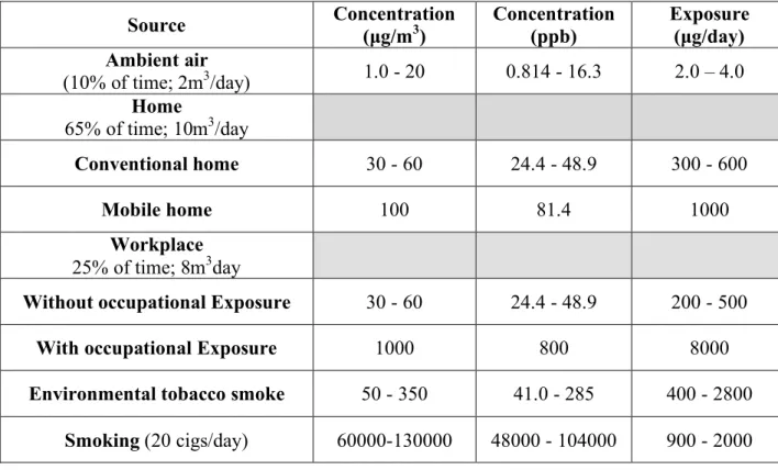
Table 1: Sources and average concentrations of formaldehyde in the environment. Source: WHO Regional Office for Europe - Air Quality Guidelines Chapter 5.8 Formaldehyde
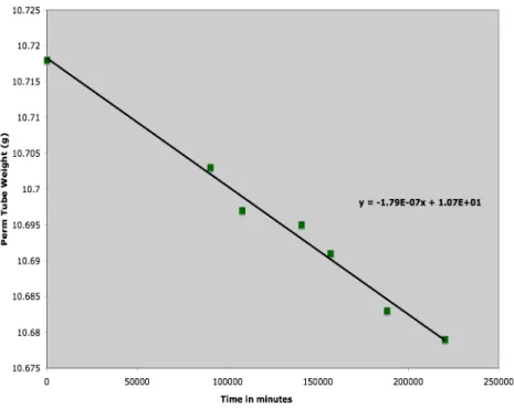
5.2 Permeation Tube Generation Rate – Weight-loss Histories
The permeation tube generation rate was tracked throughout the study by regular mass-loss determinations. Weight-loss histories of the permeation tubes were recorded to determine each permeation tube’s individual source strength.
Source Concentration (µg/m3) Concentration (ppb) Exposure (µg/day) Ambient air
(10% of time; 2m3/day) 1.0 - 20 0.814 - 16.3 2.0 – 4.0
Home
65% of time; 10m3/day
Conventional home 30 - 60 24.4 - 48.9 300 - 600
Mobile home 100 81.4 1000
Workplace
25% of time; 8m3day
Without occupational Exposure 30 - 60 24.4 - 48.9 200 - 500
With occupational Exposure 1000 800 8000
Environmental tobacco smoke 50 - 350 41.0 - 285 400 - 2800
21
Figure 7: Weight-loss history for permeation tube “89”. Concentration output is approximately 55 ppb.
22
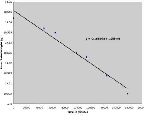
Figure 9: Weight-loss history for permeation tube "85". Concentration output is approximately 80 ppb.
23
5.3 Results for Permeation Tube Exposures of 55 ppb to 220 ppb
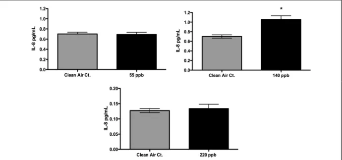
5.3.1 IL-8 Results for Concentrations 55 ppb, 140 ppb, and 220 ppb
24
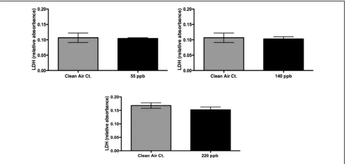
5.3.2 LDH Results for Concentrations 55 ppb, 140 ppb, and 220 ppb
Figure 12: LDH results for HCHO exposures of 55 ppb, 140 ppb, and 220 ppb. No significant differences were found between HCHO exposures and their clean air controls.
5.4 Characterization of Formaldehyde Loss
25
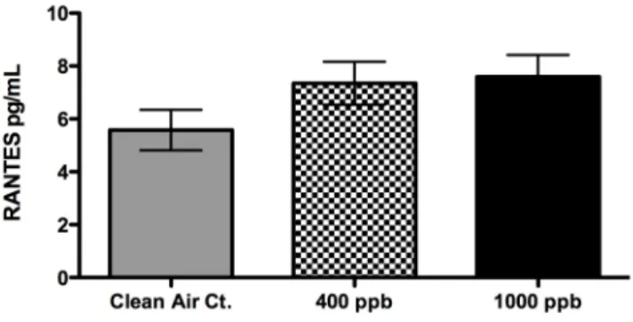
5.5 Multi-cytokine Antibody Array Results
Figure 14: Results from RANTES detection assay. No statistical differences between clean air controls and HCHO exposures, however there is a trend of increased RANTES expression from HCHO exposures.
26
6 References
1. Arenberg, D., S. Kunkel. 1996. Inhibition of Interleukin-8 Reduces Tumorigenesis of Human Non-Small Cell Lung Cancer in SCID Mice. Journal of Clinical Investigation, 97(12): 2792–2802.
2. Bakand, S., A Hayes, C Winder. 2005. In vitro Cytotoxicity Testing of Airborne Formaldehyde Collected in Serum-free Culture Media. Toxicology & Industrial Health, 21: 147-154, 2005.
3. Beasley, R., J Crane. 2000. Prevalence and Etiology of Asthma. Journal of Allergy and Clinical Immunology, 105(2): S466-S472.
4. Bender, J. 2002. The Use of Noncancer Endpoints as a Basis for Establishing a Reference Concentration for Formaldehyde. Regulatory Toxicology and
Pharmacology, 35, 23–31.
5. Chen, J. 2004. Atmospheric Formaldehyde Monitoring in the Greater Houston Area in 2002. Applied Spectroscopy, 58(2): 243-247.
6. Darveau, R. 1998. Local Chemokine Paralysis, a Novel Pathogenic
Mechanism for Porphyromonas gingivalis. Infectious Immunology, 66(4): 1660–1665.
7. Dasgupta, et al. (1988) Atmospheric Environment. 22: 949-96.
8. Dasgupta, P.K. 2005. Summertime Ambient Formaldehyde in Five U.S. Metropolitan Areas: Nashville, Atlanta, Houston, Philadelphia, and Tampa.
Environmental Science and Technology, 39(13): 4767-4783.
9. Dos Santos C. 2005. DNA Microarray Analysis of Gene Expression in Alveolar Epithelial Cells in Response to TNFα, LPS and Cyclic Stretch. Physiological Genomics, 19: 331-342.
10. Doyle M, Sexton KG, Jeffries HE, Bridge K, and Jaspers I. 2004.
Environmental Health Perspectives, 112(15): 1488-1495.
27
12. EPA’s air toxic website. Hazard Summary; formaldehyde. (Resource found on-line)
13. Hagimoto, N., K. Kuwano. 1999. Induction of Interleukin-8 Secretion and Apoptosis in Bronchiolar Epithelial Cells by Fas Ligation. Journal of Respiratory Cell and Molecular Biology, 21(3): 436-445.
14. Hiemstra, Peter, R. Bals. 2004. Innate Host Defense of the Respiratory Epithelium. Journal of Leukocyte Biology, 75: 3-4.
15. Highlights from Worldwide Scientific Studies. Formaldehyde Council, 2004. (Resource found on-line).
16. Hooper, C., D. Phillips. 1998. The Up-Regulation of IL-6 and IL-8 in Human Endothelial Cells by Activated Protein C. The Journal of Immunology, 161: 2567-2573.
17. Jaspers, Ilona, L. Chen, E. Flescher. 1998. Induction of Interleukin-8 by Ozone is Mediated by Tyrosine Kinase and Protein Kinase A, but Not by Protein Kinase C. Journal of Cellular Physiology, 177: 313-323.
18. Keping, Xie. 2001. Interleukin-8 and Human Cancer Biology. Cytokine and Growth Factor reviews, 12(4): 375-391.
19. Kim, W. J. 2002. Effect of formaldehyde on the Expression of Adhesion Molecules in Nasal Micro-vascular Endothelial Cells: the Role of Formaldehyde in the Pathogenesis of Sick Building Syndrome. Clinical & Experimental Allergy, 32: 287-295.
20. Lee, Li-Fen, R. Hellendall. 2000. IL-8 Reduced Tumorgenicity of Human Ovarian Cancer In Vivo Due to Neutrophil Infiltration. The Journal of Immunology, 164: 2769-2775.
21. Li, A., S. Dubey, M. Varney. 2003. IL-8 Directly Enhanced Endothelial Cell Survival, Proliferation, and Matrix Metalloproteinases Production and Regulated Angiogenesis. The Journal of Immunology, 170: 3369-3376.
22. Li, Guang-Young, Hye-Young Lee, Ho-Sang Shin. 2007. Identification of Gene Markers for Formaldehyde Exposure in Humans. Environmental Health Perspectives, 115 (10): 1460-1466.
23. Minisymposium 1: Formaldehyde and Cancer: Current Evidence and Future Perspectives. Occupational Environmental Medicine, 2004. 61: e17.
28
25. Reidl, MA. 2008. The Effect of Air Pollution on Asthma and Allergy. Current Allergy and Asthma Reports, 8(2): 139-146.
26. Rudack, C., S. Maune, J. Eble. 2003. The Primary Role in Biologic Activity of the Neutrophil Chemokines IL-8 and GRO-alpha in Cultured Nasal Epithelial Cells.
Journal of Interferon & Cytokine Research, 23(2): 113-123.
27. Smith, D., P. Polverini. 1994. Inhibition of Interleukin-8 Attenuates Angiogenesis in Bronchogenic carcinoma. Journal of Experimental Medicine, 179: 1409-1415.
28. Toxicological Assessment of Formaldehyde. BFR. March 30, 2006. (Resource found on-line)
29. TOx Probe; Ten Carcinogens in Toronto. Prepared by ToxProbe Inc. for Toronto Public Health. (Resource found on-line)
30. WHO Regional Office for Europe - Air Quality Guidelines for Europe Second Addition, Chapter 5.8 Formaldehyde. Internet Resource: