ISSN: 2231-3354 Received: 27-07-2011 Revised on: 29-07-2011 Accepted: 03-08-2011
A.Ashok Kumar
J.N.T.University,
Kakinada, Andhra Pradesh, India.
Putta Rajesh Kumar
V.L.College of Pharmacy, Raichur, Karnataka, India.
A.Anil Kumar
Koringa college of Pharmacy Korangi, Andhra Pradesh, India.
K.Lokeswara Reddy, T.E.G.K.Murthy and T.Venkateswara Rao
Bapatla college of Pharmacy, Bapatla, Andhra Pradesh,India.
For Correspondence:
Putta Rajesh kumar
Lecturer, Department of Pharmaceutics, V.L.College of Pharmacy,
Raichur – 584103, Karnataka, India. Phone: +91-98453 57475,
Formulation design of Aceclofenac microcapsules by
inotropic gelation technique, characterization studies
and release kinetics
A. Ashok Kumar, Putta Rajesh Kumar, A.Anil Kumar, K.Lokeswara
Reddy, T.E.G.K.Murthy and T.Venkateswara Rao
ABSTRACT
Osteoarthritis predominantly affects the large weight bearing joints and the clinical characteristics include morning stiffness of short duration, stiffness or gelling on rest, pain on use, joint inflammation and bone deformity. Microcapsules of aceclofenac were formulated with methyl cellulose, sodium CMC and hydroxyl propyl methyl cellulose by technique. The microcapsules showed excellent rheological properties for all batches. The formulations showed drug content uniformity with high drug entrapment efficiency. The in vitro wash off test revealed the mucoadhesive ability of the micro capsules. Scanning electron microscopy indicated structural and surface morphology uniformity. The in vitro release studies indicated sustained release of aceclofenac from the formulations. Kinetic study of the release data indicated the zero order release and non fickian sustained release mode of drug release. The microcapsules showed the correlation between wall thickness and drug release pattern.
Key words: Aceclofenac, Ionic gelation, Entrapment efficiency, In vitro release.
INTRODUCTION
n
nd
d
averagetreatment of pain and inflammation with a reduced side effect profile especially regarding gastrointestinal complications (Parfitt 1999, Brogden et al. 1996). To reduce the dosing frequency and adverse effects during prolong treatment it is needed to formulate in long acting dosage form. Aceclofenac directly blocks prostaglandin E2 secretion at the site of inflammation by inhibiting IL-Beta and Tumour necrosis factor in the inflammatory cells. Recommended dose of Aceclofenac is 100mg twice daily, due to short biological half-life of the drug 4h makes it suitable candidate for the modified release dosage forms. To reduce the frequency of administrations and to improve patient compliances, aceclofenac is suitable for making sustain release dosage form. (Trivedi P, 2008). Microcapsules are homogeneous, monolithic particles which improve the treatment by providing localization of the drug at the site of action and by prolonging the drug release. These techniques are widely used in pharmaceutical research (Breghausen SW., 2002). Multiparticulate delivery systems like microcapsules are prepared to obtain prolonged or controlled drug delivery, to improve bioavailability or stability and to target drug to specific sites (S Haznedar., 2004). They can distribute in the GI tract homogeneously, thus maximizing drug absorption and reducing peak plasma fluctuations, minimizing the risk of local GI tract irritation and dose dumping, decreasing dosing frequency and increasing patient compliance, improving the safety and efficacy of the active ingredient (Shavi GV., 2009).
Ionic gelation technique for microencapsulation is the method which prolongs the duration of drug effect significantly, provides symptomatic relief to the patient and improves patient compliance. Eventually the total dose and few adverse reactions may be reduced since a steady plasma concentration is maintained. Hence in the current study formulation and in vitro evaluation of sustained released Aceclofenac microcapsules with polymers like methyl cellulose, Na CMC and HPMC employing ionic gelation technique. The prepared formulations were studied for their flow properties, encapsulation efficiency, in vitro release studies and drug release kinetics. The optimized formulation was subjected for Scanning electron microscopic study.
MATERIAL AND METHODS
Aceclofenac drug was obtained as gift sample from Micro Labs, Bangalore, India. Methyl cellulose, Sodium carboxy methyl cellulose and hydroxyl propyl methyl cellulose were procured from S.D.Fine chemicals, Mumbai. All other chemicals and solvents used in the study were of LR grade.
Ionic gelation process: The ionic gelation processes (Kim C. K., 1992, Hari P. C., 1996) was used to prepare microcapsules. Sodium alginate (1.0 g) and Mucoadhesive polymer like Na CMC (1.0 g), MC (1.0 g) and HPMC (1.0 g) were dissolved in purified water (90ml) to form a homogeneous polymer solution. Core material, Aceclofenac (2.0 g) was added to the polymer solution and mixed thoroughly to form a smooth viscous dispersion. The resulting dispersion was then manually dropped into 10% w/v of 100ml Calcium Chloride solution through a syringe with a needle
of size no.18. The added droplets were retained in the Calcium Chloride solution for 15 minutes to complete the curing reaction and to produce spherical rigid microcapsules. The microcapsules were collected by decantation and the product thus separated was washed repeatedly with water and dried at 450c for 12 h.
Characterization studies of microcapsules: Particle size distribution and size analysis
For size distribution analysis, different sizes in a batch were separated by sieving, using a range of standard sieves. The amounts retained on different sieves were weighed. The mean particle size of the samples was calculated by the formula.
(Goudanavar, 2010).
Where n is the frequency weight and d is the mean size.
Determination of flow properties properties: Angle of Repose
The flow properties of different mucoadhesive microcapsules were studied by measuring the angle of repose employing fixed funnel method. The angle of repose was calculated by using the following formula (de Souza et al., 2007).
Where h = height of the pile, r = radius of the base of the pile.
Bulk density
Accurately weighed amount of the beads and transferred into 50 ml measuring cylinder. It was subjected to tapping for 3 times and the volume occupied by the beads was noted. Bulk density was estimated by using the following formula. Bulk density = Weight of the beads / Bulk volume of the beads (Ozyazici et al., 1996).
Tapped density
Accurately weighed amount of the beads and transferred into 50 ml measuring cylinder. It was subjected to tapping for 50 times and the volume occupied by the beads was noted. Tapped density = Weight of the beads / Tapped volume of the beads
(Ozyazici et al., 1996).
Hausner ratio
It can be calculated by using the formula Hausner’s ratio = Tapped density / Bulk density average of three readings were computed. (Vijay Kumar et al., 2001).
Carr’s index
It can be calculated by using the following formula
Carr’s index (%) = Tapped density – Bulk density / Tapped density X 100.
True density
It was done by using Liquid displacement method by using Specific gravity bottle. This method is possible if the beads
r
h
Tan
or
r
h
] 1 ) 1 ( 2 [ 3
1 ) 1 (
d p pd
d p r
h
were non porous. For this solvent is selected in such way a loaded beads were insoluble in it.True density = weight of sample/ weight of liquid displaced by solids (Ozyazici et al., 1996).
Determination of Moisture content
The formulations were subjected to moisture content study (Indian Pharmacopoeia., 1996); by placing the microcapsules at 600C for 10 min in an IR moisture balance.
Surface Accumulation study
This study was conducted to estimate the amount of drug present on the surface of the formulations which may shows immediate release in the dissolution media.50mg of formulation were suspended in 50ml of phosphate buffer (PH6.8). The samples were shaken vigorously for 30 min by hand shaking. The amount of drug leached out from the surface was analyzed spectrophotometrically at 275 nm. Percentage of drug released with respect to entrapped drug in the sample recorded (Abu et al., 1996).
Wall Thickness
Wall thickness of micro capsules was determined by the method of (Luu et al., 1973) using the equation. The wall thickness is correlated with the drug release to study the influence of polymer coat with drug release form microcapsules.
Where: h is the wall thickness, r is the arithmetic mean radius of the microcapsules, d1 is the density of the core material, d2 is the density of the coat material, p is the proportion of the medicament in the microcapsules.
Drug content evaluation
Aceclofenac content in the microcapsules was estimated by an UV spectrophotometric method based on the measurement of absorbance at 275 nm in phosphate buffer of pH 6.8. Estimated percent drug content was determined from the analysis of 50mg microcapsules and the theoretical percent drug content was calculated from the employed coat: core ratio in the formulation of microcapsules (Ozyazici et al., 1996).
Microencapsulation Efficiency
Microencapsulation efficiency was calculated using the following formula. It is used to find the drug entrapment capability of various polymers with respect to the ionic gelation method used for formulation (Sarfaraj et al., 2010).
Scanning Electron Microscopy (SEM)
The samples for the SEM analysis were prepared by sprinkling the microcapsules on one side of the double adhesive
stub. The stub was then coated with fine gold dust. The microcapsules were then observed with the scanning electron microscope (Leica Electron Optics, Cambridge, USA) at 15 kv (Sarfaraj et al., 2010).
In Vitro Release Studies
Microcapsules (16/22 mesh size (1141.5 µ) containing equivalent to 200 mg of aceclofenac were packed in ‘5’ size hard gelatin capsule and subjected to in vitro drug release studies. Release of aceclofenac from the capsule was studied in phosphate buffer of pH 6.8 (900 mL) using a United States Pharmacopoeia (USP) XXIV 8-station dissolution rate test apparatus (Model TDT - 08L, M/s Electro lab, Mumbai, India) with a rotating paddle stirrer at 100 rpm and 370C ± 10C. Samples of dissolution fluid were wthdrawn through a filter (0.45 µ) at different time intervals and were assayed at 275 nm for aceclofenac content using a Shimadzu UV-1700 double beam spectrophotometer (Shimadzu Corporation, Japan). The drug release experiments were conducted in triplicate (A. Hoffman et al., 1986).
Drug release kinetics
Data obtained from dissolution studies was fitted to various kinetic equations. Were kinetic model used were a zero order equation (Q=Q0-k0t), first order equation (Ln Q=LnQ0-k1t) and Higuchi equation(Q=Kht
1/2
)1/2), Korsemeyer-peppas equation log Qt vs. logt, were Qt, is the cumulative amount of drug released at time t and Q0is the initial amount the drug present in the microcapsules. k0 is the zero order release rate constant,k1 is the first order release rate constant and kh is the diffusion rate constant
(Chowdary, et al., 2003).
Permeability Studies
The permeability constant Pm of the microcapsules was calculated as described by Koida et alusing the equation.
s m
C
A
H
V
K
P
Where V is the volume of the dissolution medium (cm3), H is the wall thickness of the microcapsules (mm), A is the surface area of the microcapsules (cm2), Cs is the solubility of the core material (mg) in the dissolution medium and K is the release rate constant (mg/hr or hr-1).
For a given microcapsule and under standard testing conditions the values of V, A and Cs remain constant and hence the equation can be written as Pm = K x H where K is the release rate constant and H is the wall thickness of the microcapsule. (Koida Y. et al., 1986).
Evaluation test for mucoadhesion by In vitro Wash off test
The mucoadhesive property of the microcapsules was evaluated by an in vitro adhesion testing method known as the in vitro wash-off method. Freshly excised pieces of intestinal mucosa (2 × 2 cm) from sheep were mounted onto glass slides (3 × 1 inch) with cyanoacrylate glue. Two glass slides were connected with a
100 X content drug percent l Theoretica
content drug percent Estimated efficiency
sulation
suitable support. About 50 microcapsules were spread onto each wet rinsed tissue specimen, and immediately thereafter the support was hung onto the arm of a USP tablet disintegrating test machine. When the disintegrating test machine was operated, the tissue specimen was given a slow, regular up-and-down movement in the test fluid at 37°C contained in a 1 L vessel of the machine. At the end of 30 minutes, at the end of 1 h, and at hourly intervals up to 8 h, the machine was stopped and the number of microcapsules still adhering to the tissue was counted. The test was performed at both gastric pH (0.1N HCl, pH 1.2) and intestinal pH (phosphate buffer, pH 6.8). (Madhusudhana Rao Y. et al., 1998).
RESULTS AND DISCUSSION
Aceclofenac Methyl cellulose (MC) Microcapsules
The ionic gelation process produced uniform microcapsules. Aceclofenac was coated with the mixture of MC and calcium alginate. Microcapsules were developed with 1:1, 1:2 and 1:3 ratios to determine the affect of coating material concentration on the release rate of Aceclofenac. These microcapsules were characterized for diameter, density, flow properties, moisture content, surface accumulation study, drug content and entrapment efficiency. All the formulations indicated fairly good flow property with uniform drug content. The technique also showed good drug entrapment efficiency by the employed polymers. Low coefficient variation in the percent drug content indicated uniformity of the drug content in each batch of microcapsules (Table 1 and 2).
Table 1: Rheological parameters of Aceclofenac microcapsules prepared by ionic gelation method. Key Polymer used Core: Coat Ratio Angle of Repose Bulk Density
(g/cm3)
Carr’s Index Hausner’s Ratio True Density
(g/cm3)
Average Particle Size (µ)
Methyl cellulose
1:1 25.16 0.29 25.64 1.34 3.63 1073.1 1:2 19.74 0.43 17.30 1.20 6.45 1127.8 1:3 15.13 0.53 8.62 1.09 7.63 1224.4 Sodium
CMC
1:1 27.49 0.29 32.55 1.45 4.50 1350.58 1:2 25.13 0.38 26.92 1.36 7.91 1394.51 1:3 21.43 0.43 23.21 1.28 9.76 1435.51
HPMC
1:1 28.01 0.31 34.0 1.49 6.23 1085.7 1:2 25.87 0.37 28.8 1.39 10.15 1337.38 1:3 22.66 0.44 23.2 1.32 11.67 1414.11
Table 2: Drug content, Entrapment efficiency of Aceclofenac microcapsules prepared by ionic gelation method.
Key Polymer used Core: Coat Ratio Moisture content (%) Surface Accumulation study (%) Drug content Entrapment efficiency (%) Wall thickness (m) Permeability Coefficient (m\hr) Methyl cellulose
1:1 2.0 3.68 43.24 86.48 68.6 1808.98 1:2 1.0 2.66 41.23 82.47 84.27 1745.23 1:3 1.6 1.55 38.2 76.40 90.33 1708.14 Sodium
CMC
1:1 1.0 4.61 45.68 91.36 60.00 1699.32 1:2 0.8 3.62 44.08 88.17 72.56 1696.2 1:3 1.2 3.00 42.36 84.72 81.26 1686.9
HPMC
1:1 1.0 4.96 45.68 91.36 51.36 1488.5 1:2 1.2 3.93 44.08 88.17 60.12 1484.5 1:3 1.8 3.15 42.36 84.72 67.45 1451.9
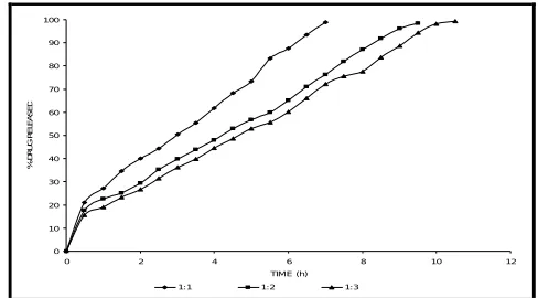
The microcapsules were subjected to in vitro release studies by employing 6.8 pH phosphate buffer (Figure 1) and when amount of drug release values were plotted against time straight lines were obtained in all the cases indicating that the rate of drug release from these microcapsules followed zero order kinetics. To ascertain the mechanism of drug release from various microcapsules studied in the present investigation, plots of log
fraction of drug released vs log time (peppas plots) were drawn. The plots were found to be linear with all the microcapsules and the exponential coefficient values >0.5 indicating the non-fickian controlled release mechanism (Table 3).
0 10 20 30 40 50 60 70 80 90 100
0 2 4 6 8 10 12
TIME (h) % D R U G R E L E A S E D
1:1 1:2 1:3
Fig 1. In vitro drug release from aceclofenac microcapsule formulations with MC.
Table 3. In vitro drug release kinetics from aceclofenac microcapsule formulations prepared by ionic gelation method.
Technique Core: coat
ratio
Correlation coefficient Values Release
Rate ConstantK0 (mg/hr) T50 (hr) T90
(hr) n
Zero order
First
order Matrix Peppas
MC
1:1 0.9573 0.8389 0.9826 0.9927 26.37 2.7 7.2 0.5958 1:2 0.9816 0.8670 0.9608 0.9832 20.17 3.8 9.5 0.6470 1:3 0.9853 0.8179 0.9571 0.9856 18.91 4.3 10.3 0.6701
Na CMC
1:1 0.9433 0.8947 0.9883 0.9949 28.27 2.3 5.9 0.6124 1:2 0.9405 0.8649 0.9882 0.9961 23.42 2.6 7.1 0.5925 1:3 0.9715 0.8989 0.9730 0.9917 20.86 3.6 8.9 0.6527
HPMC
1:1 0.9751 0.875 0.9557 0.9686 28.27 2.6 6.5 0.6447 1:2 0.9775 0.8612 0.9673 0.9861 24.76 3.0 7.5 0.6466 1:3 0.9725 0.8412 0.9716 0.9917 22.01 3.3 8.5 0.6333
The corresponding correlation coefficients and other release parameters indicated that the release rate was found to decrease with increase in the concentration of coating material applied. The wall thickness of microcapsules was found to be increased with the increase in concentration of coating material applied. There exists a good correlation ship in between wall thickness and release rate constant. The formulation was also subjected to In-vitro wash off test in presence of 0.1N HCL and pH 6.8 phosphate buffer. The wash off was relatively rapid in phosphate buffer than in acidic fluid. The results of wash off test indicated fairly good mucoadhesive property of the microcapsules (Table 4).
Table 4: In vitro wash-off test to assess mucoadhesive property of aceclofenac microcapsules formulated by employing ionic gelation technique.
Microcapsules
Percent of microcapsules adhering to tissue at 5 times (h)
0.1 N HCl, pH 1.2 Phosphate buffer, pH 6.8
1 2 4 6 8 1 2 4 6 8
MC 84 81 76 73 71 88 76 66 59 52 NaCMC 78 72 62 57 56 71 62 51 40 33 HPMC 70 64 58 56 54 68 53 41 34 26
Aceclofenac Sodium caboxy methyl cellulose (NaCMC) Microcapsules
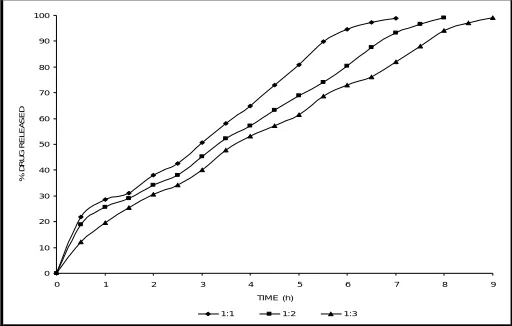
coating material concentration on the release rate of Aceclofenac. These microcapsules were characterized for diameter, density, flow properties, moisture content, surface accumulation study, drug content and entrapment efficiency. The results indicated microcapsules good flow property. The technique also showed good entrapment efficiency. Low coefficient variation in the percent drug content indicated uniformity of the drug content in each batch of microcapsules (Table 1 and 2). The microcapsules were subjected to in vitro release studies by employing 6.8 pH phosphate buffer and the when amount of drug release values were plotted against time straight lines were obtained in all the cases indicating that the rate of drug release from these microcapsules followed zero order kinetics. To ascertain the mechanism of drug release from various microcapsules studied in the present investigation, plots of log fraction of drug released vs. log time (peppas plots) were drawn. The plots were found to be linear with all the microcapsules and the exponential coefficient values >0.5 indicating the non-fickian controlled release mechanism (Table 3). The corresponding correlation coefficients and other release parameters indicated that the release rate was found to decrease with increase in the concentration of coating material applied. The wall thickness of microcapsules was found to be increased with the increase in concentration of coating material applied. There exists a good correlation ship in between wall thickness and release rate constant. The formulation was also subjected to In-vitro wash off test in presence of 0.1N HCL and pH 6.8 phosphate buffer. The wash off was relatively rapid in phosphate buffer than in acidic fluid. The results of wash off test indicated mucoadhesive property of the microcapsules (Table 4).
0 10 20 30 40 50 60 70 80 90 100
0 1 2 3 4 5 6 7 8 9 10
TIME (h)
%
D
R
U
G
R
E
L
E
A
S
E
D
1:1 1:2 1:3
Fig 2. In vitro drug release from aceclofenac microcapsule formulations with NaCMC
Aceclofenac Hydroxy propyl methyl cellulose (HPMC) Microcapsules
This process produced uniform microcapsules. Aceclofenac was coated with the mixture of HPMC and calcium alginate. Microcapsules were developed with 1:1, 1:2 and 1:3 ratios to determine the affect of coating material concentration on the release rate of Aceclofenac. These microcapsules were characterized for diameter, density, flow properties, moisture content, surface accumulation study, drug content and entrapment
efficiency. The results showed micro capsules with good flow property. The technique also showed good entrapment efficiency. Low coefficient variation in the percent drug content indicated uniformity of the drug content in each batch of microcapsules (Table 1 and 2). The microcapsules were subjected to In vitro
release studies by employing 6.8 pH phosphate buffer and when amount of drug release values were plotted against time straight lines were obtained in all the cases indicating that the rate of drug release from these microcapsules followed zero order kinetics. To ascertain the mechanism of drug release from various microcapsules studied in the present investigation, plots of log fraction of drug released vs. log time (peppas plots) were drawn. The plots were found to be linear with all the microcapsules and the exponential coefficient values >0.5 indicating the non-fickian controlled release mechanism (Table 3). The corresponding correlation coefficients and other release parameters were indicated that the release rate was found to decrease with increase in the concentration of coating material applied.
0 10 20 30 40 50 60 70 80 90 100
0 1 2 3 4 5 6 7 8 9
TIME (h)
%
D
R
U
G
R
E
L
E
A
S
E
D
1:1 1:2 1:3
Fig 3. In vitro drug release from aceclofenac microcapsule formulations with HPMC
The wall thickness of microcapsules was found to be increased with the increase in concentration of coating material applied. There exists a good correlation ship in between wall thickness and release rate constant. The formulation was also subjected to In-vitro wash off test in presence of 0.1N HCL and pH 6.8 phosphate buffers. The wash off was relatively rapid in phosphate buffer than in acidic fluid. The results of wash off test indicated fairly good mucoadhesive property of the microcapsules (Table 4).
Table 5. Comparison of drug release from MC, NaCMC and HPMC Microcapsules formulated by ionic gelation technique.
Polymer Regression Equation Release Rate Constant ko (mg/hr)
for a wall thickness of 10µ
MC Y=-0.3469X+50.117 46.6 NaCMC Y=-0.6274X+66.489 60.12 HPMC Y=-0.6857X+69.291 62.43
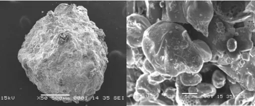
Microcapsules with a coat consisting of alginate and a Mucoadhesive polymer exhibited good Mucoadhesive property in the in vitro wash off test .The was-off was relatively rapid in phosphate buffer than in acid fluid. The results represented in (table 4) for wash off test indicated fairly good Mucoadhesive property of the prepared microcapsules. The SEM of the optimized microcapsule batch showed the uniform surface with their morphology (Figure 4 and 5).
Fig 4 and 5. SEM Photographs of Aceclofenac Microcapsules optimized batch formulated with ionic gelation method.
CONCLUSION
The observations made during study and results obtained showed the suitability of the investigated polymers for microencapsulation of aceclofenac for its sustained release. The ionic gelation method was easy to adopt and also to achieve high drug entrapment efficacy. All the formulations indicated accurate drug content and content uniformity. The in vitro drug release studies revealed that the drug release by zero order and non-fickian controlled release mechanismfor all the formulations studied. The wash off test showed the mucoadhesive nature of microcapsules that in turn helps for steady and slow drug release which is essential for sustain release dosage form. The studies have further scope for investigation of formulation and process variables with polymer to formulation method to study their influence on drug release at desired rate.
REFERENCES
Abu IK, Gracia CL, Robert LD. Preparation and evaluation of zidovudine-loaded sustained-release microspheres. J Pharm Sci. 1996; 85(6): 575-576.
Breghausen SW, Schote U, Frey M and Schmidt F. Comparison of microencapsulation techniques for the water soluble drugs nitenpyram and clomipramine HCl. J. Control. Release., 2002; 85: 35-43.
Brogden, R. N. and Wiseman, L. R. Aceclofenac: A review of
its pharmacodynamic properties and therapeutic potential in the treatment of rheumatic disorders and in pain management. Drug 1996;52:113-124.
Chowdary KPR and Srinivasa RY. Preparation and evaluation of mucoadhesive microcapsules of Indomethacin. Indian J. of Pharm Sci. 2003; 65(1): 49-52
Goudanavar P. S., M.Patil S., Manvi F. V. Design and Characterisation of Sustained Release Microcapsules of Salbutamol Sulphate. International Journal of Pharm Tech Research. 2010; 2(2): 1144-1149.
Hari, P. C., Chandy, T and Sharma, C. P., Chitosan/calcium alginate microcapsules for intestinal delivery of nitrofurantoin. J Microencapsulation. 1996; 13: 319 - 329
Haznedar S, Portune B. Preparation and in vitro evaluation of eudragit microspheres containing acetazolamide. International Journal of Pharmaceutics 2004; 269: 131‐140.
Hoffman A, M. Donbrow, S.T. Gross, S. Benita, R. Bahat. Fundamentals of release mechanism interpretation in multiparticulate systems: determination of substrate release from single microcapsules and relation between individual and ensemble release kinetics. International Journal of Pharmaceutics. 1986; 29(3): 195-211.
Indian Pharmacopoeia. 5th ed. Controller of Publication. (1996) 328-329, 393-395.
Kim, C. K. and Lee, E. J., The controlled release of blue dextran from alginate beads. Int. J. Pharm. 1992; 79: 11-19.
Koida, Y, Kobayashi, M and Samejima, M. Studies on microcapsules. IV: Influence of properties of drugs on microencapsulation and dissolution behavior. Chem. Pharma. Bull. 1986; 34(8): 3354-3361.
Legrand E. Aceclofenac in the management of inflammatory pain. Expert Opinion on. Pharmacotherapy. 2004; 5 (6): 1347-1357.
Luu Si Nang, Patrick F. Carlier, Pierre Delort, Jean Gazzola and Didier Lafont. Determination of coating thickness of microcapsules and influence upon diffusion. J Pharm Sci. 1973;62(3): 452–455.
Madhusudan Rao Y., Vani G., Ramesha Chary R. Bala. Design and evaulation of mucoadhesive drug delivery systems. Indian Drugs 1998; 35(9)558–565
Mine ozyazici, Ferhan Sevgi and Gokhan Ertan. Micromeritic studies on nicardipine hydro chloride microcapsules. International Journal of Pharmaceutics. 1996; 138 (1): 25-35.
Pareek A, Chandanwale AS, Oak J, Jain UK, Kapoor S. Efficacy and safety of aceclofenac in the treatment of osteoarthritis: a randomized double‐blind comparative clinical trial versus diclofenac an Indian experience. Current Medical Research and Opinion 2006; 22 (5): 977‐988. Parfitt, K. Martindale: The Complete drug reference, 32nd ed., Massachusetts, (1999) 2-12.
Sarfaraz MD., Hiremath D., and. Chowdary K. P. R. Formulation and Characterization of Rifampicin Microcapsules. Indian J Pharm Sci. 2010; 72(1): 101–105.
Shavi GV, Nayak U, Averineni RK, Arumugam K, Meka SR, Nayanabhirama U, Sureshwar P. Multiparticulate drug delivery system of aceclofenac: development and in vitro studies. Drug Dev Ind Pharm. 2009; 35(2):252-8.
Tatiane Pereira de Souza, Ramon Martinez-Pacheco, Jose Luiz Gomez Amoza., and Pedro Ros Petrovick. Eudragit E as excipient for production of granules and tablets from phyllanthus niruri spray-dried extract. AAPS Pharm Sci Tech. 2007; 8 (2): E1 –E7.
Trivedi P, Verma AML and Garud N. Preparation and characterization of aceclofenac microspheres. Asian Journal of Pharmaceutical Sciences. 2008; 2 (2):110-115.