R E S E A R C H A R T I C L E
Open Access
Efficacy and potential use of novel
sustained release fillers as intracanal
medicaments against
Enterococcus faecalis
biofilm in vitro
Bernhard Funk
1*†, David Kirmayer
2†, Sharonit Sahar-Heft
3, Irith Gati
2, Michael Friedman
2and Doron Steinberg
1,4Abstract
Background:Enterococcus faecalisis a bacterium frequently isolated after failed root canal therapy. This study analyzed the antibacterial and antibiofilm effects in vitro of sustained-release fillers (SRF) containing cetylpyridinium chloride (CPC) against vancomycin resistantE. faecalis.
Methods:First, the solidification capability was tested by introducing liquid SRF into phosphate buffered saline, followed by 30 s of vortexing. The antimicrobial effects of SRF-CPC against static monospecies biofilms were analyzed with a metabolic assay. Inhibition of biofilm formation was tested by exposing daily refreshedE. faecalis suspensions to SRF-CPC for 9 weeks. To evaluate the effects of SRF-CPC against preformed biofilms, biofilms were grown for 1, 3 and 7 days, and then treated with SRF-CPC for 24 h. Biofilm kill time was tested by applying SRF-CPC to a 3-day-old biofilm and measuring its viability at different time points. All experiments were compared to Placebo SRFs and to untreated control biofilms. Data were analyzed with two-way ANOVA followed by Tukey’s test. Results were considered significant atP< 0.05.
Results:The liquid SRF solidified within seconds and no structural changes were observed after 30 s of vortexing at maximum speed. SRF-CPC inhibitedE. faecalisbiofilm formation for 7 weeks and significantly reduced its viability in weeks 8 and 9. Mature biofilms grown for 1, 3 and 7 days were destructed by SRF-CPC in less than 24 h. Fifty percent of a 3-day-old biofilm was destructed in 2 h and complete destruction occurred in less than 12 h. (P< 0.05 in all cases, compared to SRII-Placebo).
Conclusions:SRF-CPC’s physical properties and long-lasting anti-biofilm effects make it a promising coadjuvant medication for endodontic therapy.
Keywords:Cetylpyridinium chloride,E. faecalisbiofilm, Sustained release, Intracanal medication
Background
Enterococcus faecalisis the most commonly recovered spe-cies from the root canals after failed root canal therapy (RCT) [1]. The primary goal of RCT is to remove all micro-organisms from the inner surface of the root canal system, to prevent reinfection and to establish or maintain healthy periapical tissues [2]. Modern techniques and equipment
have greatly contributed to increased clinical success rates and significantly shortened the time needed to complete the RCT, yet there are still limitations to the endodontic disinfection process; viable biofilm cells can persist in undertreated and untreated locations of the root canal sys-tem due to the inherent challenges associated with its com-plex anatomy [3–5]. Difficult clinical cases require more time, better skills of the practitioner and modern instru-ments for their treatment, and this can lead to the necessity of performing the RCT in more than one appointment [5]. Multiple patient visits might also be required to avoid the risk of flare-ups when periapical lesions are present [6].
© The Author(s). 2019Open AccessThis article is distributed under the terms of the Creative Commons Attribution 4.0 International License (http://creativecommons.org/licenses/by/4.0/), which permits unrestricted use, distribution, and reproduction in any medium, provided you give appropriate credit to the original author(s) and the source, provide a link to the Creative Commons license, and indicate if changes were made. The Creative Commons Public Domain Dedication waiver (http://creativecommons.org/publicdomain/zero/1.0/) applies to the data made available in this article, unless otherwise stated.
* Correspondence:[email protected]
†Bernhard Funk and David Kirmayer contributed equally to this work. 1The Institute of Dental Sciences, Faculty of Dental Medicine, The Hebrew University of Jerusalem, Jerusalem, Israel
Hence the use of intracanal medication is still a widely spread clinical practice to attain disinfection and prevent reinfection between appointments [7].
Ideally, an intracanal medicament should eliminate any remaining bacteria, reduce inflammation of periapical tissues, render canal contents inert and neutralize debris, act as a barrier against leakage from temporary filling, and help drying persistently wet canals [8]. Thus, the widely ex-tended use of calcium hydroxide (CH) is reasonable [7,9]. Still, despite its good physical, biological and pharmaceut-ical properties, CH cannot completely eradicate resilient intracanal microorganisms asE. faecalis[10]. One alterna-tive to CH is chlorhexidine (CHX) in gel which was found to be very effective againstE. faecalis[11].However, CHX gel does not act as a physical barrier when injected into the root canal, nor does it inactivate lipopolysaccharides (LPS), a wall component and degradation product of gram-nega-tive cells, which, when persisting, causes periapical inflam-mation, bone resorption and pain [12–14].
Another antibacterial agent, only recently tested for endodontic purposes, is cetylpyridinium chloride (CPC), a cationic quaternary compound, commonly found in mouthwashes [15]. Incorporated to dental cements and gutta-percha points or used as irrigation solution, it proved to be very effective againstE. faecalis[16–18]. Furthermore, a recent study showed that repeated exposure ofE. faecalis to CHX led to its resistance, whereas CPC did not elicit such deleterious response [19]. Another important feature that favors CPC over CHX for endodontic applications is its inhibiting effect on LPS binding to the toll-like receptor 4 involved in inflammatory cytokine production [20].
Nevertheless, good antibacterial effects of a drug alone are not sufficient against endodontic biofilms. The penetra-tion of a drug into deeper layers of the biofilm is limited by extracellular polysaccharides (EPS) that act as an affinity matrix, increase viscosity of the medium, and thus slow down the diffusion of antimicrobials [21]. Nevertheless, higher concentrations of antimicrobial agents at the outer layer of the biofilm should increase the overall diffusion rates into the deeper layers [22]. To achieve high local con-centrations over an extended period of time, a drug can be incorporated into a locally-administrable type of formula-tion or device that controls its local release at the site of action [23, 24]. Numerous sustained release devices have already been developed, tested and commercialized in different fields of dentistry [22]. This concept of controlled release can also be applied to the root canal system [25].
Phaechamud et al. [26] have recently described a solv-ent exchange-induced in situ forming gel, based on pharmaceutical ammoniomethacrylate type b, for peri-odontitis treatment. However, to be applicable to RCT, the formulation should remain in the area for the period sufficient to eradicate the infection, have sufficient pene-tration power, yet seal off the canal and fill it completely.
Hence, in this study we tested a novel concept of sus-tained-release fillers (SRFs) containing CPC (SRF-CPC), based on the polymers used by Phaechamud et al. [26]. Our approach encompasses the use of pharmaceutical polymers dissolved in acceptable solvents thus providing injectability into the root canal. Due to carefully chosen additives, unlike in-situ gels, our SRF solidifies very rap-idly upon contact with aqueous medium. Our main goal for the current study was to analyze the sustained anti-bacterial effects of SRF-CPC againstE. faecalisbiofilm in vitro. The null hypothesis of the study was that a con-trolled-release filler comprising CPC cannot effectively eradicate biofilms or inhibit growth of microbiota im-plied in root canal infections for extended time intervals.
Methods
Preparation of the SRF
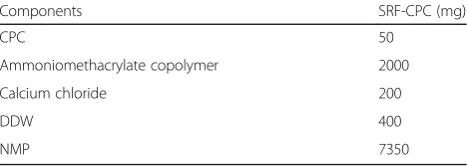
The detailed preparation procedure is set forth in the Add-itional file1. Briefly, the heat-sterilized ammoniomethacry-late copolymer, type A, according to the United States Pharmacopeia 41–National Formulary 36, was dissolved in sterile-filtered N-methyl pyrrolidone - water mixture with CPC and a small amount of calcium chloride. The resulting formulation contained 0.5% of CPC. The formula of the composition is summarized in Table1. The pharma-ceutical release profile of CPC for the SRF can be found in Additional file2.
Solidification of SRFs
Solidification of the SRF was performed by injecting about 200μL of the composition into a scintillation vial containing about 10 mL of phosphate buffer according to the USP (i.e. as defined in the United States Pharma-copeia), at pH 6.8. The immediately solidified filler was vortexed for about 30 s at maximal velocity, to observe minimal or no shape change. The solidified residue was soft spongiform solid, maintaining the original shape upon flow but yielding to mechanical tools.
Handling of SRFs
SRFs were solidified on agar plates to allow consistent shape and ease of handling during experimentation, as follows: polypropylene tubes of 6 mm diameter were segmented into 2-mm pieces, sterilized and placed on small 60-mm petri dishes (Miniplast, Ein Shemer, Israel)
Table 1Composition of the tested SRF-CPC (10-g formulation)
Components SRF-CPC (mg)
CPC 50
Ammoniomethacrylate copolymer 2000
Calcium chloride 200
DDW 400
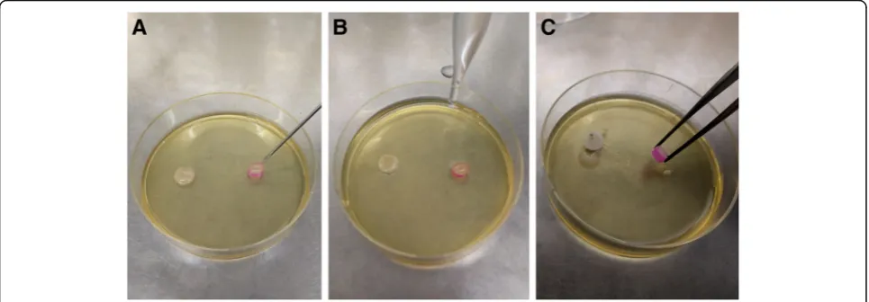
containing 9 mL brain heart infusion agar (BHI agar; Neo-gen Corporation, Lansing, Michigan, USA). Aliquots of 30μL of SRF-CPC (or SRF-Placebo if needed) were then separately dispensed into their respective reservoirs using syringes and 23-Gauge needles. Contact of the SRF with the agar initiated solidification. To complete solidification, 9 mL of sterile phosphate buffered saline (PBS; Sigma Life Sciences, St. Louis, MO, USA) were carefully added to the agar plate to assure full immersion of the SRFs. After 5 min SRFs were removed and used immediately for the experi-ments. The steps can be seen in Fig.1.
Antibiofilm effects of SRF-CPC Biofilm growth
To growE. faecalisbiofilms we followed the initial steps of a protocol presented by Kwasny and Opperman [27] for the growth of static biofilm of non-motile gram-posi-tive bacteria, with some minor modifications. The method of biofilm growth described here, was used for all following experiments in the current study. Briefly, cultures of E. faecalis V583 grown in an orbital shaker overnight in aerobic conditions at 37 °C were diluted 1: 50 (OD600 ~ 0.1) with fresh BHI broth containing 2%
glucose [28]. Aliquots of 270μL of this bacterial suspen-sion were used for biofilm formation inside the individ-ual wells of a sterile polystyrene 48-well tissue culture plate (TCP; SPL Life Sciences, Pocheon-si, Gyeonggi-do, Korea). TCPs were then incubated at 37 °C in aerobic conditions for 24 h.
Inhibition of biofilm formation
To evaluate the long-term antibacterial effects of SRFs against biofilm formation de novo, daily renewed bac-terial suspensions intended for biofilm growth were exposed to specimens of either CPC or
SRF-Placebo for 9 weeks. SRF specimens were prepared as described in the section above “Handling of SRFs”
and placed into the individual wells of a 48-well TCP containing 270μL of bacterial suspension. Each day during the test period, the supernatant-fluid sur-rounding the SRFs was removed and replaced by 270μL of fresh bacterial suspension for biofilm growth. Specimens of SRF-CPC and SRF-Placebo were transferred to the wells of a new TCP under sterile conditions every 7 days. The grown biofilms located in the wells of the used plate were then analyzed with the MTT assay (3-(4,5-dimethylthiazol-2-yl)-2,5-diphe-nyltetrazolium bromide reduction assay) described in a section below. Control biofilms in this experiment were grown without addition of SRFs.
Biofilm destruction
This experiment was performed to assess the ability of SRF-CPC to destroy pre-grown biofilms. Biofilms were grown in separate TCPs for either 1, 3 or 7 days. Media, 270μL of BHI with 2% glucose, was replaced every day. The pre-grown biofilms inside the individual wells were then exposed to either one specimen of SRF-CPC or SRF-Placebo for 24 h. Next, the biofilms were analyzed with an MTT assay to measure the antibiofilm effects of the SRFs. Control biofilms were grown without addition of SRFs.
Biofilm kill time
To determine the time necessary to destroy a mature existing biofilm, pre-grown mature biofilms were ex-posed to SRFs and evaluated at different time points. The experiment was performed as follows. Biofilms were grown for 3 days and media replaced every 24 h with 270μL of fresh BHI broth supplemented with 2%
glucose. Mature biofilms were then exposed to speci-mens of SRF-CPC or SRF-Placebo for either 1/2, 1, 2, 6 or 12 h. Next, an MTT assay was performed. Control biofilms were not exposed to SRFs.
MTT assay
This method allows to measure the metabolic activity of viable cells that compose the biofilm. The protocol employed by Walencka et al. [29] was slightly modified as follows. In brief, at the end of each experiment, after microscopically confirming the absence of contaminating microorganisms, SRFs were removed with sterile tweezers and the wells were washed twice with 200μL of PBS solu-tion to remove planktonic cells. Aliquots of 50μL of 0.1% w/v MTT (Calbiochem, Darmstadt, Germany) were then added to each well to cover the biofilm. After 1 h of incu-bation at 37 °C in aerobic conditions, the excess of MTT was washed with 150μL of PBS. Next, 200μL of dimethyl sulfoxide (DMSO; Bio-Lab, Hayetzira, Jerusalem, Israel) were added to each well and the plate was placed on an orbital shaker (S-3.02.10 M, ELMI, Riga, Latvia) for 10 min at room temperature. Aliquots of 150μL of the solubilized MTT solution were transferred to unused wells of a 96-well plate (SPL Life Sciences, Pocheon-si, Gyeonggi-do, Korea). Great care was taken to avoid aspiration of poly-mer debris and biofilm masses on the bottom of the wells. Finally, absorbance was measured at 540 nm with a refer-ence wavelength of 630 nm using an Infinite 200 PRO spectrophotometer (Tecan Austria, Grödig, Austria).
Statistical analysis
Statistical analysis was performed using GraphPad Prism version 7.04 (GraphPad Software, La Jolla, CA, USA). Results from the biofilm experiments were analyzed with a two-way ANOVA test to detect overall statistical dif-ferences between test groups, followed by Tukey’s test to identify specific differing groups. All data were expressed as mean with standard deviation and considered signifi-cant at P< 0.05. Supporting data were provided in Additional file3.
Results
Solidification of SRF-CPC ensues quickly
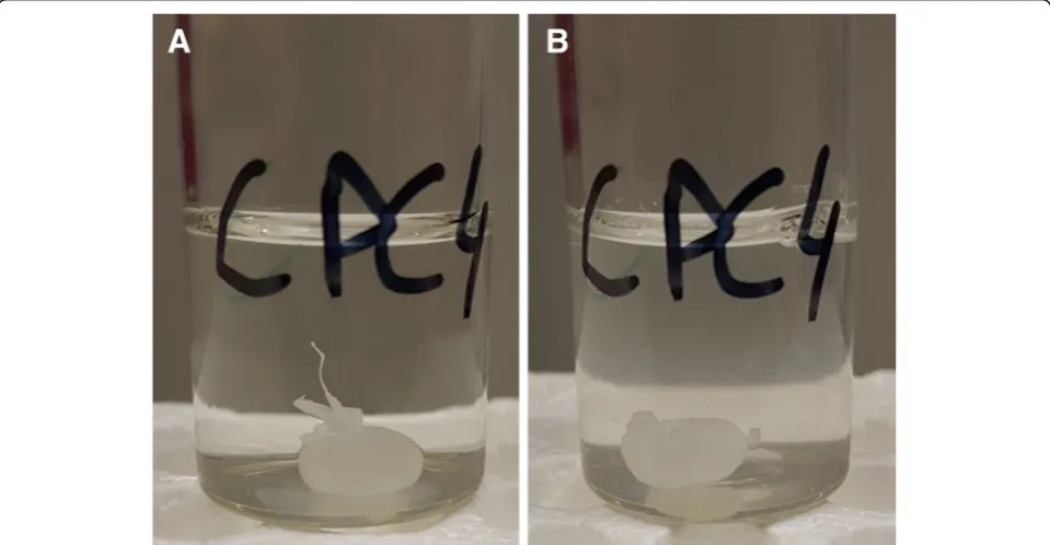
The formulation solidified within several seconds upon contact with phosphate buffer USP, pH 6.8. Mixing thoroughly for 30 s resulted in no structural change of the of the solidified medicament. The photographs are shown in Fig.2.
SRF-CPC inhibits biofilm formation for 7 weeks in multi-well plates
To assess the long-term effects of CPC and SRF-Placebo on biofilm formation, metabolic activity was measured once a week with the MTT assay. SRF-CPC inhibited biofilm formation for 7 weeks and during the following 2 weeks a significant reduction of biofilm ac-tivity was observed when compared to the placebo and control wells (Fig.3). SRF-Placebo produced a significant decrease in biofilm formation on the bottom surface of
the microwells compared to the control sample after the first week.
SRF-CPC terminates metabolic activity of biofilms at different maturity stages
SRF-CPC was able to destruct biofilms that where pre-grown for either 1, 3 or 7 days, regardless of their matur-ity stage, whereas SRF-Placebo alone did not signifi-cantly reduce biofilm viability at any stage (Fig.4).
SRF-CPC terminates metabolic activity of mature biofilms in less than 12 h
The results displayed in Fig. 5 show a clear decrease in viability of the biofilm as time progresses. Around 50% of the biofilm was destructed by SRF-CPC after only 2 h and no metabolic activity was detected after 12 h. SRF-Placebo failed to destruct E. faecalis biofilm viability, however a significant reduction in metabolic activity was observed after 1 h onwards in comparison to the control sample.
Fig. 3Percentages of biofilm viability in presence of SRF-CPC and SRF-Placebo. Data expressed as mean with standard deviation. *P< 0.05, compared with SRF-Placebo and control sample;n= 6
Discussion
The present results indicate that the null hypothesis must be rejected.
Many improvements have been made lately in the che-mico-mechanical preparation of the root canal. Rotary files with optimized taper sizes remove more infected dentin and make it possible to prepare the root canals that favor effective irrigation [30,31]. The removal of biofilm from the inner walls of the canal is further enhanced when irrigation is assisted by ultrasonic or laser activation [32,
33]. However, several clinical studies have shown no sig-nificant clinical advantage of these procedures over con-ventional protocols [34–36] and the extent in which they affect biofilms located in remote regions of oval-shaped canals, lateral canals and tubules, is still unknown [37]. Hence, intracanal medicaments are inserted into root ca-nals intending to restrict bacterial regrowth and supply continued disinfection [38]. Ideally such medicaments should deliver and maintain significant concentrations of antimicrobial agent during the time period between RCT sessions; this is the role that the sustained-release formu-lations can readily play.
Due to the versatility of pharmacopeial methacrylate polymers it is possible to design sustained release formu-lations with tailor-made release profiles, to which a drug of choice can be added, in our case, CPC. The efficacy of CPC against dental plaque as component of sustained release films was already confirmed in a clinical study published by Friedman et al. [39]; here, for the first time we tested CPC as component of a novel sustained re-lease solidifying filler intended for endodontic use. Using the currently reported approach, we have successfully
reached drug release at controllable rate of CPC. The initial drug concentration of 0.5% is in line with the present practice; however when the SRF solidifies and the solvent leaches out from SRF and partially switches for water, this initial concentration in the solution becomes higher in the solid SRF. Nevertheless, due to the controlled release properties of the SRF, particularly in restrictive conditions where the extraction solvent is not readily available, the amounts of active agent re-leased in that solvent are relatively low, allowing for the pronounced inhibition period demonstrated in the present work.
As with any antiseptic, also the use of CPC may raise a question of possible cytotoxicity. The general outnum-bering principle of antiseptic use, which stipulates that when killing one tissue cell, the tissue has one cell less, but when killing one bacterium cell, the bacterium is dead, generally proves true, which lead the regulators already over two decades ago to believe that antiseptics, inter alia CPC, are safe for use on oral mucosa [40]. Yet, the cytotoxicity of CPC cannot be completely ignored. Depending on the tested cell type, the half-population cytotoxic concentration of CPC (CC50) was found to
vary between 0.003 and 0.001% [41, 42]. On the other hand, the minimum inhibitory concentration (MIC) and the minimal biocidal concentration (MBC) of CPC forE.
faecalisin our experimental conditions was estimated as 0.0003 and 0.0006%, respectively (data not shown), whereas Tomino et al. [18] reported the MIC value of 0.0001% with CPC incorporated into agar plates. This means that there is a window of effective concentrations between 3 × 10⁻⁴% and 10−3% before any cytotoxic
effect could even potentially be seen. Due to flexibility of the pharmaceutical formulation, the concentration of CPC in practice could be readily adjusted to any desire value. Despite the total concentration of CPC in SRF-CPC being 0.5%, it is impossible to know at this stage what concentrations of CPC could be obtained in the root canal throughout the drug release period, which we have seen can be as long as 7 weeks at effective antibac-terial concentrations; the dose spread over such time interval effectively brings the amounts of CPC released to the safe range. At the minute amounts of solvents that would be available in the root canal, it is believed that there will be even less release of CPC.
The cationic nature of the polymer used in the formu-lation may play a certain role in its antimicrobial effect. It was demonstrated that ammoniomethacrylates poly-mers USP, are capable of promoting cell growth on sur-face, in particular the adhesion, proliferation and differentiation of human mesenchymal stem cells [43]. We have also seen that certain other types of adhering cells attach to the polymer, regardless of the physical form, i.e. either as a film or on supported particles. Therefore, we believe that the viable bacteria may be attracted to the constitutive positive charge on the sur-face of the SRF-CPC, and thus be exposed to higher concentration of CPC – at the surface of the SRF. This is supported by the fact that placebo filler decreased the amount of biofilm grown on the TCP well, which can only be explained that the surface of the filler attracts the viable bacteria, until it is completely covered with biofilm. This decoy mechanism may explain the long duration efficacy of the formulation against E. faecalis, but comparative studies with other polymers may be needed to prove unequivocally that the surface charge may attract bacteria to these drug delivery systems.
The long-lasting antibacterial efficacy of SRF-CPC is fundamental for clinical applicability as the time be-tween endodontic appointments can range from a few days to several weeks, since maintaining a clean and dis-infected root canal after initial treatment until the next visit is one of the main concerns in endodontic therapy. Our results show that single doses of SRF-CPC were able to inhibit E. faecalis growth and biofilm formation for 7 weeks in vitro, in a biological set-up where media was changed every day and fresh bacterial cells were added. In complex root canals, biofilms can persist in areas that escape instrumentation and irrigation. Our findings suggest that any survivingE. faecalisbiofilm left inside the root canal system would be exposed to high local concentrations of CPC during the first days. SRF-CPC destructed 50% of mature 3-day-oldE. faecalis bio-films in only 2 h and 7-day old biobio-films in less than 24 h. During the following months CPC concentrations would be reinforced and maintained by a continuous drug
release from the SRF-CPC. An initial burst effect followed by a more graduate release rate of CPC would drastically decrease the probability ofE. faecalis biofilm survival.
Besides its significant antimicrobial/antibiofilm effects, SRF-CPC also presents good physical properties. Its vis-cosity allows it to be easily ejected through a 23-Gauge needle. Humidity does not constitute an obstacle and only contributes to SRF-CPC’s solidification. In a clinical setting, humidity present on the internal walls of the root canals and in the apical end of the root is expected to quickly start the solidification process of SRF-CPC and encapsulate the remaining solution, hence prevent-ing unwanted leak to the periapex. In addition, it is ex-pected to block penetration of periapical fluids into the root canal and act as a physical and pharmaceutical bar-rier against leakage coming from a poor temporary obtu-ration at the coronal end. SRF-CPC’s soft consistency should facilitate its effortless removal using endodontic files and irrigation/aspiration. Nevertheless, more testing must be done to confirm SRF-CPC’s applicability for RCT in clinical settings.
The concept of using sustained release drugs as an ad-junctive to the intracanal procedure has been reported in the past using different pharmaceutical technologies than proposed in this study. Heling et al. [44] success-fully tested a sustained release device in a form of a strip containing chlorhexidine, with the capability to swell in the presence of liquids. Their device prevented E.
structures, well beyond the contemplated drug delivery paradigm [49]. Finally, a study performed by Gandhi et al. [50] presented a sol-gel device, consisting of GEL-RITE Gellan polymer loaded with CHX. This device would release the drug only in an acidic inflammatory environment, hence preventing premature peaking of the drug associated with possible side effects. Whether such a feature would be of clinical relevance or even de-sirable inside the root canals is yet to be unequivocally determined.
The quest for sustained release alternatives to common intracanal medicaments has been ongoing for nearly 30 years. Despite its excellent antibacterial and antibiofilm ef-fects, especially against vancomycin-resistantE. faecalis, the consideration of CPC as constituent of sustained release drugs in endodontics remained mostly unexplored. The well-established use of calcium hydroxide may be appropri-ate for most clinical cases, however, when confronted with refractory periapical lesions due to the persistence ofE.
fae-calis, a different intracanal medicament should be selected. The SRF-CPC presented in this study has promising anti-microbial, pharmacological and physical properties, essen-tial against endodonticE. faecalisbiofilms.
Conclusions
Due to its good physical properties, its efficacy against mature biofilms and its long-lasting antibacterial effects against vancomycin resistant E. faecalis, SRF-CPC has the potential to become a significant coadjuvant medica-tion to root canal therapy.
Additional files
Additional file 1:Preparation and drug release of SRF-CPC. The file contains a detailed preparation procedure of the SRF, and the release of CPC measurements. (DOCX 13 kb)
Additional file 2:Release profile of SRF-CPC. The file contains a figure with the results of the release profile of CPC from SRF-CPC. (TIF 381 kb)
Additional file 3:Supporting data. The file contains data to support Figs.3,4and5. (XLSX 30 kb)
Abbreviations
BHI:Brain heart infusion; CH: Calcium Hydroxide; CHX: Chlorhexidine; CPC: Cetylpyridinium chloride; EPS: Extracellular polysaccharides; LPS: Lipopolysaccharide; MBC: Minimum bactericidal concentration; MIC: Minimum inhibitory concentration; MTT: 3-(4,5-dimethylthiazol-2-yl)-2,5-diphenyltetrazolium bromide reduction assay; PBS: Phosphate buffered saline; RCT: Root Canal Therapy; SRF: Sustained release filler; CPC: Sustained release filler containing cetylpyridinium chloride; SRF-Placebo: Placebo sustained release filler; TCP: Tissue culture plate; USP: United States Pharmacopeia; ZOI: Zone of inhibition
Acknowledgements
The research was partially supported by the BECAL scholarship program of the Paraguayan Government.
Authors’contributions
BF: study design and performance of biological experiments, data collection and analysis, major contributions to the writing, critical revision and approval
of the manuscript. DK: formulation of the sustained release fillers, design and performance of non-biological experiments, data collection and analysis, major contributions to the writing, critical revision and approval of the manuscript. SSH: contributions to the study design and supervision of the biological experiments, critical revision and approval of the final manuscript. IG: preparation of the drug, non-biological experiments, data collection, ap-proval of the final manuscript. MF: Design, formulation and preparation of the drug, critical revision and approval of the final manuscript. DS: Study con-cept and design, major contributions in the writing, critical revision and ap-proval of the final manuscript. BF and DK contributed equally.
Funding
Not applicable.
Availability of data and materials
The datasets supporting the conclusions of this articles are included within the article and its additional files.
Ethics approval and consent to participate
Not applicable.
Consent for publication
Not applicable.
Competing interests
The authors declare that they have no competing interests.
Author details
1The Institute of Dental Sciences, Faculty of Dental Medicine, The Hebrew University of Jerusalem, Jerusalem, Israel.2The Institute of Drug Research, School of Pharmacy, Faculty of Medicine, The Hebrew University of Jerusalem, Jerusalem, Israel.3Department of Endodontics, Faculty of Dental Medicine, The Hebrew University of Jerusalem, Jerusalem, Israel.4Faculty of Dentistry, Hebrew University–Hadassah, PO Box 12272, 91120 Jerusalem, Israel.
Received: 17 September 2018 Accepted: 6 August 2019
References
1. Gulabivala K, Ng Y-L. Biofilm-associated infections in root canals: treatment and outcomes. In: de Paz LEC, Sedgley CM, Kishen A, editors. The root canal biofilm. Berlin Heidelberg: Springer; 2015. p. 191–258.
2. Versiani MA, Ordinola-Zapata R. Root canal anatomy: implications in biofilm disinfection. In: de Paz LEC, Sedgley CM, Kishen A, editors. The root canal biofilm. Berlin Heidelberg: Springer; 2015. p. 155–87.
3. Kishen A. Advanced therapeutic options for endodontic biofilms. Endod Top. 2010;22:99–123.
4. Nair PN, Henry S, Cano V, Vera J. Microbial status of apical root canal system of human mandibular first molars with primary apical periodontitis after “one-visit”endodontic treatment. Oral Surg Oral Med Oral Pathol Oral Radiol Endod. 2005;99:231–52.
5. Wong AW, Zhang C, Chu CH. A systematic review of nonsurgical single-visit versus multiple-visit endodontic treatment. Clin Cosmet Investig Dent. 2014; 6:45–56.
6. Schwendicke F, Gostemeyer G. Single-visit or multiple-visit root canal treatment: systematic review, meta-analysis and trial sequential analysis. BMJ Open. 2017;7:e013115.
7. Madarati AA, Zafar MS, Sammani AMN, Mandorah AO, Bani-Younes HA. Preference and usage of intracanal medications during endodontic treatment. Saudi Med J. 2017;38:755–63.
8. Chong BS, Pitt Ford TR. The role of intracanal medication in root canal treatment. Int Endod J. 1992;25:97–106.
9. Itoh A, Higuchi N, Minami G, Yasue T, Yoshida T, Maseki T, et al. A survey of filling methods, intracanal medications, and instrument breakage. J Endod. 1999;25:823–4.
11. Gomes BP, Souza SF, Ferraz CC, Teixeira FB, Zaia AA, Valdrighi L, et al. Effectiveness of 2% chlorhexidine gel and calcium hydroxide against
enterococcus faecalis in bovine root dentine in vitro. Int Endod J. 2003;36:267–75. 12. Gomes BP, Vianna ME, Zaia AA, Almeida JF, Souza-Filho FJ, Ferraz CC.
Chlorhexidine in endodontics. Braz Dent J. 2013;24:89–102. 13. Hong CY, Lin SK, Kok SH, Cheng SJ, Lee MS, Wang TM, et al. The role of
lipopolysaccharide in infectious bone resorption of periapical lesion. J Oral Pathol Med Off Publ Int Assoc Oral Pathol Am Acad Oral Pathol. 2004;33:162–9. 14. Jacinto RC, Gomes BP, Shah HN, Ferraz CC, Zaia AA, Souza-Filho FJ.
Quantification of endotoxins in necrotic root canals from symptomatic and asymptomatic teeth. J Med Microbiol. 2005;54(Pt 8):777–83.
15. Haps S, Slot DE, Berchier CE, Van der Weijden GA. The effect of cetylpyridinium chloride-containing mouth rinses as adjuncts to toothbrushing on plaque and parameters of gingival inflammation: a systematic review. Int J Dent Hyg. 2008;6:290–303.
16. Estrela C, Sousa-Neto MD, Alves DR, Alencar AH, Santos TO, Pecora JD. A preliminary study of the antibacterial potential of cetylpyridinium chloride in root canals infected by E. faecalis. Braz Dent J. 2012;23:645–53. 17. Gjorgievska E, Apostolska S, Dimkov A, Nicholson JW, Kaftandzieva A.
Incorporation of antimicrobial agents can be used to enhance the antibacterial effect of endodontic sealers. Dent Mater. 2013;29:e29–34. 18. Tomino M, Nagano K, Hayashi T, Kuroki K, Kawai T. Antimicrobial efficacy of
gutta-percha supplemented with cetylpyridinium chloride. J Oral Sci. 2016; 58:277–82.
19. Kitagawa H, Izutani N, Kitagawa R, Maezono H, Yamaguchi M, Imazato S. Evolution of resistance to cationic biocides in Streptococcus mutans and enterococcus faecalis. J Dent. 2016;47:18–22.
20. Haught JC, Xie SC, Circello B, Tansky CS, Khambe D, Sun YP, et al. Lipopolysaccharide and lipoteichoic acid binding by antimicrobials used in oral care formulations. Am J Dent. 2016;29:328–32.
21. Kreth J, Herzberg MC. Molecular principles of adhesion and biofilm formation. In: de Paz LEC, Sedgley CM, Kishen A, editors. The root canal biofilm. Berlin Heidelberg: Springer; 2015. p. 23–53.
22. Steinberg D, Friedman M. Sustained-release drug delivery of antimicrobials in controlling of supragingival oral biofilms. Expert Opin Drug Deliv. 2017;14:571–81. 23. Feldman M, Shenderovich J, Al-Quntar AA, Friedman M, Steinberg D.
Sustained release of a novel anti-quorum-sensing agent against oral fungal biofilms. Antimicrob Agents Chemother. 2015;59:2265–72.
24. Gefter Shenderovich J, Zaks B, Kirmayer D, Lavy E, Steinberg D, Friedman M. Chlorhexidine sustained-release varnishes for catheter coating - Dissolution kinetics and antibiofilm properties. Eur J Pharm Sci. 2018;112(Supplement C):1–7.
25. Tronstad L, Yang ZP, Trope M, Barnett F, Hammond B. Controlled release of medicaments in endodontic therapy. Endod Dent Traumatol. 1985;1:130–4. 26. Phaechamud T, Jantadee T, Mahadlek J, Charoensuksai P, Pichayakorn W.
Characterization of antimicrobial agent loaded Eudragit RS solvent exchange-induced in situ forming gels for periodontitis treatment. AAPS PharmSciTech. 2017;18:494–508.
27. Kwasny SM, Opperman TJ. Static biofilm cultures of Gram-positive pathogens grown in a microtiter format used for anti-biofilm drug discovery. Curr Protoc Pharmacol. 2010;13:Unit 13A 8.
28. Seneviratne CJ, Yip JW, Chang JW, Zhang CF, Samaranayake LP. Effect of culture media and nutrients on biofilm growth kinetics of laboratory and clinical strains of enterococcus faecalis. Arch Oral Biol. 2013;58:1327–34. 29. Walencka E, Sadowska B, Rozalska S, Hryniewicz W, Rózalska B. Lysostaphin
as a potential therapeutic agent for staphylococcal biofilm eradication. Pol J Microbiol. 2005;54:191–200.
30. Boutsioukis C, Gogos C, Verhaagen B, Versluis M, Kastrinakis E, Van der Sluis LW. The effect of root canal taper on the irrigant flow: evaluation using an unsteady computational fluid dynamics model. Int Endod J. 2010;43:909–16. 31. Paraskevopoulou MT, Khabbaz MG. Influence of taper of root canal shape
on the Intracanal bacterial reduction. Open Dent J. 2016;10:568–74. 32. Layton G, Wu WI, Selvaganapathy PR, Friedman S, Kishen A. Fluid dynamics
and biofilm removal generated by syringe-delivered and 2 ultrasonic-assisted irrigation methods: a novel experimental approach. J Endod. 2015; 41:884–9.
33. Sahar-Helft S, Stabholtz A, Moshonov J, Gutkin V, Redenski I, Steinberg D. Effect of Er:YAG laser-activated irrigation solution on enterococcus Faecalis biofilm in an ex-vivo root canal model. Photomed Laser Surg. 2013;31:334–41. 34. Hertel M, Sommer K, Kostka E, Imiolczyk SM, Ballout H, Preissner S.
Outcomes of endodontic therapy comparing conventional sodium
hypochlorite irrigation with passive ultrasonic irrigation using sodium hypochlorite and Ethylenediaminetetraacetate. A Retrospective Analysis. Open Dent J. 2016;10:375–81.
35. Liang YH, Jiang LM, Jiang L, Chen XB, Liu YY, Tian FC, et al. Radiographic healing after a root canal treatment performed in single-rooted teeth with and without ultrasonic activation of the irrigant: a randomized controlled trial. J Endod. 2013;39:1218–25.
36. Martins MR, Carvalho MF, Pina-Vaz I, Capelas JA, Martins MA, Gutknecht N. Outcome of Er,Cr:YSGG laser-assisted treatment of teeth with apical periodontitis: a blind randomized clinical trial. Photomed Laser Surg. 2014;32:3–9.
37. van der Sluis L, Boutsioukis C, Jiang L-M, Macedo R, Verhaagen B, Versluis M. Root canal irrigation. In: de Paz LEC, Sedgley CM, Kishen A, editors. The root canal biofilm. Berlin Heidelberg: Springer; 2015. p. 259–301.
38. Peters OA, Peters CI, Basrani B. Cleaning and Shaping the Root Canal System. In: Cohen’s Pathways of the Pulp. 11th ed. Philadelphia: Mosby; 2016. p. 209–79.
39. Friedman M, Harrari D, Rimer A, Stabholz A. Inhibition of plaque-formation by a sustained-release delivery system for Cetylpyridinium chloride. Int J Pharm. 1988;44:243–7.
40. Federal Register. Oral health care drug products for over-the-counter human use; Tentative Final Monograph for Oral Antiseptic Drug Products. Food and Drug Administration. 1994. https://washingtonsafewater.com/wp-content/uploads/21-cfr-356.pdf. Accessed 14 Aug 2018.
41. Fromm-Dornieden C, Rembe JD, Schafer N, Bohm J, Stuermer EK. Cetylpyridinium chloride and miramistin as antiseptic substances in chronic wound management -prospects and limitations. J Med Microbiol. 2015;64(Pt 4):407–14.
42. Popkin DL, Zilka S, Dimaano M, Fujioka H, Rackley C, Salata R, et al. Cetylpyridinium chloride (CPC) exhibits potent, rapid activity against influenza viruses in vitro and in vivo. Pathog Immun. 2017;2:252–69. 43. Grin A, Sasson Y, Beyth S, Mosheiff R, Rachmilewitz J, Friedman M. In vitro
study of a novel polymeric mesenchymal stem-cell coated membrane. J Drug Deliv Sci Technol. 2009;19:241–6.
44. Heling I, Sommer M, Steinberg D, Friedman M, Sela MN. Microbiological evaluation of the efficacy of chlorhexidine in a sustained-release device for dentine sterilization. Int Endod J. 1992;25:15–9.
45. Huang J, Wong HL, Zhou Y, Wu XY, Grad H, Komorowski R, et al. In vitro studies and modeling of a controlled-release device for root canal therapy. J Control Release. 2000;67:293–307.
46. Sousa FF, Luzardo-Alvarez A, Perez-Estevez A, Seoane-Prado R, Blanco-Mendez J. Development of a novel AMX-loaded PLGA/zein microsphere for root canal disinfection. Biomed Mater. 2010;5:055008.
47. Fan W, Li Y, Sun Q, Ma T, Fan B. Calcium-silicate mesoporous nanoparticles loaded with chlorhexidine for both anti- enterococcus faecalis and mineralization properties. J Nanobiotechnol. 2016;14:72.
48. Makkar H, Patri G. Fabrication and appraisal of poly (lacticcoglycolic acid) -moxifloxacin nanoparticles using vitamin E-TPGS: a potential Intracanal drug delivery agent. J Clin Diagn Res. 2017;11:5–8.
49. Wolfram J, Zhu M, Yang Y, Shen J, Gentile E, Paolino D, et al. Safety of nanoparticles in medicine. Curr Drug Targets. 2015;16:1671–81. 50. Gandhi K, Maganti RS, Kaur H, Vinod KS, Verma P. Formulation and
evaluation of sol-gel drug delivery system for Intracanal pH sensitive controlled delivery of chlorhexidine. J Clin Diagn Res. 2017;11:68–72.
Publisher’s Note