Volume 8, No. 5, May – June 2017
International Journal of Advanced Research in Computer Science RESEARCH PAPER
Available Online at www.ijarcs.info
ISSN No. 0976-5697
A Robust Automatic Vascular and Non-Vascular based Retina Recognition system
Using PSO
Rupinder Kaur
Department of Computer Science and Engineering Shaheed Bhagat Singh State Technical Campus
Ferozepur, India
Sonika Jindal
Department of Computer Science and Engineering Shaheed Bhagat Singh State Technical Campus
Ferozepur, India
Abstract:Retinal recognition is the most accurate, stable and reliable identification method in biometric system. Unique vascular pattern of retinal fundus image is used to perform person’s identification. In this paper, two different retinal recognition methods, vascular-based feature extraction and non-vascular based feature extraction with a PSO-based feature selection approach is presented. In our first method bifurcation or minutiae points are extracted from the retinal image and in second method structural features like luminance, contrast and structure are extracted instead of taking bifurcation points. After extraction of features, feature selection process has been applied to select a subset of features that efficiently represents original extracted features. To perform classification, the optimized feature vector is passed to the support vector machine (SVM) classifier to find the fitness values. Our proposed work is implemented on RIDB and DRIDB retinal image databases. Experimental results for both vascular and non-vascular methods are measured with 99% accuracy with false acceptance rate (FAR) and false rejection rate (FRR) parameters on both databases.
Keywords: biometric, retina recognition, vascular based retina recognition, non-vascular based retina recognition, PSO Algorithm.
I. INTRODUCTION
Biometric system is used in personal recognition methods in which physical and behavioral traits are used to identify human. Characteristics which are used in the biometric recognition methods must have two significant properties i.e. uniqueness and repeatability. Biometric characteristics are divided into physical and behavioral categories based on their essence. Using physical trait is one of the oldest identification methods which get more diverse by technological advancements. Fingerprint, face, iris, and retina are examples of the most popular physical biometrics. The most important advantages of this category are their high uniqueness and their stability over time. Behavioral techniques are evaluated by doing some task by the user. Signature modes, walking style, writing style, voice recognition and expression style are examples of these features. Lack of stability is a large drawback of these features because people's habits and behaviors are being changed over time and therefore these characteristics will be changed accordingly [1] [2].
Blood vessel's pattern of retina is unique and forms a good differentiation among people. Having this property, recognition using retinal images is one of the best choice for biometric system. Retina is one of the most trustworthy biometric feature because of its usual characteristics. Pattern of human's retinas rarely changes during their life and also it is stable, could not be manipulated and low chance of fraud. Retina-based identification and recognition systems include properties like individuality and stability for the reason that pattern of retina's vessels is unique and stable [3] [4].
Retina recognition method firstly starts with the acquisition phase in which retinal image is taken using fundus camera. Blood vessels are captured from fundus retinal image when green light moves in eye in a circle [5]. Retinal recognition mostly consists of three main steps
which are: Preprocessing, Segmentation and matching. Captured fundus image initially goes through the preprocessing phase in which noise and all unwanted structures are removed. After first step, image is segmented into true and false vessels and features are extracted in next phase. Matching phase is used to find out the true identity of individual by comparing an image with stored patterns of database [6].
Two different feature extraction methods are used in retinal recognition. At first in vascular feature extraction method, vascular properties are extracted from retinal image for recognition. This method is an improved vessel segmentation algorithm. Secondly, in non-vascular based feature extraction method structural features like luminance, contrast and structure are extracted from image to do matching [7]. After extracting features from retinal image in both methods, feature selection process is applied to remove irrelevant and noisy images. In this paper, particle swarm optimization (PSO) technique is used for feature selection and support vector machine (SVM) is used as classifier to evaluate the PSO fitness function [32]. The remaining paper is ordered as follows: Section 2 discusses some previous techniques done on retina recognition to extract retinal features. Section 3 explains the proposed methodology in detail. Experimental results are presented in section 4. At last, Section 5 gives the conclusion of this paper.
II. RELATED WORK
A. Vascular based feature extraction research work Hichem et al. [8] evaluated a retinal identification algorithm on retina vascular properties. In this paper, watershed technique is used for segmentation to extract features from retinal fundus image. This technique extracts the vascular skeleton for detecting biometric attributes like crossover and bifurcation points.
In this paper [10], Sahinaz et al. presented a method for segmenting blood vessels in retinal fundus images automatically. Five algorithms are used to segment out retinal blood vessels based on different image processing techniques in this paper. Hybrid algorithm for retinal blood vessel segmentation is proposed to combine the result of five methods.
For the detection and measurement of blood vessels of the retina, a minutiae technique for finding the bifurcation points of blood vessels for personal identification is proposed by Patwari et al. [11]
Bevilacque et al. [12] derived a new approach in which six steps are defined. In first five steps five different operators are represented by a combined application: Naka-Rushton filter, cluster filter, hyperbole filter, median filter and a skeleton process. These computational steps procedure is applied on retinal images in order to remove noise and then they can generate an optimized skeleton version of the vessels. In last step bifurcation and crossover points are detected from retinal vessels.
R.S Choras et al. [13] used the set of geometrical and texture features based on the complex vessel structure of the retina. In this feature extraction is done with image preprocessing, locating and segmentation of the region of interest and then the feature vector is determined for the recognition.
This paper [14] includes feature extraction, template generation and finally matching the patterns of retina. Segmentation process is used to identify blood vessel intersection points in the retina, and then the generated template consist the bifurcation points in the blood vessels and finally matching is done on the intersection points in various different patterns.
An automatic person identification algorithm is proposed by Islam et al. [15] based on retina recognition. In this paper, first of all reference point are detected to compensate the translational and rotational displacements, secondly blood vessel segmentation is used to identify blood vessel intersection points properly and finally matching the intersection points.
The supervised method is presented by Wang et al. [16] to deal with the difficulty of retinal blood vessel segmentation in which two superior classifiers are combined: Convolutional Neural Network and Random Forest. CNN classifier is used as a trainable hierarchical feature extractor and RFs used as a trainable classifier. By combining these two classifiers the merits of feature learning and traditional classifier is able to automatically find out features from the unprocessed images and calculate the patterns.
Imani et al. [17] proposed a morphological component analysis algorithm based on sparse representation of signals to extract retinal blood vessels and separate these vessels and lesions from each other in this paper. Each signal in this algorithm is a linear combination of several morphologically distinct components. To enhance the retinal vessels, Morlet
Wavelet Transform is used and the final vessel map is obtained by adaptive thresholding.
B. Non-vascular based feature extraction research work In this paper, [18] the biometric graph matching algorithm is used for an automatic retina verification framework. A family of matched filters is used to extract vascular pattern in the frequency domain and morphological operators and formal spatial graphs called retina templates are derived from this retinal vascular pattern. This BGM algorithm is a noisy graph matching algorithm which is robust to translation, nonlinear distortion, and small rotations, is used to compare retinal templates.
Wavelet Energy Feature (WEF) is a multi-resolution analysis tool which is defined in this paper by Vora et al. [19]. WEF can reflect the wavelet energy distribution of vessels with different thickness and width in several directions at different wavelet decomposition levels, so its ability to discriminate retinas is very strong. This paper also presents new and faster type of wavelets called Kekre's wavelets for creating WEF and extracting retinal feature vector. Wavelet energy entropies based on Kekre wavelets are calculated for retina features and used for match using Euclidian distance.
Abu Hasnat et al. [9] proposed a simple biometric method based on RGB retinal images. Fast normalized cross-correlation based feature matching is used in this paper to identify person after constructing feature vectors using prominent vessel energy from retinal fundus images. A new biometric identification system is defined by Zahedi et al. [21] which based on the Fourier transform with special partitioning. In this paper, template matching is used to localize the optical disc and the retinal image is rotate to reference position. Features of retinal images are defined with Fourier transform coefficient and angular partitioning of these coefficients. Feature matching is completed with Manhattan distance.
A robust non-vascular Retina recognition system using structural features of retinal image is proposed by Waheed et al. [22]. In this paper, matching is performed in retina recognition on the basis of non-minutiae points and only structural information like contrast and luminance is required to extract the features from particular image.
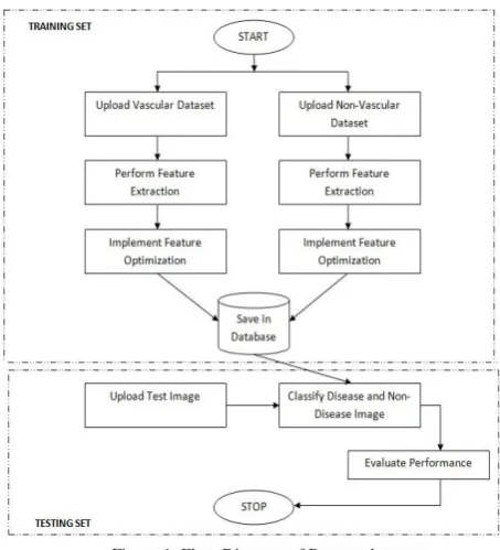
III. PROPOSED METHOD
Figure 1. Flow Diagram of Proposed system
A. A Vascular Based Feature Extraction using PSO optimization
In vascular-based feature extraction system, vessel properties of retinal image are used to perform identification. This method uses the extraction of bifurcation and ending points for recognition. Vessel properties of retinal image make better performance of retinal recognition system. So this method has an improved vessel segmentation algorithm which works on true vessels as well as it will consider pathological signs as false vessels which removes the effect of lesions to obtain good result. A vascular based feature extraction method is further divided into preprocessing, segmentation and feature points extraction phases [23].
1) Preprocessing: The Preprocessing phase is used to enhance the image by removing noise or pixel whose color is vague from background [24]. In first step green channel is taken out from fundus image because green channel displays the best vessel background contrast as compare to red and blue channels. Mathematical procedure for finding green channel is as follows in (1):
𝑔𝑔=𝐺𝐺/𝑅𝑅+𝐺𝐺+𝐵𝐵 (1)
Here g is a Green channel and R, G and B are Red, Green and Blue respectively. The green channel image is changed into gray scale image. The preprocessing of fundus image is shown in Fig. 2 (a) and (b).
Figure. 2 (a) Fundus Image of Retina (b) Preprocessed Image
2) Blood Vessel Segmentation: Blood vessel segmentation of an original image is an important step in feature extraction. The proposed method foremost enhance and sharpen the vascular pattern of retinal blood vessels using Gabor wavelets. The Gabor filter was originally proposed by Dennis Gabor and it have been broadly used in various applications of image processing, like object recognition, edge detection and texture classification. Gabor Wavelets filter out structure of retinal image at different magnitude and orientation, for every scale and orientation there is a pair of odd and even wavelets. This technique locates and segments the blood vessels of retinal image using region based edge detection
algorithm and morphological properties of true and false vessels. The thin and less visible vessels are enhanced using 2-D Gabor wavelet [25] [26]. Before extracting vessels from enhanced retinal image, the blood vessels are sharpened using sharpening filter. After preprocessing segmentation phase applied on retinal image is shown in Fig. 3 (a) and (b).
Figure 3. (a) Preprocessed Image (b) Segmented Image
3) Feature Point Extraction: In retinal recognition, performance of any system highly depends on the selected features. So a good feature extraction method is used to extract bifurcation points or minutiae points using properties of blood vessels. Crossing number technique (CN) is used commonly to detect these feature points from image. The retinal bifurcation points are distinct for each human being, so they are used for succeeding procedure of identification of person. Bifurcation points are those points where a single vessel splits into two vessels in retina image shown in fig. 4(b). Ending points are those points where these blood vessels finish or terminate [27] [28]. The extracted vascular pattern consists of blood vessels of variable thickness. In order to generate thinned image, the breadth of blood vessels are equivalent to single pixel, we apply morphological thinning operation shown in fig. 4(a) [11]. The valid feature set formation is oriented with the help of thinned image. Extracted feature points (bifurcation or ending points) are used to produce feature vectors for each retinal image which are further stored in the database to perform matching for the identification of an individual person [23].
Figure. 4 (a) Thinned Image (b) Bifurcation Points
[image:3.595.319.561.294.387.2] [image:3.595.317.561.674.755.2] [image:3.595.37.283.676.752.2]4) Matching: In our proposed system, data is divided into two different phases: training phase and testing phase. In training phase, feature vectors generated from the retinal images are stored in database after applying pre-processing; segmentation, feature extraction and feature selection methods on input image. During testing phase, a query image is also goes through the same procedure as input image. After applying all steps on query image, a feature vector is generated and then this feature vector is compared with the feature vectors stored in database. Matching score attained from the image find out the individual identity [23] [29].
B. A Non-Vascular Based Feature Extraction using PSO Optimization
Non-vascular based feature extraction method is known as a minutia based retinal recognition in which non-vessel properties of retinal images are evaluated. In this method only structural features are extracted from retinal images instead of minutiae point extraction. As vessel segmentation and bifurcation point extraction done in our previous method is a very time consuming process. So, this method decreases the overall execution time of system while maintaining its good performance. In this method, an original color retinal image is taken as input image and structural features like luminance, contrast and structure are extracted from the given input image and combine these extracted features by using an empirically optimized function to generate a similarity score from two different candidate images at same time and on the base of highest score value matching result is taken. This method works into two phases: Feature Extraction and Matching [22] [29].
1) Feature Extraction: In non-vascular based feature extraction method, recognition is done using structural features of retinal image instead of extracting minutiae points. In this method, structural features like luminance, contrast and structure are assembled into three different categories. An original color retinal image is taken as input and query image also features are extracted from both the images at same time. This process decreases the overall execution time of the system. Values generated after performing feature extraction process is known as feature vector having three different lengths for luminance, contrast and structure features and these features vectors are further stored in database for next phase called matching.
2) Matching: After extracting features from input and query images, three structural features are taken from these two retinal images. These three features are then combined using a function named as an empirically optimized function. In matching phase, to attain a similarity score in the form of 0 and 1, these all extracted features are joined into a single unit. For matching, in each comparison similarity score is calculated and true identification is done on the basis highest score matching value.
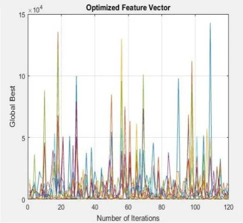
C. Feature Selection using PSO-SVM
After performing feature extraction in both the methods of retinal recognition system, some irrelevant and noisy
features are found during extraction process. To improve the accuracy in recognition method these useless features have to be removed and optimal features have to be chosen for better recognition or accuracy. So the main purpose of feature selection phase is to decrease the noisy data and eliminate the inappropriate features. Particle Swarm Optimization (PSO) is an optimization technique which is used to locate optimal characteristics from all the extracted features with local and global searches in an iterative way from the feature search space.
PSO is a computational intelligence oriented, stochastic and population based scheme used for optimization presented by Dr. Eberhart and Dr. Kennedy in 1995 [30] [31]. PSO is formed by flash of birds herd and group of fishes. This swarm consists a group of random particles are called potential solutions used to solve the optimization problems. Every particle makes use of its own individual memory and information achieved by the flock as a whole to get the best solution. All particles have a location, speed, and a memory to keep its best location from the beginning of the procedure. During movement, each particle changes its place according to its personal knowledge and the experience of a nearest particle, and uses the best position encountered by itself and its neighbor. A track of all coordinates is maintained by each particle in the problem space related with the best solution and this value is known as
In PSO, computation time is much less because all the particles in PSO tend to unite to the best solution rapidly. In order to perform classification, support vector machine (SVM) classifier is used after selecting features from retinal image with the help of optimization technique. This classification procedure is applied to classify vessels of retinal image as true and false vessels and then all false vessels will be removed after keeping only true vessels to increase the recognition rate and accuracy.
[image:4.595.317.560.549.771.2]pbest. An additional lbest value is tracked by the element swarm optimizer is obtained by any particle in the neighbors of the particle. The global best value (gbest) is tracked while a particle takes all the population as its topological neighbors. The global best particle found along with the swarm is the only information shared among particles is shown in Fig. 5 [32][33].
Particle swarm optimization is useful for a lot of engineering problems for its exclusive searching method, easy concept, computational effectiveness and easy implementation. Another reason is that in PSO very few parameters need to be adjusted. Due to this, PSO has been widely used in a variety of applications.
IV. EXPERIMENTAL RESULTS
A. Databases and Performance measures
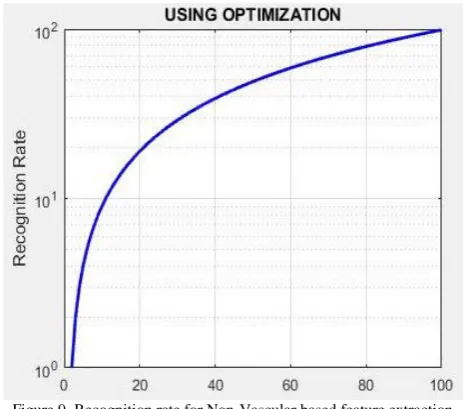
The performance of the proposed system is evaluated using two locally captured databases; RIDB and DRIDB. RIDB is a Retinal Identification Database having 40 images of healthy person in JPEG format with 784x590 resolutions and DRIDB is a Diseased Retinal Identification Database contains 40 images of diseased person with 565x584 resolutions and in TIFF format. To check the authority of person identification, the system is tested on both databases. Dataset for both databases is divided into training and testing set. In our system, feature vectors generated after performing various operations like preprocessing, segmentation feature extraction and feature selection of all training data are stored in the database. In testing dataset, query image is also processed in the same way and feature vector created from query image is compared with the ones stored in database. Finally, recognition rate (RR) for both databases is calculated using (2)
RR= Number of images matched / Total number of images (2)
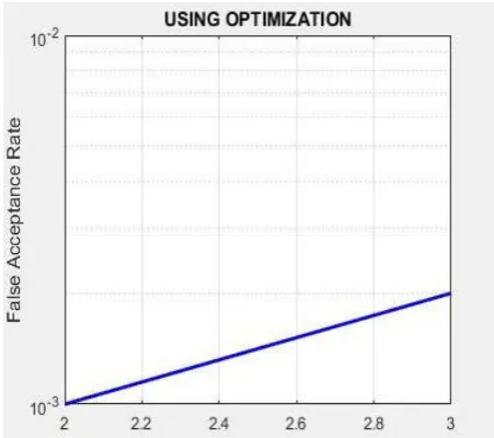
Validity or strength of our proposed system is measured by calculating two following performance parameters: False Acceptance Rate (FAR) and False Rejection Rate (FRR) shown in (3) and (4).
FAR= Total false acceptances/ total identification attempts (3)
[image:5.595.324.554.55.258.2]FRR= Total false rejections/ total identification attempts (4)
[image:5.595.324.551.280.480.2]Figure 6. Recognition rate for Vascular based feature extraction
[image:5.595.321.553.504.708.2]Figure 7. FAR curve for Vascular based feature extraction method
Figure 8. FRR curve for Vascular based feature extraction method
[image:5.595.42.276.536.730.2]Figure 10. FAR curve for Non-Vascular based feature extraction method Figure 11. FRR curve for Non-Vascular based feature extraction method
Table I. Evaluation Result of Proposed System
Methods Database Recognition Rate (RR)
False Acceptance Rate (FAR)
False Rejection Rate (FRR)
Vascular Based Feature
Extraction Method DRIDB RIDB 99.95 99.99 0.0040 0.0168 0.0002 0.0008
Non-Vascular Based Feature Extraction
Method
RIDB DRIDB
99.96 99.98
0.0366 0.0129
0.0008 0.0002
V. CONCLUSION
Retinal based recognition and identification system is the most reliable and accurate biometric system. In this paper, we present two different retinal recognition methods to identify the identity of a particular person using retinal image. Firstly, in vascular based method bifurcation points are extracted from vascular pattern of retinal image and in second method structural features like luminance, contrast and structure are extracted from the color retinal image. In our proposed system, a Particle Swarm Optimization based feature selection algorithm is efficiently used for both these retinal recognition methods. Our experiments performed on training and testing module aimed to increase the recognition rate for both methods. The utmost success of 99.99 % accuracy has been achieved using PSO for diseased and non- diseased images in both methods.
VI. REFERENCES
[1] N. K. Sandhu and R. Kaur, “Biometric Security Technique: A Review,” Indian Journal of Science and Technology, Vol 9(47), December 2016.
[2] Duarte, Tiago and joao Paulo Pimentao, Pedro Sousa and Sergio Onofre, “Biometric access control systems: A review on technologies to improve their efficiency,” In IEEE, 2016. [3] Kuppusamy, P and Divya, B, “A Survey of retina based
disease identification using Blood vessel segmentation,” In ICTACT Journal on Image & Video Processing, 2016. [4] Wafa Barkhoda1, Fardin Akhlaqian, Mehran Deljavan Amiri
and Mohammad Sadeq Nouroozzadeh, “Retina identification based on the pattern of blood vessels using fuzzy logic,” EURASIP Journal on Advances in Signal Processing, 2011.
[5] Hannu Oinonen, Heikki Forsvik, Pekka Ruusuvuori, Olli Yli-Harja, Ville Voipio and Heikki Huttunen, “IDENTITY VERIFICATION BASED ON VESSEL MATCHING FROM FUNDUS IMAGES,” Proceedings of 2010 IEEE 17th International Conference on Image Processing, September 26-29, 2010.
[6] Sana Qamber, Zahra Waheed, and M. Usman Akram, “Personal Identification System Based on Vascular Pattern of Human Retinal,” In 6th
[7] Zahra Waheed, M. Usman Akram, Amna Waheed, Muazzam A. Khan, Arslan Shaukat, Mazhar Ishaq, “Person identification using vascular and non-vascular retinal features,” In Elsevier, 2016.
Cairo International Biomedical Engineering Conference, 2012.
[8] Hichem Betaouaf, Abdelhafid Bessaid, “A Biometric Identification algorithm based on retinal blood vessels segmentation using watershed transformation,” 2013 IEEE. [9] Abu Hasnat, Mohammad Rubaiyat, Shubhra Aich, Tanjin
Taher Toma, Abidur Rahman Mallik and Rafat-Al-Islam, “Fast Normalized Cross-Correlation Based Retinal Recognition,” In 17th
[10]Sahinaz Safari Sanjani, Jean-Baptiste Boin, Karianne Bergen, “Blood Vessel Segmentation in Retinal Fundus Images”.
International conference on Computer and Information Technology (ICCIT) 2014.
[11]Manjiri B. Patwari, Ramesh R. Manza, Yogesh M. Rajput, Manoj Saswade, Neha Deshpande, “Personal Identification algorithm based on Retinal Blood Vessels Bifurcation,” In 2014, International conference on Intelligent Computing Applications.
[12]Bevilacqua S. Cambò L. Cariello G. Mastronardi, “A Combined method to detect retinal fundus features,”
[13]R.S Choras, “Retinal Recognition for biometrics,” in ICDIM, 2012.
[image:6.595.326.551.54.255.2][14]L.Latha, M.Pabitha and S.Thangasamy, “A Novel Method for Person Authentication using Retinal Images,” In International Conference on Innovative Computing Technologies, (ICICT) 2010, Tamil Nadu.
[15]Muhammad Nazrul Islam, Md. Amran Siddiqui and Samiron Paul, “An Efficient Retina Pattern Recognition Algorithm (RPRA) towards Human Identification,” 2nd
[16]Shuangling Wang, YilongYin, GuibaoCao, BenzhengWei, YuanjieZheng, Gongping Yang, “Hierarchical retinal blood vessel segmentation based on feature and ensemble learning,” In Elsevier 2014.
international conference on Computer Control and Communication 2009.
[17]Elaheh Imani, Malihe Javidi and Hamid-Reza Pourreza, “Improvement of Retinal Blood Vessel Detection Using Morphological Component Analysis,” In Computer Methods and Programs in Biomedicine, Vol.118, Issue 3, March 2015. [18]Seyed Mehdi Lajevardi, Arathi Arakala, Stephen A. Davis,
and Kathy J. Horadam, “Retina Verification System Based on Biometric Graph Matching,” In IEEE Transaction on Image processing, September 2013.
[19]Rita A. Vora, Dr. V A Bharadi, Dr. H B Kekre, “Retinal Scan Recognition using Wavelet Energy Entropy,” In International conference in 2012 on Communication, Information & Computing Technology (ICCICT).
[20]Zahedi A , Sadjedi H , Behrad A ., “A new retinal image processing for human identification using radon transform,” In: 6th Iranian conference on machine vision and image processing (MVIP), Isfahan; Oct 2010.
[21] Zahedi A , Sadjedi H ., “A human identification system based on retinal image, processing using partitioned fourier spectrum,” In: International conference on audio language and image processing (ICALIP), Shangai; Nov 2010 .
[22]Waheed, Zahra and Waheed, Amna and Akram, M Usman, “A robust non-vascular retina recognition system using structural features of retinal image,” In Applied Sciences and Technology (IBCAST), 13th International Bhurban Conference on IEEE, 2016.
[23]Zahra Waheed, M. Usman Akram, Amna Waheed, Arslan Shaukat, “Robust Extraction of Blood Vessels for Retinal Recognition,” 2015 IEEE.
[24] V. P. Patil , P. R. Wankhede,” Pre-Processing Steps for Segmentation of Retinal Blood Vessels,” International Journal of Computer Applications, May 2014.
[25]Mohamed A. El-Sayed, M. Hassaballah, Mohammed A. Abdel-Latif, “Identity Verification of Individuals Based on Retinal Features Using Gabor Filters and SVM,” Journal of Signal and Information Processing, 2016.
[26]Shuangling Wang, YilongYin, GuibaoCao, BenzhengWei, YuanjieZheng, Gongping Yang, “Hierarchical retinal blood vessel segmentation based on feature and ensemble learning,” In Elsevier 2014.
[27]Joddat Fatima, Adeel M. Syed and M. Usman Akram, “A Secure Personal Identification System Based on Human Retina,” 2013 IEEE Symposium on Industrial Electronics & Applications (ISIEA2013), September 22-25, 2013.
[28]Ryszard S. Chora, “Retina recognition for biometrics,” In ICDIM 2012, IEEE.
[29]Zahra Waheed, M. Usman Akram, Amna Waheed, Muazzam A. Khan, Arslan Shaukat, Mazhar Ishaq, “Person identification using vascular and non-vascular retinal features,” In Elsevier, 2016.
[30]Yudong Zhang, Shuihua Wang, and Genlin Ji, “A Comprehensive Survey on Particle Swarm Optimization Algorithm and Its Applications,” Hindawi Publishing Corporation Mathematical Problems in Engineering Volume 2015,
[31]Rabab M. Ramadan and Rehab F. Abdel – Kader, “Face Recognition Using Particle Swarm Optimization-Based Selected Features,” International Journal of Signal Processing, Image Processing and Pattern Recognition Vol. 2, No. 2, June 2009
[32]Chung-Jui Tu, Li-Yeh Chuang, Jun-Yang Chang, and Cheng-Hong Yang, “Feature Selection using PSO-SVM,” IAENG International Journal of Computer Science, 33:1, IJCS. [33]A. Zabidi, L. Y. Khuan, W. Mansor, I. M. Yassin, and R.