International Journal of Emerging Technology and Advanced Engineering
Website: www.ijetae.com (
ISSN 2250-2459,
ISO 9001:2008 Certified Journal,
Volume 3, Issue 5, May 2013)
776
Wavelet and Canny Based Edge Detection Method for Noisy
Lung CT Image
Bhadauria H S
1, Singh A
2 1Electrical Engineering Depatment., IIT Roorkee, Roorkee
2Computer Engineering Department, G. B. Pant Engineering College
Abstract-The development of computer–aided diagnosis (CAD) has been extended to various medical imaging modalities including computer Tomography (CT), magnetic resonance imaging, nuclear medicine etc. Medical image edge detection is an important work for diagnosis of various lung diseases in thoracic CT images. When detecting the edge based on wavelet transform, the edge detection results are poor because of the noise influence. This paper proposed a new edge detection algorithm based on wavelet transform and canny operator. In the wavelet domain, the low-frequency edges are detected by canny operator, while the high-frequency edges are detected by solving the maximum points of local wavelet coefficient model to restore edges after reducing the noise by wavelet. Then, both sub-images edges are fused according to fusion rules. A noisy CT image of abnormal lung infected by Honeycombing is used to evaluate the performance of algorithms.
Keywords- Lung CT images, edge detection, Wavelet Transform, Canny operator
I. INTRODUCTION
Recent advances in Computed Tomography (CT) technology have enabled it to be widely used in diagnosing and quantifying different diseases [1]. Several lung diseases are diagnosed by investigating the patterns of lung tissue in pulmonary CT images, therefore texture segmentation and analysis is one of the important parts of CAD systems [5]. Edge information in thoracic CT image is an important characteristic because lung region contains an abundance of edges like vessels, air tree, air sacs, artery branches etc. and conventional edge detection algorithms are typically based on differential operators, such as the Sobel, Prewitt, and Roberts operators [6][7][8] .Traditional differential operators work well with edge detection of noiseless images, however in the presence of noise these may miss the edges or detect the false edges due to the intensity discontinuities. Medical images are noisy in nature due to limitations of imaging techniques, devices and health constraints. Therefore it may be difficult to distinguish the exact edges from the noise.
Wavelet analysis developed rapidly as a useful research method.
The traditional wavelet transform based edge detection methods perform the wavelet multi-resolution decomposition for image firstly, and then extract the low-frequency sub-image to further process, which may discard some important details and the edge extraction will be affected by lots of noise in the high-frequency sub-images [2]. This paper, proposed a new fusion algorithm based on wavelet transform and canny operator to detect image edges, which may reduce the noise and obtain the continuous and distinct edges.
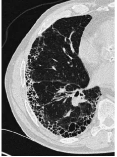
A noisy CT image of abnormal lung infected by honeycombing is taken. Honeycombing is a set of cysts filled with air, whereas the vessel branches are denser than air. In other words in a lung CT image honeycombing is seen as dark holes but vessel branches are seen as brighter area [12][13][14].
II. IMAGE DECOMPOSITION BASED ON WAVELET
TRANSFORM
Based on image decomposition model of wavelet transform, the original image can be divided into low-frequency information and high-low-frequency information. After two-dimensional frequency decomposition of wavelet transform, low-frequency information can be decomposed low-frequency area LL and frequency area LH, high-frequency information can be decomposed low-high-frequency area HL and high-frequency area HH. LL shows the smoothing image of the original image which contains the most information of the original image. LH preserves the vertical edge details. HL preserves the horizontal edge details. HH preserves the diagonal details which are influenced by noise greatly. The process is shown in Figure.1
International Journal of Emerging Technology and Advanced Engineering
Website: www.ijetae.com (
ISSN 2250-2459,
ISO 9001:2008 Certified Journal,
Volume 3, Issue 5, May 2013)
777
III. METHODOLOGY
Though the edge extracted by wavelet transform can reduce the most noise of the image, the real edges of the image are also mixed with much noise, especially HH areas are affected by the noise greatly. Thus many methods of edge detection will discard these HH areas [9]. The four areas obtained from two-dimensional decomposition of wavelet transform contain the useful information of the original image, so these areas should be used completely when detecting the image edges. This paper presents a new fusion algorithm based on wavelet transform and canny operator to detect image edges, which can reduce the noise and obtain the continuous and distinct edges.
3.1 Low-frequency sub-image edge detection based on canny operator
The LL area shows the smooth image of the original image which contains the most information of the original image. Canny algorithm is an optimal edge detection method based on a specific mathematical model for edges. The Canny edge detector was devised to be an optimal edge detector, which minimize the situations of detecting false edges and missing actual edges, minimize the distance between the detected edges and actual edges and minimize multiple responses to an actual edge, i.e. to ensure there is only one response for an actual edge point. So canny operator [7,10] are adopted to detect the edges of the low-frequency sub-image.
Through adopting the canny operator to detect the low-frequency sub-image can obtain clear edge image which will miss some real edges and. Thus the edges of the high-frequency sub-images should be fused.
3.2 High-frequency sub-image edges detection based on wavelet transform
After two-dimensional frequency decomposition of wavelet transform, there will be three high-frequency sub-images LH、HL and HH which contain the noise and the details of the image. When extracting the edges from the high-frequency sub-images, the result will be affected because of much noise. Thus, noise should be reduced before extracting the edges from the high-frequency sub-images. The algorithm can not only keep the important details of the high-frequency sub-images but also remove the noise.
3.3 Denoising algorithm of the high-frequency sub-images based on wavelet transform
Among the wavelet coefficients of the high-frequency sub-image, the wavelet coefficients which have smaller amplitude present the most noise part, and the wavelet coefficients which have larger amplitude present the details of the image [2,3]. Based on the characteristic of the high-frequency sub-images, we proposed a method based on wavelet transform to reduce the noise in the high-frequency sub-images. The wavelet coefficients are multiplied by a denoising factor which is relative to their own coefficients’ value. This denoising factor is less than 1 and will decrease if the absolute value of the wavelet coefficients increases. This algorithm considers energy distribution property of the wavelet decomposition of the image globally, and obtains a better denoising result, which support the foundation for extracting the edge of the high-frequency sub-images.
f(x,y) = (3)
w(x,y) denotes the high frequency coefficients, f(x,y) denotes coefficients after denoising ,σ and η are the variance and mean value of the high frequency coefficients in different wavelet decomposition levels. k is a function given as
k = -1 (4)
When w(x,y) 3σ, w(x,y) is considered as the signal. Therefore k=1
-1=1 (5) When w(x,y) , k=0, Therefore for k=0
-1=0 (6) From the above two equations we found
a= and b = aη (7)
After substituting a and b to equation 4 we obtain the value of k as
International Journal of Emerging Technology and Advanced Engineering
Website: www.ijetae.com (
ISSN 2250-2459,
ISO 9001:2008 Certified Journal,
Volume 3, Issue 5, May 2013)
778
Denoising algorithm will be computed when k substituted into equation (3). This algorithm is aiming for the wavelet coefficients multiplied by different denoising factors from different wavelet decomposition levels and different high-frequency sub-image, which can reduce the image noise and keep useful details
3.4 Edge detection of denoised high-frequency sub-image based on wavelet transform
After eliminating the noise from the high-frequency sub-image, the edges of the high-frequency sub-image can be detected using wavelet modulus maxima algorithm [11]. The wavelet modulus maxima algorithm is obtained from an irregular sampling based on multiscale wavelet transform which can describe mechanism singularity of the signal. The wavelet modulus maxima algorithm can describe multiscale edges of the target in the image which has translation, scale and rotation-invariant performance. Thus the wavelet modulus maxima algorithm is an effective algorithm to detect the edges.
After these processes, we can get the edges of the low-frequency sub-image and the weighting edges of the high-frequency sub-images. The final edge images are obtained through wavelet composition from the fusion edge sub-images [15].
IV. EXPERIMENTS RESULTS
This section presents the performance evaluation of proposed method on lung CT image having simulated additive Gaussian noise of standard deviation 10 and 20. As noise suppression and edge preservation are conflicting objectives, thus simultaneous comparison of these objectives becomes necessary. To analyze the denoising capabilities of proposed method on lung CT image, two different quantitative measures are used. These are (i)- the measure of the noise suppression by using Signal-to-Noise-Ratio (SNR), Peak-Signal-to-Noise-Signal-to-Noise-Ratio (PSNR) and Universal Quality Index (UQI), and (ii)- the measure of the edge or fine details preservation by using Edge Keeping Index (EKI). In this section in order to verify the efficiency and accuracy of the proposed algorithm, the test has been applied on abnormal lung CT image as shown in figure 1.
The abnormal lung is infected by honeycombing which is seen as dark holes but vessel branches are seen as brighter area in CT image. Figure 2 is the original image with additive Gaussian noise with zero mean and σ= 10.
In this experiment we also compare the proposed approach with wavelet algorithm and LOG operator. Figure 3 shows the comparison between operators on Gaussian noised lung CT image.
The quantitative evaluation of these methods on lung CT image for noise suppression and edge preservation has been carried out and are presented for comparison in Table 1. The Table 1 provides the values of different performance indices like SNR, PSNR and UQI for noise suppression and EKI for edge preservation with two different noise levels (σ=10 and 20). The value of GAE for all the denoising methods is found to be 0.00, which shows the fact that the information contents between the original and processed images remain same. Further from Table 1, it is observed that the proposed method yields the higher values of SNR, PSNR, UQI and EKI as 29.36, 40.05, 0.2238, and 0.9271 respectively for σ=10, and 25.56, 36.44, 0.1853, and 0.8565 respectively for σ=20 and as compared to other denoising methods. This is an evidence of the maximum noise suppression with significant edges and fine details preservation.
V. CONCLUSION
Noise removal is an important pre-processing step in most of the image processing applications especially when any relevant information could be extracted from the images.
International Journal of Emerging Technology and Advanced Engineering
Website: www.ijetae.com (
ISSN 2250-2459,
ISO 9001:2008 Certified Journal,
Volume 3, Issue 5, May 2013)
[image:4.612.151.267.137.292.2]779
Figure.1 Original image Figure.2 Noisy image
[image:4.612.120.497.343.511.2]
(a) LOG (b) Wavelet algorithm (c) Proposed algorithm
[image:4.612.153.465.557.713.2]Figure.3 Comparison between edge detection operators with Gaussian noise (σ=10)
Table 1
Quality assessment parameters on lung CT image
Std. dev.
Assessment
Parameters LOG Wavelet Proposed
10
GAE 0.00 0.00 0.00
SNR (db) 27.19 22.07 29.36
PSNR (db) 37.87 32.75 40.05
UQI 0.1848 0.2110 0.2238
EKI 0.9165 0.6112 0.9271
20
GAE 0.00 0.00 0.00
SNR (db) 23.01 18.92 25.56
PSNR (db) 35.12 29.60 36.44
UQI 0.1746 0.1616 0.1853
EKI 0.8487 0.4478 0.8565
International Journal of Emerging Technology and Advanced Engineering
Website: www.ijetae.com (
ISSN 2250-2459,
ISO 9001:2008 Certified Journal,
Volume 3, Issue 5, May 2013)
780
REFERENCES
[1] Sluimer IC, van Waes PF, Viergever MA, et al: Computer-aided diagnosis in high resolution CT of the lungs. Med Phys 30:3081-3090,2003
[2] MALLAT S, HW ANG W L. ―Singularity Detection and Processing with Wavelets‖. IEEE Trans 2002, IT-38(2):617-643.
[3] MALLAT S. ―Multi-frequency Channel Decomposition of Images and Wavelet Models‖. IEEE Trans 1998, AS-SP-37(12) :2091-2110 [4] Roushdy M. ―Comparative Study of Edge Detection Algorithms
Applying on the Grayscale Noisy Image Using Morphological Filter‖ GVIPJournal, Volume 6, Issue 4, pp 18-23,December, 2006
[5] R. Uppaluri, E.A. Hoffman, M. Sonka, P.G. Hartley, G.W Hunninghake, and G. McLennan, ―Computer recognition of regional lung disease patterns,‖ American Journal of Respiratory and Critical Care Medicine, vol. 160, no. 2, pp. 648–654, August 1999
[6] Huertas, A. an9d Medioni, G., ―Detection of intensity changes with sub pixel accuracy using Laplacian-Gaussian masks,‖ IEEE Trans. On Pattern Analysis and Machine Intelligence, PAMI, vol. 8, pp.651–664, 1986.
[7] Canny, J., "A Computational Approach to Edge Detector", IEEE Transactions on PAMI, pp679-698, 1986
[8] Maini R, Aggrawal H.‖ Study and Comparison of Various Image Edge Detection Techniques‖ International Journal of Image Processing (IJIP), Volume (3) : Issue (1).
[9] Musheng-Chen. ―A New Method of Image Denoising Based on Wavelet Transform‖. Optical Technique, 2006, vol.32(5):796-798. [10] Yanlong-Chen, Chenghu-Zhu. ―Improved Edge Detection Algorithm
Based on Canny Operator‖. Computer Applications and Software. 2008, vol.25(8):51-53.
[11] Zhu-lihua, Ji-xiaoping. ―The application of wavelet modulus maxima algorithm in the image retrieval‖. SCI-tech information development and economy. 2007, vol.17(3): 169-170
[12] Webb WR (1989) High-resolution CT of the lung parenchyma. Radiol Clin North Am 27:1085–1097
[13] Webb WR, Stein MG, Finkbeiner WE et al (1988) Normal and diseased isolated lungs: high-resolution CT. Radiology 166:81–87
[14] Zerhouni E (1989) Computed tomography of the pulmonary parenchyma: an overview. Chest 95:901–907