Journal of Chemical and Pharmaceutical Research, 2017, 9(6):120-126
Research Article
CODEN(USA) : JCPRC5
ISSN : 0975-7384
120
Ab Initio and DFT Study of Uracil-Water Complexes
Neha Shukla, Brijesh Kumar Sharma, Achchhe Lal and Devendra K Singh
*Department of Physics, Udai Pratap (Autonomous) College, Varanasi,Uttar Pradesh, India _____________________________________________________________________________
ABSTRACT
Uracil is a naturally occurring pyrimidine derivative and is one of the four nitrogenous bases found in RNA. Hydrogen bond plays a significant role in the structure determination of nucleic acids. Various types of hydrogen bond formed by purine and pyrimidine bases have been investigated and are of great significance. The effect of hydrogen bond on various Uracil-Water complexes has been investigated. In this paper four cyclic isomers of uracil forming hydrogen bonds with the water molecule have been discussed. Optimized geometries of all the isomers of uracil-water complex have been obtained at MP2/6-311++G (d,p), B3LYP/6-311++G (d,p), B3LYP/Aug-CC-pVDZ levels for the first time upto this level. Structural parameters of the optimized geometries, total energies and the APT charges of uracil-water complex have been computed. We show that addition of water molecules in uracil, the strength of the binding energy decreases i.e. stability increases. The optimized bond length and bond angles are in agreement with the corresponding experimental results.
Keywords: Uracil-water complex; Ab-initio and DFT calculations; Molecular structure; Optimized geometry _____________________________________________________________________________
INTRODUCTION
The overall structure of RNA (ribonucleic acid) consists of four nitrogenous bases adenine (A), cytosine (C), guanine (G), and uracil (U). These bases compose the backbone of their strands. In RNA, uracil binds to adenine via two hydrogen bonds. Uracil is a common and naturally occurring pyrimidine.
121
Four stable structure for uracil-water complex has been found in this work. The four isomers of uracil-water reported here have been reported earlier [2,8,10]. Optimized geometries of all the four isomers of uracil-water complex have been obtained at MP2/6-311++(d,p), B3LYP/6-311++ G (d,p), B3LYP/Aug-CC-pVDZ levels. Structural parameters of the optimized geometries, total energies and the APT charges of uracil-water complex have been computed. We show that addition of water molecules in uracil, the strength of the binding energy decreases i.e. stability increases. The optimized bond length and bond angles are in agreement with the corresponding experimental results.
COMPUTATIONAL METHODOLOGY
The ground state geometries and vibrational spectra for free Uracil and its hydrogen-bonded complexes with molecules of water have been optimized. The total energies, structural parameters of the optimized geometries and the APT charges of have been computed using ab initio method:-(i) MP2 [11]and hybrid density functional theory (DFT) methods- (ii) B3LYP which uses Becke’s three-parameter functional [12-14] with nonlocal correlation provided by Lee–Young–Parr expression [15] with 6-311++ G (d,p) and Aug-CC-pVDZ basic sets. For theoretical study, we have used GAUSSIAN 09 [16] package of programs without any constraint on the geometry.
Initially Ab initio calculations were done using MP2/6-311++G (d,p) level. The optimized geometry at the
MP2/6-311++ G (d,p) level was taken as the input structure for the DFT calculation using B3LYP/6-MP2/6-311++ G (d,p) level. Similarly, the optimized geometry at the B3LYP/6-311++ G (d, p) level was used as the input structure for the calculation at the B3LYP/ Aug-CC-pVDZ level. The geometries were optimized by minimizing the energies without imposing any constraint on the geometry. It has well known that this level of theory is sufficient to reliably predict molecular geometries hydrogen bonded systems.
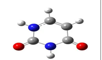
[image:2.612.224.391.375.474.2]The geometry of the free uracil and uracil-water complex have been fully optimized by ab-initio using MP2/6-311++G (d,p) and the density functional theory using B3LYP/6-311++ G (d,p), B3LYP/Aug- CC-pVDZ levels for the first time. The optimized structure of free uracil and four isomers at B3LYP/6-311++ G (d,p) along with atomic numbering have been shown in Figures 1 and 2 (a-d) respectively.
Figure 1: Optimized structure of free Uracil at B3LYP/6-311++G (d,p)
[image:2.612.202.410.493.680.2]122
RESULT AND DISCUSSIONS
Total energies (Hartree) for free Uracil and Uracil-Water complexes
[image:3.612.148.465.209.274.2]Table 1 shows energies of free uracil and uracil-water complexes. From Table 1 we see that stability of uracil molecule increases when we use DFT method B3LYP than the ab-initio method MP2. Also in four isomers of Uracil-Water complexes, the isomer 1 is most stable having least optimized energy while isomers 2 and 3 are always higher in energies and very similar energies. Isomer 4 has higher energy to all isomers as weak hydrogen bond is involved in this isomer. We also see that as uracil-water complex is more stable than the free uracil molecule.
Table 1: Total energies for free Uracil and Uracil-Water complexes
Complex MP2/6-311++G (d,p) B3LYP/6-311++G (d,p) B3LYP/Aug-CC-pVDZ
Uracil -413.849589 -414.9461817 -414.8842574
Isomer 1 -490.1425106 -491.4215643 -491.3447523
Isomer 2 -490.1393317 -491.4180688 -491.3413123
Isomer 3 -490.1399392 -491.4191045 -491.3424643
Isomer 4 -490.1369726 -491.4160114 -491.3398372
Structural parameters
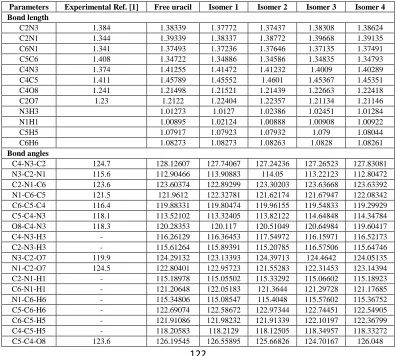
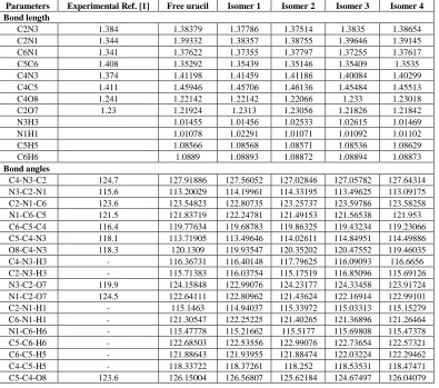
[image:3.612.108.505.359.715.2]The optimized geometrical parameters namely bond lengths (in Å) and bond angles (in Degree) of the uracil and uracil-water complexes computed at the B3LYP/ 6-311++G (d,p) and B3LYP/Aug-CC-pVDZ levels are collected in Tables 2 and 3.
Table 2: Optimized Bond lengths (Å) and Bond angles (Degree) of free Uracil and Uracil-Water complexes at B3LYP/ 6-311++ G (d,p) level
Parameters Experimental Ref. [1] Free uracil Isomer 1 Isomer 2 Isomer 3 Isomer 4 Bond length
C2N3 1.384 1.38339 1.37772 1.37437 1.38308 1.38624
C2N1 1.344 1.39339 1.38337 1.38772 1.39668 1.39135
C6N1 1.341 1.37493 1.37236 1.37646 1.37135 1.37491
C5C6 1.408 1.34722 1.34886 1.34586 1.34835 1.34793
C4N3 1.374 1.41255 1.41472 1.41232 1.4009 1.40289
C4C5 1.411 1.45789 1.45552 1.4601 1.45367 1.45351
C4O8 1.241 1.21498 1.21521 1.21439 1.22663 1.22418
C2O7 1.23 1.2122 1.22404 1.22357 1.21134 1.21146
N3H3 1.01273 1.0127 1.02386 1.02451 1.01284
N1H1 1.00895 1.02124 1.00888 1.00908 1.00922
C5H5 1.07917 1.07923 1.07932 1.079 1.08044
C6H6 1.08273 1.08273 1.08263 1.0828 1.08261
Bond angles
C4-N3-C2 124.7 128.12607 127.74067 127.24236 127.26523 127.83081
N3-C2-N1 115.6 112.90466 113.90883 114.05 113.22123 112.80472
C2-N1-C6 123.6 123.60374 122.89299 123.30203 123.63668 123.63392
N1-C6-C5 121.5 121.9612 122.32781 121.62174 121.67947 122.08342
C6-C5-C4 116.4 119.88331 119.80474 119.96155 119.54833 119.29929
C5-C4-N3 118.1 113.52102 113.32405 113.82122 114.64848 114.34784
O8-C4-N3 118.3 120.28353 120.117 120.51049 120.64984 119.60417
C4-N3-H3 - 116.26129 116.36453 117.54972 116.15971 116.52173
C2-N3-H3 - 115.61264 115.89391 115.20785 116.57506 115.64746
N3-C2-O7 119.9 124.29132 123.13393 124.39713 124.4642 124.05135
N1-C2-O7 124.5 122.80401 122.95723 121.55283 122.31453 123.14394
C2-N1-H1 - 115.18978 115.05502 115.33292 115.06602 115.18923
C6-N1-H1 - 121.20648 122.05183 121.3644 121.29728 121.17685
N1-C6-H6 - 115.34806 115.08547 115.4048 115.57602 115.36752
C5-C6-H6 - 122.69074 122.58672 122.97344 122.74451 122.54905
C6-C5-H5 - 121.91086 121.98232 121.91339 122.10197 122.36799
C4-C5-H5 - 118.20583 118.2129 118.12505 118.34957 118.33272
123
Also the Optimized bond lengths (in Å) and bond angles (in Degree) of free uracil and Isomer 1 (most stable isomer) of uracil-water complexes at MP2/6-311++ G (d,p) level are collected in Table 4. For atomic numbering scheme, see Figures 1 and 2(a-d). The magnitudes of the bond lengths between the pairs (C2=O7) and (C4=O8) in uracil are found
to be nearly equal and the slightly difference is due to the higher electro-negativity of the O7 atom which attracts
more the C atom than the O8 atom in their respective bonds with the resulting a little smaller bond length than other
bond in the all molecules. Here, it is noticeable that the N1–C6 bond is found to be the smallest bond due the
formation of the lone pair bond between N1 and C6 atoms as shown in Figure 2a uracil as accordance to the uracil
[image:4.612.112.507.218.567.2]molecule due to one with single bonding and another localized double bond respectively. The bond lengths and bond angles obtained theoretically are in better agreement to their corresponding experimental values [1].
Table 3: Optimized Bond lengths (Å) and Bond angles (Degree) of free Uracil and Uracil-Water complexes at B3LYP/ Aug-CC-pVDZ level
Parameters Experimental Ref. [1] Free uracil Isomer 1 Isomer 2 Isomer 3 Isomer 4 Bond length
C2N3 1.384 1.38379 1.37786 1.37514 1.3835 1.38654
C2N1 1.344 1.39332 1.38357 1.38755 1.39646 1.39145
C6N1 1.341 1.37622 1.37355 1.37797 1.37255 1.37617
C5C6 1.408 1.35292 1.35439 1.35146 1.35409 1.3535
C4N3 1.374 1.41198 1.41459 1.41186 1.40084 1.40299
C4C5 1.411 1.45946 1.45706 1.46136 1.45484 1.45513
C4O8 1.241 1.22142 1.22142 1.22066 1.233 1.23018
C2O7 1.23 1.21924 1.2313 1.23056 1.21826 1.21842
N3H3 1.01455 1.01456 1.02533 1.02615 1.01469
N1H1 1.01078 1.02291 1.01071 1.01092 1.01102
C5H5 1.08566 1.08568 1.08571 1.08536 1.08629
C6H6 1.0889 1.08893 1.08872 1.08894 1.08873
Bond angles
C4-N3-C2 124.7 127.91886 127.56052 127.02846 127.05782 127.64314
N3-C2-N1 115.6 113.20029 114.19961 114.33195 113.49625 113.09175
C2-N1-C6 123.6 123.54823 122.80735 123.25737 123.59786 123.58258
N1-C6-C5 121.5 121.83719 122.24781 121.49153 121.56538 121.953
C6-C5-C4 116.4 119.77634 119.68783 119.86325 119.43234 119.23066
C5-C4-N3 118.1 113.71905 113.49646 114.02611 114.84951 114.49886
O8-C4-N3 118.3 120.1309 119.93547 120.35202 120.47552 119.46035
C4-N3-H3 - 116.36731 116.40148 117.79625 116.09093 116.6656
C2-N3-H3 - 115.71383 116.03754 115.17519 116.85096 115.69126
N3-C2-O7 119.9 124.15848 122.99076 124.23177 124.33458 123.91724
N1-C2-O7 124.5 122.64111 122.80962 121.43624 122.16914 122.99101
C2-N1-H1 - 115.1463 114.94037 115.33972 115.03313 115.15279
C6-N1-H1 - 121.30547 122.25225 121.40265 121.36896 121.26464
N1-C6-H6 - 115.47778 115.21662 115.5177 115.69808 115.47378
C5-C6-H6 - 122.68503 122.53556 122.99076 122.73654 122.57321
C6-C5-H5 - 121.88643 121.93955 121.88474 122.03224 122.29462
C4-C5-H5 - 118.33722 118.37261 118.252 118.53531 118.47471
C5-C4-O8 123.6 126.15004 126.56807 125.62184 124.67497 126.04079
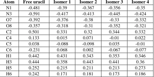
Atomic Polar Tensor (APT) charges
The atomic polar tensor (APT) charges for the uracil and uracil-water complexes computed at the B3LYP/ 6-311++ G (d,p) and B3LYP/Aug-CC-pVDZ levels are collected in Tables 5 and 6. Also the APT charges for free uracil and Isomer 1 (most stable isomer) of uracil-water complexes at MP2/6-311++ G (d,p) level are collected in Table 7. For atomic numbering scheme, see Figures 1 and 2(a-d) respectively. In term of the electron charge 1e =1.602188x10⁻¹⁹ C. From Tables 5-7, we see that due to high negativity of the respective atoms compared to the other atoms results enhancement of the bond length. In free uracil we see that the bond length of the magnitudes of the bond lengths between the pairs (C2=O7) and (C4=O8) are found to be differed due as O7 is more negative than the O8. Thus O7
attracts more to C atom than O8 which gives difference in their bond lengths. Hence the bond length of (C2=O7) is
shorter than the bond length of (C4=O8). Similarly, with the help of APT charges we can explain the difference in
124
Table 4: Optimized Bond lengths(Å) and Bond angles (Degree) of free Uracil and Isomer 1 (most stable isomer) of Uracil-Water complexes at MP2/6-311++ G (d,p) level
Parameters Experimental Ref. [1] Free uracil Isomer 1 Bond length
C2N3 1.384 1.38596 1.38107
C2N1 1.344 1.39037 1.382
C6N1 1.341 1.37657 1.3748
C5C6 1.408 1.35421 1.35569
C4N3 1.374 1.40878 1.41191
C4C5 1.411 1.46014 1.45757
C4O8 1.241 1.22016 1.22027
C2O7 1.23 1.21643 1.22658
N3H3 1.01447 1.01461
N1H1 1.0102 1.02044
C5H5 1.08144 1.08152
C6H6 1.08483 1.08492
Bond angles
C4-N3-C2 124.7 128.66806 128.04385
N3-C2-N1 115.6 112.64157 113.54913
C2-N1-C6 123.6 123.77855 122.84186
N1-C6-C5 121.5 121.82799 122.23064
C6-C5-C4 116.4 119.79445 119.70211
C5-C4-N3 118.1 113.28939 113.03578
O8-C4-N3 118.3 120.61166 120.41584
C4-N3-H3 - 116.13808 115.97318
C2-N3-H3 - 115.19386 115.20727
N3-C2-O7 119.9 124.16814 123.07981
N1-C2-O7 124.5 123.19028 123.3458
C2-N1-H1 - 115.15105 114.93917
C6-N1-H1 - 121.0704 121.57557
N1-C6-H6 - 115.44958 115.17097
C5-C6-H6 - 122.72243 122.57592
C6-C5-H5 - 121.63621 121.62585
C4-C5-H5 - 118.56934 118.60949
[image:5.612.175.440.511.645.2]C5-C4-O8 123.6 126.09895 126.54145
Table 5: APT Charges at various atomic sites of free Uracil molecule and Uracil-Water Complexes at B3LYP /6-311++G (d,p) Level
Atom Free uracil Isomer 1 Isomer 2 Isomer 3 Isomer 4
N1 -0.481 -0.39 -0.367 -0.356 -0.35
N3 -0.591 -0.417 -0.413 -0.445 -0.424
O7 -0.392 -0.376 -0.38 -0.33 -0.332
O8 -0.357 -0.318 -0.31 -0.352 -0.321
C2 0.501 0.331 0.32 0.344 0.332
C4 0.133 0.015 0.071 -0.01 0.022
C5 0.038 -0.088 -0.098 0.035 -0.01
C6 -0.231 0.068 0.002 -0.067 -0.077
H1 0.442 0.431 0.343 0.353 0.346
H3 0.444 0.358 0.443 0.441 0.36
H5 0.252 0.215 0.211 0.213 0.273
125
Table 6: APT Charges at various atomic sites of free Uracil molecule and Uracil-Water Complexes at B3LYP / Aug-CC-pVDZ Level
Atom Free uracil Isomer 1 Isomer 2 Isomer 3 Isomer 4
N1 -0.092 0.107 -0.043 -0.103 -0.074
N3 -0.03 -0.001 0.105 0.096 -0.086
O7 -0.513 -0.571 -0.575 -0.517 -0.519
O8 -0.56 -0.574 -0.561 -0.645 -0.621
C2 0.562 0.5 0.512 0.535 0.543
C4 0.296 0.295 0.229 0.203 0.352
C5 1.376 1.37 1.4 1.55 1.637
C6 0.56 0.572 0.591 0.536 0.447
H1 -0.148 -0.086 -0.151 -0.146 -0.142
H3 -0.146 -0.15 -0.123 -0.127 -0.144
H5 -0.681 -0.69 -0.687 -0.727 -0.767
H6 -0.623 -0.712 -0.654 -0.621 -0.627
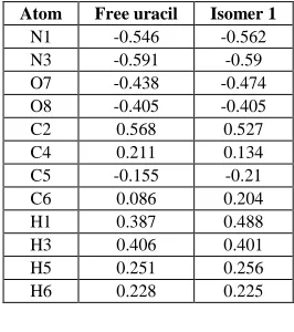
Table 7: APT Charges at various atomic sites of free Uracil and Isomer 1 (most stable isomer) of Uracil-Water complexes at Mp2/6-311++G (d, p) level
Atom Free uracil Isomer 1
N1 -0.546 -0.562
N3 -0.591 -0.59
O7 -0.438 -0.474
O8 -0.405 -0.405
C2 0.568 0.527
C4 0.211 0.134
C5 -0.155 -0.21
C6 0.086 0.204
H1 0.387 0.488
H3 0.406 0.401
H5 0.251 0.256
H6 0.228 0.225
CONCLUSIONS
The optimized geometries of uracil and four isomers of uracil-water complexes have been calculated employing ab-initio with MP2/6-311++G (d,p) level and DFT with the hybrid functional B3LYP/6-311++G (d,p) and B3LYP /Aug-CC-pVDZ levels. The most of the geometrical parameters for uracil-water complexes remains the same as uracil excepted the geometry of the site of the water bonded atom. Structural parameters of the optimized geometries, total energies and the APT charges of uracil-water complex have been discussed in detail. We show that addition of water molecules in uracil, the strength of the binding energy decreases i.e. stability increases. The optimized bond length and bond angles are in agreement with the corresponding experimental results.
ACKNOWLEDGEMENT
Authors are grateful to Secretary and Principal, Udai Pratap Autonomous College for providing necessary facilities.
REFERENCES
[1] GS Parry. Acta Cryst. 1954, 7, 313.
[2] T van Mourik; SL Price; DC Clary. J Phys ChemA. 1999, 103, 1611-1618. [3] S Rybak; K Szalewicz; B Jeziorski; G Corongiu. Chem Phys Lett. 1992, 199, 567. [4] J Smets; WJ McCarthy; LJ Adamowicz. Phys Chem. 1996, 100, 14655.
[image:6.612.239.372.299.449.2]126
[7] MY Choi; RE Miller. Phys Chem Chem Phys. 2005, 7, 3565-3573. [8] A Yoshikawa; S Matsika. Chem Phys. 2008, 347, 383-404.
[9] AK Chandra; MT Nguyen; T Zeegers-Huyskens. J Phys Chem A. 1998, 102, 6010-6016. [10] RB Zhang; T Zeegers-Huyskens; A Ceulemans; MT Nguyen. ChemPhys. 2005, 316,35-44. [11] C Møller; MS Plesset. Phys Rev. 1934, 46, 618-622.
[12] AD Becke. J Chem Phys. 1992, 97, 9173. [13] AD Becke. J Chem Phys. 1993, 98, 5648. [14] AD Becke. J Chem Phys. 1996, 104, 1040.
[15] C Lee; W Yang; RG Parr. Phys Rev B. 1988, 37, 785.