Butallylonal 1,4-dioxane hemisolvate
Thomas Gelbrich,* Denise Rossi and Ulrich J. Griesser
Institute of Pharmacy, University of Innsbruck, Innrain 52, 6020 Innsbruck, Austria Correspondence e-mail: [email protected]
Received 15 September 2010; accepted 27 September 2010
Key indicators: single-crystal X-ray study;T= 293 K; mean(C–C) = 0.009 A˚; Rfactor = 0.064;wRfactor = 0.145; data-to-parameter ratio = 14.4.
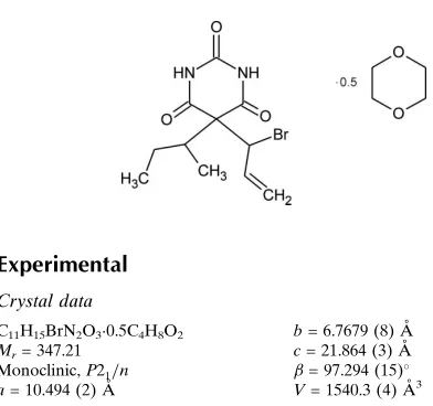
The asymmetric unit of the title compound [systematic name: 5-(1-bromoprop-2-en-1-yl)-5-sec-butylpyrimidine-2,4,6-trione 1,4-dioxane hemisolvate], C11H15BrN2O30.5C4H8O2, contains
one half-molecule of 1,4-dioxane and one molecule of butallylonal, with an almost planar barbiturate ring [largest deviation from the mean plane = 0.049 (5) A˚ ]. The centro-symmetric dioxane molecule adopts a nearly ideal chair conformation. The barbiturate molecules are linked together by an N—H O hydrogen bond, giving a single-stranded chain. Additionally, each dioxane molecule acts as a bridge between two antiparallel strands of hydrogen-bonded
barbi-turate molecules via two hydrogen bonds, N—
H O(dioxane)O H—N. Thus, a ladder structure is obtained, with the connected barbiturate molecules forming the ‘stiles’ and the bridging dioxane molecules the ‘rungs’.
Related literature
For the preparation of butallylonal, see: J. D. Riedel Akt.-Ges. (1924); Boedecker (1929). For related structures, see: Al-Saqqaret al.(2004); Gelbrichet al.(2007, 2010); Cravenet al. (1969); Gatehouse & Craven (1971); Lewis et al. (2004); Zencirci et al. (2009). For hydrogen-bond motifs, see: Bern-steinet al.(1995).
Experimental
Crystal data
˚
MoKradiation = 2.68 mm1
0.250.080.07 mm
Data collection
Oxford Diffraction Xcalibur Ruby Gemini ultra diffractometer Absorption correction: multi-scan
(CrysAlis PRO; Oxford Diffraction, 2007) Tmin= 0.990,Tmax= 1.000
9189 measured reflections 2714 independent reflections 1171 reflections withI> 2(I) Rint= 0.100
Refinement
R[F2> 2(F2)] = 0.064 wR(F2) = 0.145 S= 0.95 2714 reflections 189 parameters 2 restraints
H atoms treated by a mixture of independent and constrained refinement
max= 0.56 e A˚ 3
[image:1.610.47.243.568.752.2]min=0.33 e A˚ 3
Table 1
Hydrogen-bond geometry (A˚ ,).
D—H A D—H H A D A D—H A
N1—H1 O4i 0.88 (1) 1.98 (2) 2.837 (5) 166 (6) N3—H3 O1S 0.89 (4) 1.87 (4) 2.757 (6) 177 (5)
Symmetry code: (i)x;yþ1;z.
Data collection: CrysAlis PRO(Oxford Diffraction, 2007); cell refinement: CrysAlis PRO; data reduction: CrysAlis PRO; program(s) used to solve structure: SHELXS97 (Sheldrick, 2008); program(s) used to refine structure:SHELXL97(Sheldrick, 2008); molecular graphics:XPinSHELXTL(Sheldrick, 2008) andMercury
(Brunoet al., 2002); software used to prepare material for publica-tion:publCIF(Westrip, 2010).
TG acknowledges financial support from the Lise Meitner Program of the Austrian Science Fund (FWF, project LM 1135-N17).
Supplementary data and figures for this paper are available from the IUCr electronic archives (Reference: FJ2340).
References
Al-Saqqar, S., Falvello, L. R. & Soler, T. (2004).J. Chem. Crystallogr.34, 61– 65.
Bernstein, J., Davis, R. E., Shimoni, L. & Chang, N.-L. (1995).Angew. Chem. Int. Ed. Engl.34, 1555–1573.
Boedecker, F. (1929). US Patent 1739662
Bruno, I. J., Cole, J. C., Edgington, P. R., Kessler, M., Macrae, C. F., McCabe, P., Pearson, J. & Taylor, R. (2002).Acta Cryst.B58, 389–397.
Craven, B. M., Vizzini, E. A. & Rodrigues, M. M. (1969).Acta Cryst.B25, 1978–1993.
Gatehouse, B. M. & Craven, B. M. (1971).Acta Cryst.B27, 1337–1344. Gelbrich, T., Rossi, D. & Griesser, U. J. (2010).Acta Cryst.E66, o1219. Gelbrich, T., Zencirci, N. & Griesser, U. J. (2007).Acta Cryst.C63, o751–o753. J. D. Riedel Akt.-Ges. (1924). GB Patent 244122.
Lewis, T. C., Tocher, D. A. & Price, S. L. (2004).Cryst. Growth Des.4, 979–987. Oxford Diffraction (2007).CrysAlis PRO. Oxford Diffraction Ltd, Abingdon,
England.
Sheldrick, G. M. (2008).Acta Cryst.A64, 112–122. Westrip, S. P. (2010).J. Appl. Cryst.43, 920–925.
Structure Reports
Online
supporting information
Acta Cryst. (2010). E66, o2688 [doi:10.1107/S1600536810038651]
Butallylonal 1,4-dioxane hemisolvate
Thomas Gelbrich, Denise Rossi and Ulrich J. Griesser
S1. Comment
5,5-Dihydroxybarbituric acid (alternative names: butallylonal, butylalylonal, pernocton, pernoston, sonbutal; CAS
number 1142–70-7) has been used as a sedative drug since the 1920s, mainly as an anaesthetic in veterinary medicine.
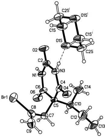
The asymmetric unit of the title compound contains one butallylonal molecule exhibiting an almost planar barbiturate
ring [where atom C6 shows the largest deviation from the mean plane, 0.049 (5) Å] and half a molecule of 1,4-dioxane.
The two torsion angles of the C8—C7—C5—C10—C14 chain are trans, C7—C5—C10—C12 is gauche and C5—C10
—C14 trans (see Fig. 1). The centrosymmetric dioxane molecule adopts a near-to-ideal chair conformation.
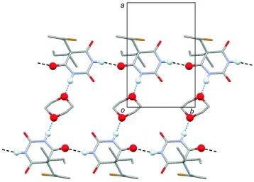
The barbiturate molecules are linked together by one N—H···O bond to give a single-stranded chain. Additionally, each
dioxane molecule acts as a bridge between two antiparallel strands of H-bonded barbiturate molecules. This interaction
involves two hydrogen bonds, N—H···O(dioxane)O···H—N. Overall, a ladder structure is generated, which propagates
parallel to the b axis (see Fig. 2). The stiles of the ladder are formed by the connected barbiturate molecules and its rungs
by the bridging dioxane molecules. This H-bonded structure is reminiscent of the ladder motif observed in single
component structures of several barbiturates, see Craven et al. (1969); Gatehouse & Craven (1971); Lewis et al. (2004);
Gelbrich et al. (2007); Zencirci et al. (2009). The main difference to the title structure is that in these cases, a second
C=O group participates in hydrogen bonding so that two antiparallel strands of H-bonded barbiturate molecules are
linked together directly via centrosymmetric R22(8) rings (Bernstein et al., 1995).
S2. Experimental
A solution of butallylonal ("Pernocton"; J. D. Riedel - E. de Haën AG, Berlin) in 1,4-dioxane was filled into an NMR tube
and left for evaporation. Colourless crystals of the title compound were obtained after several weeks.
S3. Refinement
All H atoms were identified in a difference map. H atoms bonded to C atoms were positioned geometrically and refined
with Uiso(H) = 1.2 Ueq(C). Hydrogen atoms attached to N were refined with restrained distances [N—H = 0.88 (2) Å], and
Figure 1
The molecular structures of (I) with displacement ellipsoids drawn at the 30% probability level. Hydrogen atoms are
Figure 2
A portion of the one-dimensional hydrogen bonded ladder structure which consists of two antiparallel strands of singly
N–H···O bonded barbiturate molecules, which are bridged by dioxane molecules, N–H···O(dioxane)O···H–N. The
structure is viewed parallel to the c-axis. O and H atoms involved in hydrogen bonding ar drawn as balls.
5-(1-bromoprop-2-en-1-yl)-5-sec-butylpyrimidine-2,4,6-trione 1,4-dioxane hemisolvate
Crystal data
C11H15BrN2O3·0.5C4H8O2
Mr = 347.21 Monoclinic, P21/n Hall symbol: -P 2yn
a = 10.494 (2) Å
b = 6.7679 (8) Å
c = 21.864 (3) Å
β = 97.294 (15)°
V = 1540.3 (4) Å3
Z = 4
F(000) = 712
Dx = 1.497 Mg m−3
Mo Kα radiation, λ = 0.71073 Å Cell parameters from 837 reflections
θ = 2.5–28.5°
µ = 2.68 mm−1
T = 293 K Prism, colourless 0.25 × 0.08 × 0.07 mm
Data collection
Oxford Diffraction Xcalibur Ruby Gemini ultra diffractometer
Radiation source: Enhance Ultra (Cu) X-ray Source
Mirror monochromator
Detector resolution: 10.3575 pixels mm-1
ω scans
Absorption correction: multi-scan
(CrysAlis PRO; Oxford Diffraction, 2007)
Tmin = 0.990, Tmax = 1.000 9189 measured reflections 2714 independent reflections 1171 reflections with I > 2σ(I)
Rint = 0.100
θmax = 25.1°, θmin = 3.2°
h = −10→12
k = −8→8
Refinement on F2 Least-squares matrix: full
R[F2 > 2σ(F2)] = 0.064
wR(F2) = 0.145
S = 0.95 2714 reflections 189 parameters 2 restraints
Primary atom site location: structure-invariant direct methods
Secondary atom site location: difference Fourier map
Hydrogen site location: inferred from neighbouring sites
H atoms treated by a mixture of independent and constrained refinement
w = 1/[σ2(F
o2) + (0.0409P)2] where P = (Fo2 + 2Fc2)/3 (Δ/σ)max < 0.001
Δρmax = 0.56 e Å−3 Δρmin = −0.33 e Å−3
Special details
Experimental. CrysAlisPro, Oxford Diffraction Ltd., Empirical absorption correction using spherical harmonics, implemented in SCALE3 ABSPACK scaling algorithm.
Geometry. All e.s.d.'s (except the e.s.d. in the dihedral angle between two l.s. planes) are estimated using the full covariance matrix. The cell e.s.d.'s are taken into account individually in the estimation of e.s.d.'s in distances, angles and torsion angles; correlations between e.s.d.'s in cell parameters are only used when they are defined by crystal symmetry. An approximate (isotropic) treatment of cell e.s.d.'s is used for estimating e.s.d.'s involving l.s. planes.
Refinement. Refinement of F2 against ALL reflections. The weighted R-factor wR and goodness of fit S are based on F2, conventional R-factors R are based on F, with F set to zero for negative F2. The threshold expression of F2 > σ(F2) is used only for calculating R-factors(gt) etc. and is not relevant to the choice of reflections for refinement. R-factors based on F2 are statistically about twice as large as those based on F, and R- factors based on ALL data will be even larger.
Fractional atomic coordinates and isotropic or equivalent isotropic displacement parameters (Å2)
x y z Uiso*/Ueq
Br1 0.64263 (9) 0.24500 (13) 0.04609 (4) 0.0951 (4)
N1 0.4336 (5) 0.4954 (6) 0.1405 (2) 0.0504 (15)
H1 0.435 (6) 0.6246 (17) 0.138 (2) 0.061*
N3 0.3225 (5) 0.2096 (6) 0.1096 (2) 0.0468 (14)
H3 0.253 (3) 0.163 (7) 0.087 (2) 0.056*
O2 0.2482 (5) 0.5076 (6) 0.0762 (2) 0.0786 (16)
O6 0.6264 (5) 0.4897 (5) 0.1981 (2) 0.0752 (16)
O4 0.3920 (4) −0.0903 (5) 0.13692 (18) 0.0579 (13)
C2 0.3306 (7) 0.4133 (8) 0.1065 (3) 0.0519 (18)
C4 0.4088 (6) 0.0884 (8) 0.1410 (3) 0.0450 (16)
C5 0.5229 (6) 0.1748 (6) 0.1812 (3) 0.0386 (15)
C6 0.5334 (7) 0.4010 (8) 0.1731 (3) 0.0505 (18)
C7 0.6478 (6) 0.0760 (8) 0.1667 (3) 0.0501 (17)
H7A 0.7191 0.1616 0.1817 0.060*
H7B 0.6588 −0.0463 0.1899 0.060*
C8 0.6567 (6) 0.0305 (9) 0.1013 (3) 0.0606 (19)
C9 0.6782 (7) −0.1521 (11) 0.0779 (3) 0.084 (2)
H9A 0.6886 −0.2609 0.1040 0.101*
H9B 0.6823 −0.1675 0.0360 0.101*
C10 0.5050 (7) 0.1407 (8) 0.2512 (3) 0.0605 (19)
H10 0.5815 0.1959 0.2756 0.073*
H12A 0.5544 −0.1461 0.2499 0.117*
H12B 0.4099 −0.1140 0.2593 0.117*
C13 0.5331 (10) −0.0833 (13) 0.3418 (3) 0.129 (4)
H13D 0.6067 −0.0020 0.3546 0.193*
H13E 0.5530 −0.2184 0.3525 0.193*
H13F 0.4622 −0.0404 0.3622 0.193*
C14 0.3888 (8) 0.2595 (10) 0.2694 (3) 0.103 (3)
H14A 0.4032 0.3982 0.2640 0.154*
H14B 0.3793 0.2338 0.3117 0.154*
H14C 0.3120 0.2201 0.2436 0.154*
O1S 0.1014 (4) 0.0764 (5) 0.04030 (19) 0.0650 (14)
C1S 0.0085 (7) 0.2010 (7) 0.0056 (3) 0.072 (2)
H1S1 0.0491 0.3243 −0.0036 0.086*
H1S2 −0.0595 0.2313 0.0303 0.086*
C2S 0.0465 (7) −0.1081 (8) 0.0515 (3) 0.066 (2)
H2S1 −0.0200 −0.0899 0.0781 0.080*
H2S2 0.1119 −0.1938 0.0727 0.080*
Atomic displacement parameters (Å2)
U11 U22 U33 U12 U13 U23
Br1 0.1010 (8) 0.1162 (6) 0.0699 (6) 0.0078 (6) 0.0173 (5) 0.0284 (5)
N1 0.050 (4) 0.025 (2) 0.070 (3) −0.006 (3) −0.019 (3) 0.003 (3)
N3 0.048 (4) 0.025 (2) 0.062 (3) −0.006 (2) −0.014 (3) 0.002 (2)
O2 0.068 (4) 0.046 (2) 0.112 (4) 0.003 (2) −0.028 (3) 0.021 (2)
O6 0.072 (4) 0.051 (2) 0.092 (4) −0.017 (2) −0.032 (3) −0.008 (2)
O4 0.063 (3) 0.027 (2) 0.078 (3) −0.0022 (19) −0.011 (2) 0.0016 (19)
C2 0.056 (5) 0.037 (3) 0.061 (4) 0.004 (3) 0.000 (4) 0.009 (3)
C4 0.057 (5) 0.036 (3) 0.042 (4) −0.002 (3) 0.006 (3) 0.006 (3)
C5 0.043 (4) 0.027 (3) 0.044 (4) 0.001 (3) −0.003 (3) 0.005 (2)
C6 0.059 (5) 0.042 (3) 0.046 (4) 0.009 (4) −0.011 (4) 0.002 (3)
C7 0.044 (5) 0.047 (3) 0.059 (4) 0.000 (3) 0.003 (3) −0.001 (3)
C8 0.049 (5) 0.066 (4) 0.068 (5) −0.003 (4) 0.009 (4) −0.002 (4)
C9 0.091 (7) 0.098 (5) 0.068 (5) 0.003 (5) 0.031 (5) 0.005 (4)
C10 0.082 (6) 0.050 (3) 0.052 (4) 0.020 (4) 0.017 (4) 0.006 (3)
C12 0.107 (8) 0.084 (5) 0.103 (7) −0.001 (5) 0.023 (6) 0.008 (5)
C13 0.144 (9) 0.186 (8) 0.060 (6) 0.060 (8) 0.029 (6) 0.072 (6)
C14 0.132 (8) 0.114 (6) 0.067 (5) 0.072 (6) 0.032 (5) 0.019 (5)
O1S 0.058 (3) 0.046 (2) 0.080 (3) −0.003 (2) −0.031 (3) 0.001 (2)
C1S 0.071 (5) 0.041 (3) 0.097 (6) 0.006 (3) −0.016 (4) −0.002 (4)
C2S 0.073 (6) 0.056 (4) 0.063 (5) 0.004 (4) −0.014 (4) 0.012 (3)
Geometric parameters (Å, º)
Br1—C8 1.881 (6) C10—C12 1.466 (8)
N1—C6 1.351 (7) C10—C14 1.553 (9)
N1—C2 1.351 (7) C10—H10 0.9800
N3—C2 1.384 (7) C12—H12B 0.9700
N3—H3 0.89 (4) C13—H13D 0.9600
O2—C2 1.203 (6) C13—H13E 0.9600
O6—C6 1.215 (6) C13—H13F 0.9600
O4—C4 1.224 (5) C14—H14A 0.9600
C4—C5 1.510 (7) C14—H14B 0.9600
C5—C7 1.540 (8) C14—H14C 0.9600
C5—C6 1.547 (7) O1S—C2S 1.410 (7)
C5—C10 1.583 (8) O1S—C1S 1.431 (7)
C7—C8 1.479 (8) C1S—C2Si 1.452 (8)
C7—H7A 0.9700 C1S—H1S1 0.9700
C7—H7B 0.9700 C1S—H1S2 0.9700
C8—C9 1.366 (8) C2S—C1Si 1.452 (8)
C9—H9A 0.9300 C2S—H2S1 0.9700
C9—H9B 0.9300 C2S—H2S2 0.9700
C6—N1—C2 127.5 (5) C14—C10—C5 111.4 (5)
C6—N1—H1 119 (4) C12—C10—H10 106.3
C2—N1—H1 113 (4) C14—C10—H10 106.3
C4—N3—C2 126.3 (5) C5—C10—H10 106.3
C4—N3—H3 121 (3) C10—C12—C13 110.3 (6)
C2—N3—H3 112 (3) C10—C12—H12A 109.6
O2—C2—N1 123.6 (5) C13—C12—H12A 109.6
O2—C2—N3 120.8 (6) C10—C12—H12B 109.6
N1—C2—N3 115.6 (5) C13—C12—H12B 109.6
O4—C4—N3 118.9 (5) H12A—C12—H12B 108.1
O4—C4—C5 121.4 (5) C12—C13—H13D 109.5
N3—C4—C5 119.6 (4) C12—C13—H13E 109.5
C4—C5—C7 110.2 (4) H13D—C13—H13E 109.5
C4—C5—C6 112.3 (5) C12—C13—H13F 109.5
C7—C5—C6 109.4 (5) H13D—C13—H13F 109.5
C4—C5—C10 108.8 (5) H13E—C13—H13F 109.5
C7—C5—C10 110.2 (5) C10—C14—H14A 109.5
C6—C5—C10 105.9 (4) C10—C14—H14B 109.5
O6—C6—N1 121.9 (5) H14A—C14—H14B 109.5
O6—C6—C5 120.2 (5) C10—C14—H14C 109.5
N1—C6—C5 117.8 (5) H14A—C14—H14C 109.5
C8—C7—C5 116.7 (5) H14B—C14—H14C 109.5
C8—C7—H7A 108.1 C2S—O1S—C1S 110.4 (4)
C5—C7—H7A 108.1 O1S—C1S—C2Si 111.7 (5)
C8—C7—H7B 108.1 O1S—C1S—H1S1 109.3
C5—C7—H7B 108.1 C2Si—C1S—H1S1 109.3
H7A—C7—H7B 107.3 O1S—C1S—H1S2 109.3
C9—C8—C7 125.7 (6) C2Si—C1S—H1S2 109.3
C9—C8—Br1 117.5 (6) H1S1—C1S—H1S2 107.9
C7—C8—Br1 116.8 (4) O1S—C2S—C1Si 111.1 (5)
C8—C9—H9B 120.0 C1Si—C2S—H2S1 109.4
H9A—C9—H9B 120.0 O1S—C2S—H2S2 109.4
C12—C10—C14 110.0 (7) C1Si—C2S—H2S2 109.4
C12—C10—C5 116.0 (5) H2S1—C2S—H2S2 108.0
Symmetry code: (i) −x, −y, −z.
Hydrogen-bond geometry (Å, º)
D—H···A D—H H···A D···A D—H···A
N1—H1···O4ii 0.88 (1) 1.98 (2) 2.837 (5) 166 (6)
N3—H3···O1S 0.89 (4) 1.87 (4) 2.757 (6) 177 (5)