University of South Carolina
Scholar Commons
Theses and Dissertations
1-1-2013
Decoding PRMT1: Studies On the Catalytic
Mechanism, Regulation, Inhibition, and Crosstalk
of PRMT1-Dependent Methylation
Heather L. Rust
University of South Carolina
Follow this and additional works at:https://scholarcommons.sc.edu/etd
This Open Access Dissertation is brought to you by Scholar Commons. It has been accepted for inclusion in Theses and Dissertations by an authorized administrator of Scholar Commons. For more information, please [email protected].
Recommended Citation
D
ECODINGPRMT1:
S
TUDIES ON THEC
ATALYTICM
ECHANISM,
R
EGULATION,
I
NHIBITION,
ANDC
ROSSTALK OFPRMT1-D
EPENDENTM
ETHYLATIONby
Heather Lynn Rust
Bachelor of Science Saint Francis University, 2008
Submitted in Partial Fulfillment of the Requirements
For the Degree of Doctor of Philosophy in
Chemistry and Biochemistry
College of Arts and Sciences
University of South Carolina
2013
Accepted by:
Paul Thompson, Major Professor
Caryn Outten, Committee Member
Sheryl Wiskur, Committee Member
Lorne Hofseth, Committee Member
ii
iii
Dedication
I dedicate this dissertation to my Mom, Dad, and brother Ryan for the
unconditional love and support that they have given me throughout my entire life.
Mom, I am so proud to be your daughter and so grateful that I can call you best friend.
You have always been there for me and believed in me no matter what and I couldn’t
have made it this far without you. Dad, you have always put our family first and
therefore have given me the chance to have a wonderful life, and for that I will always be
grateful. You have always pushed me to be the best that I can be and without your
support I would have never succeeded in high school, college, graduate school, and in
life. I am so proud and thankful to be your daughter. All I ever needed was your love,
but both of you have given me so much more! Ryan, I know that the age difference made
it hard for us to be close growing up and the distance didn’t help either, but I want you to
know that I will always be here for you and that I hope we can grow closer in the years to
come. You have brought a lot of laughs to our family and I hope that never changes.
Your journey is just beginning and I can’t wait to see who you become! I love you all,
and thank you for loving me!
“Other things may change us but we start and end with family.” – Anthony Brandt
iv
Acknowledgements
I would first like to thank my advisor, Dr. Paul Thompson for giving me the
opportunity to become a member of his lab. Looking back, I can’t believe how much I
have learned from you and the lab, not only in regards to techniques, but in thinking,
problem solving, and writing. I am also grateful for your advice and criticisms for they
have made me a better scientist.
I would also like to thank my committee members, Dr. Caryn Outten, Dr. Sheryl
Wiskur, Dr. Lorne Hofseth, and Dr. Dan Dixon, for not only participating on my
committee, but for their advice and encouragement thus far.
Thank you to the past and present members of the Thompson Lab for your
support and friendship, as well as sharing your knowledge and ideas to help me become a
successful scientist. Thank you Venkat Subramanian for specifically helping me with the
synthesis of pCMF and PRMT1 inhibitors discussed in this dissertation.
Next, I would like to thank Pablo Martínez Acedo for his time, patience, mass
spectrometry expertise, and friendship. You have helped make my thesis complete and
also opened new doors for our research, which is truly appreciated.
I would also like to thank Thu Truong for both her friendship and scientific advice
over the past couple years. I am grateful that The Vampire Diaries brought us together.
v
I would like to thank my long-distance best friend, Tammie Sikora, for being
there for me over the past five years. You are the sister that I never had I would have
never made it through graduate school without you!
I have dedicated this dissertation to my parents, however, I can never thank them
enough, so thank you again for all that you are and all that you do! I would also like to
thank all of my other family members (aunts, uncles, cousins, etc.) for their love and
support.
I would like to thank anyone else that I may have forgotten! There are countless
people who have helped along the way and I am very grateful for that.
Last, but most important, I would like to thank God for all of the blessings he has
given me. You have always pointed me in the right direction and given me the strength
vi
Abstract
Arginine methylation is catalyzed by the protein arginine methyltransferase
(PRMT) family of enzymes, which transfer a methyl group from S-adenosylmethionine
(SAM) to the guanidinium group of an arginine residue. This reaction first produces
monomethylated arginine (MMA) that can then be further methylated to produce either
asymmetrically dimethylated arginine (ADMA) or symmetrically dimethylated arginine
(SDMA). There are nine PRMT family members described to date, with PRMT1 as the
predominant member, suggested to be responsible for ~85% of asymmetric
dimethylation. In addition, PRMT1-dependent methylation likely plays a significant role
in a plethora of diseases (e.g., cancer, heart disease, and ALS). These observations
render it imperative that the isozyme be more thoroughly characterized and suggests that
potent and selective inhibitors may be useful as therapeutics.
Herein we describe our efforts to decode PRMT1-dependent methylation by
investigating the catalytic mechanism, the effects of post-translational modifications and
protein-protein interactions on activity, the development of potent and selective inhibitors
and inactivators, as well as examining crosstalk between arginine methylation and
phosphorylation. Using site-directed mutagenesis and unnatural amino acid
incorporation, we have identified key active site residues that are critical for catalysis
and/or substrate binding, and have determined the effects of phosphorylation, if any, on
vii
knowledge of the regulation of PRMT1 activity by protein-protein interactions. The use
of MS/MS analysis aided in the identification of the site of modification for a potent
inactivator of the isozyme, C21, and has led to the design of new inhibitors and
inactivators that will likely be more potent and selective for not only PRMT1, but
PRMT5 as well. Finally, using a peptide based model, we began to investigate crosstalk
between arginine methylation and serine/threonine phosphorylation within kinase
consensus sequences and hypothesize that it is an important means of regulation in
regards to cell signaling. Overall, the results presented in the following chapters have
enhanced our understanding of PRMT1-dependent methylation and have opened doors
viii
Table of Contents
Dedication ... iii
Acknowledgements ... iv
Abstract ... vi
List of Tables ... xi
List of Figures ... xiii
Chapter 1. Introduction ...1
1.1 Post-Translational Modifications ...1
1.2 Modification of Histones ...1
1.3 Crosstalk Between Post-translational Modifications ...4
1.4 Arginine Modifications ...6
1.5 Protein Arginine Methyltransferase Family ...10
1.6 Role of PRMT1 in Disease ...20
1.7 Conclusion ...28
Chapter 2. Mechanistic Studies on PRMT1 ...29
ix
2.2 Materials and Methods ...33
2.3 Results & Discussion ...39
2.4 Conclusions ...64
Chapter 3. Regulation of PRMT1 by Post-translational Modifications ...69
3.1 Introduction ...69
3.2 Materials and Methods ...75
3.3 Results & Discussion ...81
3.4 Conclusions ...89
Chapter 4. Regulation of PRMT1 by Protein-Protein Interactions ...91
4.1 Introduction ...91
4.2 Materials and Methods ...94
4.3 Results & Discussion ...98
4.4 Conclusions ...102
Chapter 5. Development of PRMT1 Inhibitors...104
5.1 Introduction ...104
5.2 Materials and Methods ...111
5.3 Results & Discussion ...116
5.4 Conclusions ...123
Chapter 6. Crosstalk Between Methylation and Phosphorylation ...126
6.1 Introduction ...126
6.2 Materials and Methods ...143
x
6.4 Conclusions ...153
Chapter 7. Conclusions & Future Directions ...157
References ...162
xi
List of Tables
Table 2.1. Peptide Sequences...40
Table 2.2. Kinetic parameters of PRMT1 mutants for the AcH4-21 peptide. ...40
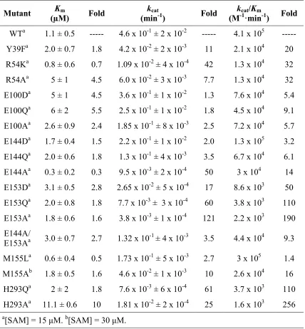
Table 2.3. Kinetic parameters of PRMT1 mutants for SAM. ...41
Table 2.4. SAH Inhibition Studies. ...42
Table 2.5. Solvent Isotope Effects (SIE) and Solvent Viscosity Effects (SVE). ...60
Table 3.1. Observed modifications of PRMT1. ...72
Table 3.2. Kinetic parameters of PRMT1 mutants for the AcH4-21 peptide. ...82
Table 3.3. Kinetic parameters of PRMT1 mutants for SAM. ...82
Table 3.4. Kinetic parameters of PRMT1 mutants for the AcH4-21 peptide. ...86
Table 3.5. Kinetic parameters of PRMT1 mutants for SAM. ...86
Table 3.6. Percent activity of select protein kinases with PRMT1. ...88
Table 5.1. IC50 values for PRMT1 inhibitors and inactivators. ...109
Table 5.2. Kinetic parameters of PRMT1 mutants for the AcH4-21 peptide.. ...118
Table 5.3. Kinetic parameters of PRMT1 mutants for SAM. ...118
Table 5.4. IC50 values for C21 ...119
xii
Table 6.1. Tested Substrates for PRMT1 and Akt. ...141
Table 6.2. PRMT1 and Akt Crosstalk Predictions. ...142
Table 6.3. Peptide Sequences...148
Table 6.4. Kinetic parameters of PRMT1 for the FOXO1 peptide substrates. ...148
Table 6.5. Kinetic parameters of PRMT1 for the FOXO1 peptide substrates. ...151
Table 6.6. Kinetic parameters of PAD4 for the FOXO1 peptide substrates. ...151
xiii
List of Figures
Figure 1.1. Selected post-translational modifications of arginine, lysine, serine, threonine,
and tyrosine. ...2
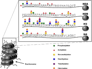
Figure 1.2. Post-translational modifications of histones. ...3
Figure 1.3. Crosstalk scenarios. ...5
Figure 1.4. PAD catalyzed reaction. ...7
Figure 1.5. Role of PAD4 in NET formation...8
Figure 1.6. PRMT catalyzed reactions. ...9
Figure 1.7. Structure of PRMT1 family members. ...11
Figure 1.8. PRMT1 splice variants. ...12
Figure 1.9. Proposed model of an ERα-dependent role of PRMT1 in breast cancer. ...22
Figure 1.10. Proposed model of the role of PRMT1v2 in breast cancer. ...23
Figure 1.11. Proposed model of the role of PRMT1 in leukemia. ...24
Figure 1.12. Proposed model of the role of PRMT1 in heart disease. ...26
Figure 1.13. Proposed model of the role of PRMT1 in ALS. ...27
Figure 2.1. Active site of PRMT1. ...31
xiv
Figure 2.3. Amino acid analysis of PRMT1-dependent methylation products...46
Figure 2.4. Processivity of WT PRMT1 and M155 mutants. ...47
Figure 2.5. D51 and H293 interaction. ...52
Figure 2.6. pH profiles of WT PRMT1 with SAM. ...55
Figure 2.7. pH profiles of WT PRMT1 and mutants with the RGG3 peptide. ...58
Figure 2.8. Processivity of WT PRMT1 and mutants. ...61
Figure 3.1. Structural comparisons between the PRMTs and contrast between amino acid side chains ...73
Figure 3.2. Structure of PRMT1 and structural contrast of amino acid side chains ...74
Figure 3.3. Immunoprecipitation of PRMT1 ...83
Figure 3.4. Synthesis of pCMF ...84
Figure 3.5. Incorporation of pCMF into PRMT1 ...85
Figure 3.6. Expression and purification of PRMT1(Y291pCMF) ...85
Figure 3.7. MS/MS Analysis of PRMT1(Y291pCMF) ...86
Figure 4.1. Enzyme regulation scenarios of PRMT1 by CAF1 and BTG1 ...93
Figure 4.2. Effect of interacting proteins on PRMT1 activity at high concentrations of substrate ...99
xv
Figure 4.4. Effect of interacting proteins on PRMT1 activity at low concentrations of
substrate ...101
Figure 4.5. Summary of the effects of CAF1 and BTG1 on PRMT1 activity ...102
Figure 5.1. Structures of selected PRMT inhibitors discovered from library screenings 105
Figure 5.2. Structures of selected PRMT inhibitors discovered from virtual screenings 106
Figure 5.3. Structures of SAM analogues and selected bisubstrate PRMT inhibitors ....107
Figure 5.4. Mechanism of PRMT1 inhibition by AAI ...108
Figure 5.5. Structures of haloacetamidine based inhibitors and inactivators ...109
Figure 5.6. Possible mechanisms of inactivation of PRMTs ...110
Figure 5.7. MS/MS of the site of modification of PRMT1 with (A) Cl-amidine and (B)
C21 ...117
Figure 5.8. Dialysis experiments of WT PRMT1 and the C101A mutant with C21 ...119
Figure 5.9. Structure of PRMT1 showing the position of the C101 residue in relation to
SAH ...120
Figure 5.10. Synthesis of N-ethyl-aminoadenosine and
Cl-acetamidino-N-ethyl-aminoadenosine...121
Figure 5.11. Dialysis experiments of WT PRMT1 with
Cl-acetamidino-N-ethyl-aminoadenosine...123
Figure 5.12. Structural representation of PRMT cysteine residues around the active site
xvi
Figure 6.1. Potential model for crosstalk between arginine methylation and
phosphorylation ...126
Figure 6.2. Serine/Threonine Protein Kinase consensus sequences ...127
Figure 6.3. Structural basis for crosstalk ...135
Figure 6.4. Processivity of PRMT1 ...150
1
CHAPTER 1
Introduction
1.1Post-Translational Modifications
Post-translational modifications (PTMs) of proteins are well-known for the
variety of roles they play in controlling cellular functions. With over 400 different
experimentally determined types (Khoury et al. 2011), PTMs add to the diversity of the
already complex nature of the proteome by influencing protein-protein interactions, the
cellular location of proteins, and protein stability through alterations to the size, charge,
and hydrogen bonding capabilities of the parent residues (Figure 1.1). These processes
are the basis for a plethora of cellular functions (e.g., transcription and signal
transduction) that are vital to the maintenance, growth, and survival of healthy cells. Due
to their fundamental roles in the cell, the dysregulation of various PTMs has been
associated with a wide range of diseases (e.g., cancer and autoimmune diseases), thus
making the responsible enzymes attractive drug targets.
1.2 Modification of Histones
The most notable and well-studied group of modified proteins are the histones.
In the nucleus, two of each of the four core histones (i.e., H2A, H2B, H3, and H4) come
together as an octamer around which DNA is wrapped and held in place by histone H1 to
form a complex known as the nucleosome. Nucleosomes are further packaged into
2
Figure 1.1 Selected post-translational modifications of arginine, lysine, serine, threonine, and tyrosine. (A) Arginine residues can be mono- and dimethylated by the PRMTs to form ω-MMA, ADMA, or SDMA. They can also be converted to citrulline by the PADs. (B) Lysine residues can be mono-, di-, and trimethylated by KMTs, acetylated by KATs, or ubiquitinated by ubiquitin ligases. (C) Serine, threonine, and tyrosine residues can be phosphorylated by kinases.
Arginine w-MMA SDMA ADMA Citrulline
PRMTs PADs
Lysine Mono-Methyl
Lysine
KMTs KATs
Di-Methyl
Lysine Tri-Methyl
Lysine Acetylated
Lysine UbiquitinatedLysine
Ubiquitin Ligases
A
B
C
Serine
Phospho-Serine
Kinase
Threonine
Phospho-Threonine Tyrosine
Phospho-Tyrosine
3
dictates whether genes are transcribed. PTMs on the unstructured N-terminal tails of
histones are responsible for switching the transcription of genes on and off using what
has been termed the histone code (Figure 1.2). For example, specific modifications can
Figure 1.2 Post-translational modifications of histones.
cause the destabilization of chromatin via the disruption of key interactions between
DNA and histones (i.e., electrostatic interactions), such as in the case of acetylation of
lysine 16 on histone H4 and the inhibition of heterochromatin formation (Shogren-Knaak
et al. 2006 & Bannister et al. 2011). They can also aid in the recruitment of the
transcriptional machinery through protein domains that are capable of binding such
PTMs (e.g., bromo-, chromo-, tudor-, and pleckstrin-homology (PH) domains (reviewed
in Kouzarides et al. 2007). In terms of blocking transcription, a modification can also
4
tri-methylated lysine 9 of histone H3 and its recruitment of heterochromatin protein 1
(HP1) (Canzio et al. 2011).
1.3 Crosstalk Between Post-Translational Modifications1
Over the last decade there have been several examples of crosstalk between two
or more different post-translational modifications (PTMs), with many of these being
observed within the context of histones. Generally, this crosstalk is thought to modulate
and fine-tune cell signaling cascades such that a desired outcome is achieved e.g.,
transcription of a particular gene or, alternatively, activation of one gene under the
control of a transcription factor and repression of another. Although crosstalk between
two or more PTMs has predominantly been studied within the context of chromatin
biology (Suganuma et al. 2008 and Lee et al. 2010), as one would expect, this type of
regulatory mechanism extends to non-histone proteins as well. Several models have been
proposed for histone crosstalk (Fischle 2003 et al., Schreiber et al. 2002, Fischle et al.
2008), and they are readily applied to non-histone proteins as well (Figure 1.3). For
example, cis crosstalk refers to communication between modifications on the same
protein (Figure 1.3A). Within cis crosstalk lies the possibility for adjacent crosstalk (i.e.,
between residues that are close to one another in both the primary and tertiary structures)
or distal crosstalk (i.e., between residues that are far apart in both the primary and tertiary
structures) (Figure 1.3A). Trans crosstalk is also possible and occurs between
modifications on two different proteins (Figure 1.3B). Functionally, direct crosstalk
refers to one PTM directly affecting the modification of a second residue (e.g.,
modification of one residue prevents the modification of another residue) (Figure 1.3C).
1 Adapted with permission from Rust, H.L.; Thompson, P.R., Kinase consensus sequences: a breeding
5
6
Indirect crosstalk involves modulating a protein-protein interaction via the presence,
or lack, of a PTM (e.g., a PTM enhances the binding of a transcription factor leading to
the recruitment of other coactivators) (Figure 1.3D). An early example of direct cis
crosstalk (Figure 1.3A) from the histone field involves the phosphorylation of H3S10 and
the acetylation of H3K14. Here, stimulation of the Ras-MAPK (mitogen activated
protein kinase) pathway (Chadee et al. 1999) 1 results in the Rsk-2 (ribosomal S6 kinase)
dependent phosphorylation of H3S10 (Sassone-Corsi et al. 1999), which enhances the
acetylation of H3K14 by generating a better substrate for the histone acetyltransferase
Gcn5 (general control non-repressed 5) (Cheung et al. 2000, Clayton et al. 2000, and Lo
et al. 2000). Although this is only one example of crosstalk from the histone field, a
plethora of others have been published (reviewed in Suganuma et al. 2008, Lee et al.
2010, and Baek et al. 2011), including several papers that describe crosstalk in
non-histone proteins, with a particular set of crosstalk examples involving serine/threonine
phosphorylation and the modification of neighboring arginine residues; and these
examples will be discussed later in Chapter 6.
1.4 Arginine Modifications
Arginine residues within proteins can undergo several different types of PTMs,
some of which are more prevalent and well-studied than others. These modifications
include enzyme mediated modifications, i.e., citrullination, methylation, phosphorylation,
and ADP-ribosylation, as well as non-enzymatic modifications, i.e., advanced glycation
end-products (AGE) and carbonylation (reviewed in Slade et al. 2013). The most
7 1.4.1 Citrullination
Citrullination, also known as deimination, is catalyzed by the protein arginine
deiminase (PAD) family of enzymes, which is comprised of PADs1-4 and PAD6. These
isozymes catalyze the conversion of the guanidium moiety of arginine to an ureido
moiety via a calcium dependent hydrolytic mechanism (Kearney et al. 2005). This PTM
alters electrostatic interactions by changing a positively charged residue to a neutral
residue (Figure 1.4). In regards to histones, the in vivo sites of citrullination have been
determined to be H2 Arg 3 (Hagiwara et al. 2005), H3 Arg 2, Arg 8, Arg 17 (Cuthbert et
al. 2004), and Arg 26 (Cuthbert et al. 2004 & Zhang et al. 2012), and H4 Arg 3 (Wang et
al. 2004). Citrullination of these sites correlate with either transcriptional repression
(e.g., citrullination of H3 Arg 17 at the pS2 promoter) (Cuthbert et al. 2004, Wang et al.
2004) or activation (e.g., citrullination of H3 Arg 26 at ERα target genes) (Zhang et al.
2012), depending upon the specific histone and residue.
Figure 1.4 PAD catalyzed reaction. PADs catalyze the conversion of the guanidinium moiety of an arginine residue to an ureido moiety via a calcium dependent hydrolytic mechanism.
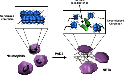
In addition to its role in transcription, histone citrullination, specifically histone
8
of the innate immune response. NETs are comprised of decondensed chromatin, with the
DNA and histones acting as traps for pathogens (Figure 1.5) (Neeli 2008 et al., Wang et
al. 2009, Li et al. 2010). Non-histone proteins, such as myelin basic protein (MPB)
(Wood et al. 2008) and antithrombin (Chang et al. 2005), are also citrullinated by
members of the PAD family, thus demonstrating the versatility of these isozymes.
Citrullination has become an increasingly important PTM because of its apparent roles in
diseases such as Rheumatoid Arthritis (RA), Multiple Sclerosis (MS), ulcerative colitis,
Alzheimers Disease (AD), and cancer (reviewed in Jones et al. 2009). In fact,
Cl-amidine, a pan-PAD inhibitor, was found to decrease disease severity in animal models
of spinal cord injury (Lange et al. 2011), collagen-induced arthritis (CIA) (Willis et al.
2011), ulcerative colitis (Chumanevich et al. 2011), and cancer (McElwee et al. 2012).
Figure 1.5 Role of PAD4 in NET formation.
PAD4 Neutrophils
Condensed Chromatin
Decondensed Chromatin Pathogen
(e.g. bacteria)
9 1.4.2 Methylation
Methylation of arginine residues is catalyzed by the protein arginine
methyltransferase (PRMT) family of enzymes. These isozymes transfer a methyl group
from S-adenosylmethionine (SAM) to the guanidinium moiety of arginine residues in
proteins, but not free arginine. This reaction first produces an ω-monomethylarginine
residue (ω-MMA), which in most cases is further methylated to produce either an
asymmetrically dimethylated arginine residue (ADMA) or a symmetrically dimethylated
arginine (SDMA) residue (Figure 1.6). The addition of one or two methyl groups does
Figure 1.6 PRMT catalyzed reactions. PRMTs catalyze the transfer of a methyl group from S-adenosylmethionine (SAM) to the guanidinium group of an arginine residue. Type I PRMTs produce asymmetric dimethyl arginine (ADMA), Type II PRMTs produce symmetric dimethyl arginine (SDMA), and Type III PRMTs only produce ω-monomethylarginine (ω-MMA).
not alter the charge of the residue; however, it decreases the number of potential
hydrogen bond donors, thus leaving the possibility of decreased intra- or intermolecular
10
arginine demethylase has yet to be discovered. Some have suggested the possibility that
PAD4 may catalyze a demethylimination reaction that would convert methylated arginine
to citrulline. This conversion would not truly reverse the modification but may have a
similar function. There is conflicting evidence in vitro and in vivo however, as to
whether this reaction actually occurs in the cell, with more evidence supporting the
notion that it does not take place (reviewed in Thompson et al. 2006).
1.5Protein Arginine Methyltransferase Family
In humans, there are nine PRMT family members including: PRMT1, 2, 3, 4,
-6, and -8 (Yang et al. 2013), which are type I PRMTs that produce ADMA; PRMT5,
which is a definitive type II PRMT and produces SDMA (Yang et al. 2013); and PRMT7,
which is a type III PRMT and generates only ω-MMA (Miranda et al. 2004 &
Zurita-Lopez et al. 2012) (Figure 1.6). Note that enzymatic activity has yet to be demonstrated
for PRMT9. All PRMTs possess a highly conserved ~310 amino acid catalytic core that
is responsible for methyltransferase activity. This core consists of a SAM binding
domain that contains a Rossmann type fold typical of Class I methyltransferases, a unique
β-barrel domain, and a dimerization arm. All family members possess an N-terminal
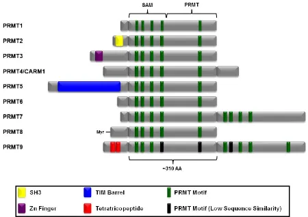
extension and several also contain C-terminal extensions (Figure 1.7) (reviewed in Yang
et al. 2013).
1.5.1 PRMT1
PRMT1 is the most prevalent PRMT isozyme and it is thought to be responsible
for ~85% of the asymmetrically dimethylated arginine residues in vivo (Tang et al. 2000
& Pawlak et al. 2000). The PRMT1 gene, located at 19q13.3 in humans (Scorilas et al.
11
Figure 1.7 Structure of PRMT family members. PRMT family members have four common motifs in their SAM binding domain and one motif in their unique PRMT domain. Each isozyme has a distinct N-terminus with some containing common protein domains such as a SH3 or a Zn finger domain. Adapted from Yang et al. 2013.
cerevisiae and over 90% between mammals, zebrafish, and Xenopus (Zhang et al. 2003).
This isozyme was originally discovered as an interacting partner of the immediate-early
gene TIS21 (Lin et al. 1996), leukemia-associated BTG1 (Lin et al. 1996), and
interferon-α receptor (IFNAR1) (Abramovich et al. 1997), as well as via sequence homology to a
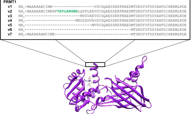
yeast homolog (Scott et al. 1998). PRMT1 is the smallest member of the PRMT family
and has three major human splice variants (i.e., PRMT1v1-v3) (Scott et al. 1998 &
Goulet et al. 2007), that translate into proteins ranging from 353-371 amino acids in
length (Scott et al. 1998 & Pawlak et al. 2000), and four minor variants (i.e.,
12
Figure 1.8 PRMT1 splice variants. The NES of PRMT1v2 is highlighted in green.
are only beginning to understand the differences between these splice variants, the
variation of which lies within the N-terminus (Figure 1.8), these variants
show different tissue expression patterns and have effects on subcellular localization. For
example, PRMT1 is expressed in all tissues studied thus far (Scott et al. 1998, Tang et al.
1998, Lin et al. 1996, Pawlak et al. 2000), with PRMT1v1 and –v2 found in several
tissues, -v4 only in the heart, -v5 predominately in the pancrease, no detection of –v6,
and -v7 mostly in the heart and skeletal muscles (Goulet et al. 2007). In regards to
subcellular localization, PRMT1 as a whole is located in both the nucleus and the
cytoplasm (Tang et al. 1998, Cote et al. 2003, Frankel et al. 2002, Goulet et al. 2007).
More specifically, PRMT1v3, -v4, -v5, and –v6 are diffuse throughout the cell (Goulet et
al. 2007), whereas PRMT1v1 and –v7 are more nuclear (Goulet et al. 2007) and
PRMT1v2 is primarily cytoplasmic (Herrmann et al. 2005 and Goulet et al. 2007). In
fact, PRMT1v2 is the only variant that contains a nuclear export signal (Goulet et al.
NH2-MAAEAANCIME---VSCGQAESSEKPNAEDMTSKDYYFDSYAHFGIHEEMLKDE
NH2-MAAEAANCIMENFVATLANGMSLQPPLEEVSCGQAESSEKPNAEDMTSKDYYFDSYAHFGIHEEMLKDE
NH2---MVGVAEVSCGQAESSEKPNAEDMTSKDYYFDSYAHFGIHEEMLKDE
NH2---MKSSSGVAVSCGQAESSEKPNAEDMTSKDYYFDSYAHFGIHEEMLKDE
NH2---MVSCGQAESSEKPNAEDMTSKDYYFDSYAHFGIHEEMLKDE
NH2---MTSKDYYFDSYAHFGIHEEMLKDE
NH2-MAAAEAANCIME---MTSKDYYFDSYAHFGIHEEMLKDE
13
2007) and was found to translocate from the nucleus, contingent on substrate methylation
status (Herrmann et al. 2005) and catalytic activity of the enzyme (Herrmann et al. 2009).
Interestingly, these different N-terminal tails are also important for the substrate
specificities of PRMT1, as demonstrated by the different methylation profiles observed
for each of the variants. For example, known PRMT substrates such as SmB and Sam68
were methylated to a greater extent by PRMT1v1 and –v2, but hnRNP A1 was a better
substrate for PRMT1v5 and -6 (Goulet et al. 2007). In addition, incubation of purified
PRMT1 variants with extracts prepared from mouse embryonic stem cell yielded distinct
visual differences in the proteins methylated by PRMT1v1 and –v2 (Goulet et al. 2007).
Similar results were also observed with purified mouse PRMT1v1 and –v2 and mouse
embryonic stem cell extracts (Pawlak et al. 2002). Surprisingly, the addition of
N-terminal His6 tags to the two main variants abolished the differences in substrate
specificity (Pawlak et al. 2002), thus demonstrating the uniqueness of the N-termini of
PRMT1. It is hypothesized that the unstructured N-terminal tail of the enzyme folds back
and interacts with its substrates, which would account for the differences in observed
substrate specificity (Goulet et al. 2007).
Although vital for early postimplantation development, PRMT1 is not essential
for cell viability (Pawlak et al. 2000). This isozyme is involved in transcriptional
regulation through both its methylation of Histone H4 at arginine 3 (Strahl et al. 2001 &
Wang et al. 2001) and other proteins involved in transcription (e.g., transcription factors
(Yamagata et al. 2008 & Jobert et al. 2009), coactivators (Teyssier et al. 2005),
elongation factors (Kwak et al. 2003)), and RNA binding proteins (Cote et al. 2003,
14
nuclear receptors (Koh et al. 2001) and transcription factors (i.e., YY1 (Rezai-Zadeh et
al. 2003), p53 (An et al. 2004), STAT5 (Kleinschmidt et al. 2008), and AE9a (Shia et al.
2012)), however, there are instances in which PRMT1 represses transcription
(Kleinschmidt et al. 2008). Interestingly, it was determined that methylation of arginine
3 of Histone H4 enhances acetylation of lysine residues on both Histone H3 (Huang et al.
2005) and Histone H4 (Wang et al. 2001 & Huang et al. 2005), but prior acetylation
prevents methylation (Huang et al. 2005). PRMT1 also plays a variety of other roles in
the cell. More specifically, it is involved in insulin signaling (Iwasaki et al.2007),
estrogen signaling (Le Romancer et al. 2008), and interferon signaling (Abramovich et al.
1997), as well as, DNA damage response pathways through methylation of MRE11
(Boisvert et al. 2005, Dery et al. 2008, Yu et al. 2009) and 53BP1 (Boisvert et al. 2005).
1.5.2 PRMT2
PRMT2, the gene of which is located at 21q22.3, was discovered by Katsanis et
al. and contains 57% nucleotide sequence homology to PRMT1 (Katsanis et al. 1997).
The translated protein is 433 amino acids in length with an N-terminal extension that
distinguishes it from PRMT1 (Krause et al. 2007). This extension contains a SRC
Homology 3 (SH3) domain, which is known to facilitate protein-protein interactions
(Pawson et al. 1992, Mayer et al. 1993). Interestingly, the SH3 domain is required for
PRMT2’s interaction with E1B-AP5 (Kzhyshkowska et al. 2001) but not estrogen
receptor alpha (ERα) (Qi et al. 2002). Originally, methylatransferase activity for this
enzyme could not be detected directly (Kzhyshkowska et al. 2001, Qi et al. 2002, Ganesh
et al. 2006, Meyer et al. 2007). It was recently discovered, however, that PRMT2 is a
15
H4 in vitro (Lakowski et al. 2009) and histone H3R8 in Xenopus (Blythe et al. 2010).
PRMT2 appears to mainly play the role of a transcriptional co-activator for a number of
nuclear hormone receptors (e.g., androgen receptor, estrogen receptor) (Meyer et al.
2007, Qi et al. 2002), which is unexpected because it is a cytoplasmic protein. However,
evidence has shown that it can be transported into the nucleus with the androgen receptor
upon hormone stimulation (Meyer et al.2007).
1.5.3 PRMT3
PRMT3 is a Type I PRMT that was identified in a yeast two-hybrid screen for
PRMT1 interacting proteins (Tang et al. 1998). The gene is located at 11p15.1 and
encodes a 531 amino acid protein that contains an N-terminal zinc finger domain (Krause
et al. 2007). This zinc finger domain was found to dictate substrate specificity and is
vital for PRMT3’s interaction with RNA-associated proteins (Frankel et al. 2000).
PRMT3 is located in the cytoplasm and, although the crystal structure suggests that it can
form homodimers (Zhang et al. 2003), it was found as a monomer using gel filtration of
rat cell extracts (Tang et al. 1998). The major substrate of PRMT3 is the 40 S ribosomal
protein S2 (rpS2), thus suggesting that this enzyme may play roles in the regulation of
protein synthesis and or ribosome assembly (Swiercz et al. 2005).
1.5.4 PRMT4/CARM1
PRMT4, more commonly known as coactivator-associated methyltransferase 1
(CARM1), was discovered during a yeast two-hybrid screen for proteins that interact
with the AD2 domain of p160 coactivators (Chen et al. 1999). The gene is located at
19p13.2 and encodes a 608 amino acid protein in humans (Krause et al. 2007). Although
16
and C-terminal extension. Neither of these extensions are required for enzymatic
activity, homo-oligomerization, or p160 binding in vitro (Teyssier et al. 2002), however,
both of them are required for the isozyme’s transcriptional coactivator function. The
N-terminus adopts a pleckstrin homology domain (PH) fold that is typically involved in the
formation of multiprotein complexes and the regulation of protein-protein interactions.
The role of that this fold plays in the function of this isozyme remains to be determined
(Troffer-Charlier et al. 2007). Importantly, it was demonstrated that CARM1 is essential
to life in that knockout mice die shortly after birth (Yadav et al. 2003). CARM1 is
known to methylate Histone H3 at arginine 2, 17, and 26 (Schurter et al. 2001), several
splicing factors (e.g., SmB and U1C) (Cheng et al. 2007), as well as transcriptional
co-activators (e.g., SRC-3 (Feng et al. 2006), CBP (Xu et al. 2001 and Chevillard-Briet et al.
2002), and p300 (Chevillard-Briet et al. 2002). The identities of these CARM1 substrates
exemplify the role of this isozyme as a coactivator involved in transcriptional regulation
(Ma et al. 2001, Schurter et al. 2001, Chevillard-Briet et al. 2002, Feng et al. 2006).
1.5.5 PRMT5
Human PRMT5, originally known as Jak-binding protein 1 (JBP1), was identified
during a search for Janus kinase 2 (Jak2) interacting proteins (Pollack et al. 1999) and
was found to be a homologue of the previously reported Skb1 from Schizosaccaromyces
pombe (Gilbreth et al. 1996) and HSL7 from Saccharomyces cerevisiae (Ma et al. 1996).
It is the only known Type II PRMT and produces SDMA (Branscombe et al. 2001).
Located at 14q11.2, the PRMT5 gene encodes a 637 amino acid protein (Krause et al.
2007) with a large N-terminal extension that contains a TIM barrel domain (Antonysamy
17
isozyme with its binding partner methylosome protein 50 (MEP50) (Antonysamy et al.
2012). MEP50 is a WD40-repeat containing protein that is required for PRMT5
methyltransferase activity and aids in substrate recognition and interactions with other
proteins, as the isozyme shows minimal activity unless it is a part of a larger multiprotein
complex (Friesen et al. 2002). Located in the nucleus and the cytoplasm, PRMT5 plays
roles in a plethora of cellular processes such as transcriptional regulation (Pal et al. 2004,
Pal et al. 2007, Wang et al. 2008), differentiation (Dacwag et al. 2007, Dacwag et al.
2009, Mallappa et al. 2011), the synthesis of ribosomes (Ren et al. 2010), and cell
proliferation (Pal et al. 2007 & Wang et al. 2008). Similar to PRMT1, PRMT5 is
essential for both embryonic development and the derivation of embryonic stem cells
(Tee et al. 2010).
1.5.6 PRMT6
Discovered during a human genome search for PRMTs (Frankel et al. 2002), the
PRMT6 gene is located at 1p13.1 and encodes a 375 amino acid protein (Krause et al.
2007). A type I PRMT, this particular isozyme is located exclusively in the nucleus and
was the first PRMT discovered to be capable of automethylation (Frankel et al. 2002).
PRMT6 plays roles in transcriptional regulation, regulation of HIV replication, base
excision repair, and cell cycle progression via methylation of histone H2A arginine 3
(Hyllus et al. 2007) and arginine 29 (Waldmann et al. 2011), histone H3 arginine 2
(Hyllus et al. 2007, Guccione et al. 2007, Iberg et al. 2008), histone H4 arginine 3 (Hyllus
et al. 2007), HIV-1 Tat (Boulanger et al. 2005), HMGA1a/b (Miranda et al. 2005 and
Sgarra et al. 2006), DNA polymerase β (El-Andaloussi et al. 2006), and tumor suppressor
18
of PRMT6 to be a rapid equilibrium random mechanism with dead end EAP and EBQ
complexes (Obianyo et al. 2012).
1.5.7 PRMT7
The PRMT7 gene, which was also discovered from computational alignments of
potential PRMT genes (Miranda et al. 2004 and Lee et al. 2005), is located at 16q22.1
(Krause et al. 2007). The gene encodes a 692 amino acid protein (Krause et al. 2007) that
uniquely contains an additional but less conserved SAM binding domain at the
C-terminus (Miranda et al. 2004). Surprisingly, the presence of both SAM binding domains
were found to be necessary for methyltransferase activity, although SAM could only
crosslink to the N-terminal SAM binding domain (Miranda et al. 2004), suggesting that
only the N-terminal half of the enzyme is active and the C-terminus acts as a regulatory
domain. Although originally there were conflicting results regarding the final product of
methylation, with one group suggesting a Type III enzyme and only the formation of
ω-MMA (Miranda et al. 2004) and another group suggesting a Type II enzyme and the
formation of both ω-MMA and SDMA (Lee et al. 2005), recent evidence has supported
the notion that it is a Type III enzyme with the latter result possibly being an artifact of
contamination by PRMT5 (Zurita-Lopez et al. 2012). Like PRMT1, PRMT7 is located in
both the nucleus and cytoplasm (Lee et al. 2005). Although one of the lesser
characterized PRMTs, it has been suggested that this isozyme plays a role in cellular
differentiation (Buhr et al. 2008) and the DNA damage response (Gros et al. 2006 and
Verbiest et al. 2008).
1.5.8 PRMT8
19
protein, was originally discovered due to its 80% sequence homology to PRMT1, with
the major variations located at the N-termini (Zhang et al. 2003). Despite the high
sequence homology between the two isozymes, PRMT8 expression is primarily restricted
to brain tissue (Lee et al. 2005, Taneda et al. 2007, Kousaka et al. 2009) and it is targeted
to the plasma membrane via myristoylation of its terminus at glycine-2 at its
N-terminus (Lee et al. 2005). Interestingly, the N-N-terminus also plays a regulatory role in
enzymatic activity in that the full length recombinant protein displays significantly
decreased activity compared to a N-terminal truncated variation that more closely
resembles PRMT1 (Sayegh et al. 2007). The isozyme is also capable of automethylation
via the production of ADMA on arginine 73 and ω-MMA on arginine 58, however, it is
still uncertain as to whether this is an intra- or inter- molecular reaction, as PRMT8 is
capable of forming homodimers (Sayegh et al. 2007). Additionally, two proline rich
sequences on the N-terminus have been found to bind SH3 domains of proteins, including
the SH3 domain of PRMT2, however, the functional significance of these interactions is
unknown as no change in PRMT8 activity was observed in in vitro assays (Sayegh et al.
2007). Several PRMT8 interacting proteins (e.g., TET-family of RNA-binding proteins,
hnRNPs, and actin) were revealed via in vitro GST-pull down experiments with
recombinant GST-PRMT8 and hypomethylated cell extracts. Specifically, the isozyme
co-localizes with Ewing’s sarcoma (EWS), independent of the binding partner’s
methylation status (Pahlich et al. 2008). The function of PRMT8 in the cell remains to be
fully defined.
1.5.9 PRMT9
20
identified by sequence homology with other PRMT family members (Lee et al. 2005).
No enzymatic activity has been reported and it has yet to be characterized. Sequence
analysis has shown that it is most closely related to PRMT7 in that it contains a second
SAM binding domain on the C-terminus. The N-terminus contains two tetratricopeptide
repeats, which based on the previously reported functions of this motif (Blatch et al.
1999), could potentially play a role in protein-protein interactions (Bedford et al. 2007).
A controversy remains in regards to a second PRMT9, PRMT9 (2p16), which is also
known as F-box only protein 11 (FBXO11). This enzyme was found to produce
ω-MMA, ADMA, and SDMA (Cook et al. 2006) despite being structurally different from
the PRMTs, which are a part of the Type I seven-β strand methyltransferase family (Katz
et al. 2003 & Bedford et al. 2009). A separate study demonstrated that this protein had
no methyltransferase activity (Fielenbach et al. 2007); the production of SDMA in the
original report may possibly be due to sample contamination by PRMT5 (Nishioka et al.
2003). Therefore, the methyltransferase classification of this protein has yet to be
confirmed.
1.6Role of PRMT1 in Disease
With the multitude and variety of roles that the PRMT isozymes play in the cell, it is
conceivable that their dysregulation would be involved in the pathogenesis of one or
more human diseases. Our research thus far has focused on PRMT1 because it is the
major Type I methyltransferase and would therefore logically play a greater role in the
onset and progression of diseases compared to the other isozymes. The following
21 1.6.1 Cancer
A recent study of the expression levels of PRMT1 in tumors from various tissues
revealed that the isozyme, and in some cases select splice variants, is/are significantly
overexpressed in a variety of cancers (Yoshimatsu et al. 2011). The results of this study
are in agreement with several more specific studies that have shown that PRMT1 is
overexpressed in breast cancer (Goulet et al. 2007)(Baldwin et al. 2012), colon cancer
(Mathioudaki et al. 2008), gliomas (Wang et al. 2012), acute lymphoblastic leukemia
(ALL) (Zou et al. 2012). In addition, higher levels of serum ADMA is observed in
cancer patients, an observation that is the first of its kind (Yoshimatsu et al. 2011).
1.6.1.A Breast Cancer
Estrogen and its receptor, estrogen receptor α (ERα), are well-known for the
genomic roles that they play in breast cancer, demonstrated by the fact that 70% of breast
cancers are estrogen dependent and ERα positive (Le Romancer et al. 2008).
Surprisingly, evidence suggests that the role that PRMT1 plays in breast cancer is of
nongenomic origin. In the cytoplasm, the DNA binding domain of ERα is methylated by
PRMT1 at arginine 260, in response to estrogen. This methylation event leads to the
formation of a multiprotein complex involving the receptor itself, Src kinase,
phosphoinositide 3-kinase (PI3K), and focal adhesion kinase (FAK). This complex
ultimately activates protein kinase B (Akt) and induces cell proliferation and survival
(Figure 1.9) (Le Romancer et al. 2008). Interestingly, in the same study, 55% of invasive
breast cancer tissues analyzed had high levels of methylated ERα and 45% had low levels
(Le Romancer et al. 2008), thus demonstrating a plausible nongenomic role for PRMT1
22
Figure 1.9 Proposed model of an ERα-dependent role of PRMT1 in breast cancer.
A second nongenomic role for PRMT1 in breast cancer has also been revealed
(Figure 1.10). The authors of this study had previously shown that the different splice
variants of PRMT1 are overexpressed in breast cancer cells to varying degrees, with
PRMT1v2 having a more significant increase than the predominant PRMT1v1 (Goulet et
al. 2007), which led them to further investigate the contribution of this particular variant
to the disease. RNA interference was used to abolish PRMT1v2 expression and yielded
an increase in apoptosis and decreased cell invasion in an aggressive cell line.
Intriguingly, overexpression of this variant caused increased invasiveness in a known
non-aggressive cell line, that was not observed with the other PRMT variants. The role
of PRMT1v2 in cell motility and invasion is dependent on the localization and activity of
the enzyme as mutations to the NES and activity site abolish the effects. Further
investigation into the role of this particular variant showed that it is likely involved in
regulating β-catenin degradation, a protein involved in cell-cell adhesion, as an increase
in phosphorylated β-catenin was observed in PRMT1v2 expressing cells (Baldwin et al.
2012) (Figure 1.10). This observation is in concordance with and in addition to another
23
cytoplasm through regulation of Wnt signaling via methylation of Axin (Cha et al. 2011).
Figure 1.10 Proposed model of the role of PRMT1v2 in breast cancer. (A) In the absence of PRMT1v2, the Wnt signaling pathway targets Axin to the membrane, which allows β-catenin to enter the nucleus. (B) In the presence of PRMT1v2, Axin is methylated, which leads to the phosphorylation of β-catenin and subsequent degradation (Cha et al. 2011 & Baldwin et al. 2012).
1.6.1.B Leukemia
The mixed lineage leukemia (MLL) gene is commonly associated with ALL and
AML in that chromosomal translocation of the gene forms fusion proteins that aberrantly
regulate and activate the expression of genes such as class I homeobox (HOX) (Daser et
al. 2005) and ultimately transform early myeloid progenitors and haematopoietic stem
cells (Cozzio et al. 2003 & So et al. 2003). PRMT1 was discovered to be a critical
component of the MLL transcriptional complex comprised of the SH3-domain containing
MLL-EEN oncogenic fusion protein, SAM68, an RNA-binding protein and substrate of
PRMT1, and CREB-binding protein (CBP), a histone acetyltransferase (Cheung et al.
24
binds SAM68 via its SH3-domain which leads to the recruitment of both PRMT1 and
CBP to downstream targets of the fusion protein, in this case Hoxa9a (Figure 1.11).
Methylation of histone H4 arginine 3 by PRMT1 leads to increased acetylation of histone
tails by CBP and activation of gene transcription (Cheung et al. 2007). This observation
is in agreement with another study demonstrating cooperativity between PRMT1 and
p300, a histone acetyltransferase related to CBP (An et al. 2004). Other studies have
shown that CBP interacts with wild type MLL (Ernst et al. 2001 & Daser et al. 2005) but
recruitment of CBP alone by another MLL fusion protein, MLL-AFX, did not induce
Figure 1.11 Proposed model of the role of PRMT1 in leukemia (Cheung et al. 2007).
cellular transformation (So et al. 2002), thus suggesting that other factors are necessary.
On the other hand, PRMT1 does not interact with wild type MLL, but the creation of a
MLL-PRMT1 fusion protein transformed primary myeloid progenitor cells, thus
demonstrating that the PRMT1 alone plays a key role in the MLL dependent leukemia
(Cheung et al. 2007).
In a separate but similar example, PRMT1 interacts with a splice form (AE9a) of
25
isoform was identified from patient samples and rapidly induces leukemia in a mouse
model (Shia et al. 2012). Recruitment of PRMT1 by AE9a to the promoters of target
genes yields activation of the genes by methylation of histone H4 and subsequent histone
acetylation (Shia et al. 2012), as observed in in the previous example. To further
demonstrate the importance of PRMT1 in this pathway, knockdown of the enzyme
decreased proliferation of AE9a leukemic cells (Shia et al. 2012).
1.6.2 Heart Disease
Heart disease is the leading cause of death in the United States for both men and
women (Hoyert et al. 2012) and PRMT1 may be a contributing factor. In addition to the
observation that the enzyme is overexpressed in tissues of patients with this disease
(Chen et al. 2006), free MMA and ADMA, which are products of the degradation of
PRMT1 substrates, are competitive inhibitors of the nitric oxide synthases (NOSs), vital
enzymes responsible for the production of nitric oxide (NO) (Valance et al. 1992). Free
MMA and ADMA are normally converted to citrulline by dimethylarginine
dimethylaminohydrolase (DDAH) and can then be further broken down and used for
protein synthesis or excreted (Vallance et al. 2004) (Figure 1.12). An increase in PRMT1
expression coupled with dysfunction of DDAH can cause an increase in free MMA and
ADMA (Vallance et al. 2004) and research efforts have focused on ADMA because its
concentration is 10 times greater than MMA in human plasma (Vallance et al. 1992 &
Tran et al. 2003). Elevation of free ADMA levels causes vasoconstriction (Achan et al.
2003 & Vallance et al. 2004), due to a decrease in NO production, and its levels are
26
mortality of patients suffering from end stage renal disease (Tran et al. 2003, Vallance et
al. 2004, Leiper et al. 2002, Landim et al. 2009, Zoccali et al. 2001).
Figure 1.12 Proposed model of the role of PRMT1 in heart disease.
1.6.3 Amyotrophic Lateral Sclerosis (ALS)
Amyotrophic lateral sclerosis is a progressive neurodegenerative disease that
affects the entire motor system and half of patients die within 3 years of onset (Mitchel et
al. 2007). In the familial form of ALS, which accounts for 10% of cases (Shaw et al.
1997), and more specifically termed ALS6, a gene fusion, i.e., fused in
sarcoma/translocated in liposarcoma (FUS/TLS), was found to be mutated (Kwiatkowski
et al. 2009 & Vance et al. 2009). FUS is normally involved in transcription, RNA
processing, and translation as a ribonuclear protein (Law 2006 et al. & Wang et al. 2008)
and plays a crucial role in neurons regarding the formation of dendritic spines (Fujii et al.
2005). Although WT FUS is found mostly in the nucleus, FUS mutants appear to be
localized in the cytoplasm (Kwiatkowski et al. 2009, Vance et al. 2009, Bosco et al.
2010, Dormann et al. 2010, Gal et al.2011, Ito et al. 2011, Kino et al. 2011) where they
have been observed to form inclusions and associate with stress granules (Dormann et al.
2010, Bosco et al. 2010, Gal et al. 2011, Ito et al. 2011, Kino et al. 2011). Two recent
27
Figure 1.13 Proposed model of the role of PRMT1 in ALS. (A) In the absence and presence of PRMT1, WT FUS localizes in the nucleus. (B) In the absence of PRMT1, mutant FUS is capable of localizing in the nucleus, however, methylation of the mutant by PRMT1 leads to aggregation and formation of inclusions in the cytoplasm (Tradewell et al. 2012).
2012, Tradewell et al. 2012) and mutant FUS (Tradewell et al. 2012) are methylated by
PRMT1, however, methylation appears to predominantly affect the function of mutant
FUS, as demonstrated by a PRMT1 knockout model in mouse embryonic stem cells
(MES) and HEK293 cells, as well as methyltransferase inhibition in motor neurons. The
results revealed that PRMT1-dependent methylation appears to likely be a defining factor
in the localization of mutant FUS to the cytoplasm (Tradewell et al. 2012). Interestingly,
the ALS linked FUS mutations are present on the C-terminus of the protein where the
nuclear localization signal (NLS) is also located and it is hypothesized that the mutations
may affect nuclear-cytoplasmic shuttling. It remains unclear as to the exact role of the
28
that ultimately lead to aggregation of mutant FUS in the cytoplasm and the formation of
inclusions (Tradewell et al. 2012).
1.7 Conclusion
The involvement of PRMT1 in the aforementioned diseases demonstrates the
likelihood that this enzyme is a worthwhile drug target. The close structural relationship
between the PRMT family members presents a challenge in designing a selective
inhibitor towards a particular isozyme. In addition, the presence of PRMT1
splice-variants with different substrate specificities and functions creates an additional level of
difficulty. There is also a lack of knowledge at this time in regards to how enzyme
activity is regulated (e.g., PTMs or protein-protein interactions). Herein, we will discuss
mechanistic studies of PRMT1, potential methods of enzyme regulation, and the
development of selective inhibitors. We will also present evidence for possible crosstalk
29
CHAPTER 2
Mechanistic Studies on PRMT1
22.1 Introduction
PRMT1 shows the widest tissue distribution and highest expression, and is
thought to be responsible for ~85% of the asymmetrically dimethylated arginine residues
in vivo (Tang et al. 2000 & Pawlak et al. 2000). It is located in both the nucleus and the
cytoplasm (Herrmann et al. 2005) and is active as a head-to-tail dimer, which is formed
by the interaction of the dimerization arm of one monomer with the SAM binding domain
of another monomer (Zhang et al. 2003). Our research thus far has focused on
developing inhibitors that target this isozyme (Osborne et al. 2007, Osborne et al.2008,
Obianyo et al. 2010, Bicker et al. 2010) due to its involvement in several diseases (e.g.,
cancer, heart disease, ALS). Previously, we demonstrated that PRMT1 preferentially
methylates a 21 residue peptide based on the N-terminus of histone H4 with comparable
kinetics to the parent protein (Osborne et al. 2007). Additionally, these studies
demonstrated that positively charged residues present in the C-terminus of this peptide,
which is denoted AcH4-21, are critical for the high rates of catalysis observed with this
substrate. We further demonstrated that PRMT1 catalyzes the methylation of the
AcH4-21 substrate in a partially processive manner, i.e., PRMT1 can rebind SAM and
2
30
subsequently produce ADMA before the first methylation product, w-MMA, is released
(Osborne et al. 2007). Because ADMA formation is not obligatory, we have suggested
that PRMT1 displays partial processivity. The partially processive nature of this reaction
is entirely consistent with the fact that PRMT1 uses a Rapid Equilibrium Random kinetic
mechanism with dead-end E•SAM•w-MMA and E•AcH4-21•SAH complexes, where the
E•SAM•w-MMA complex can undergo a second methyl transfer reaction to produce
ADMA (Obianyo et al. 2008).
To follow up on these studies and provide a mechanistic basis for the methylation
of an arginine residue, which is arguably a weak nucleophile, we examined the structure
of PRMT1 bound to SAH (Zhang et al. 2003). Based on this structure, there are a
number of highly conserved active site residues that likely play key roles in SAM
recognition, substrate binding, and catalysis (Figure 2.1). For example, in PRMT1 it has
been suggested that R54 and E100 are involved in SAM binding by hydrogen bonding
and forming electrostatic interactions with the carboxylate group and ribose moiety of
SAM, respectively (Zhang et al. 2000 & Zhang et al. 2003). The R54 residue also likely
hydrogen bonds with the side chain of E144 to orient the γ-carboxylate of this residue for
optimal electrostatic and hydrogen bond interactions with the Nη2 of a substrate arginine
residue. This interaction likely helps position Nη2 for attack on the methyl group of
SAM. The γ-carboxylate of E153 also likely contributes to the alignment of the substrate
guanidinium via electrostatic and two hydrogen bond interactions with Nη1 and Nδ
(Zhang et al. 2000 & Zhang et al. 2003), although, it should be noted that, in structures of
PRMT1, the position of this residue does not appear to be catalytically competent as it is
31
CARM1 also identified Y154, a conserved tyrosine residue that corresponds to Y39 in
PRMT1, as potentially playing a role in PRMT catalysis. Although Y39 is not visible in
the crystal structure of PRMT1, the side chain phenol of this residue forms the top of the
SAM binding pocket and is likely important for cofactor binding. Additionally, based on
the CARM1 structure, the phenol appears to interact with E153 (PRMT1 numbering) and
help orient this residue, and, as a consequence, the substrate guanidinium to promote
catalysis (Yue et al. 2007 & Troffer-Charlier et al. 2007).
Figure 2.1 Active site of PRMT1. (A) Structure of PRMT1 (white) highlighting key residues in the active site believed to play roles in substrate binding and/or catalysis. Note that the PRMT1 structure is overlaid with PRMT3 (teal) because electron density of Y39 is not present in the crystal structure of PRMT1 and the positioning of E153 in PRMT1 is different from PRMT3, which is likely due to the crystallization conditions. This figure was prepared with UCSF Chimera using the coordinates from PRMT1 (PDBID 1ORI) and PRMT3 (PDBID 1F3L).
R54
E144 H293
SAH
E100
M155 E153
Y39 Y39
H293 Y39
M155
R54
E144 E100
A
B
32
Also present in the active site is M155. Although this residue is not thought to
play a direct role in rate acceleration, it has been suggested (Branscombe et al. 2001) that
M155 is responsible for the formation of ADMA as the end product of dimethylation, as
opposed to SDMA, due to steric hindrance that would prevent the transfer of a methyl
group to Nη1 after methylation of Nη2 (Zhang et al. 2000 & Branscombe et al. 2001).
This hypothesis is supported by the fact that PRMT5, a Type II PRMT, has a serine
residue in this position that presumably creates a more open pocket that enables
symmetric dimethylation (Branscombe et al. 2001).
Given that the guanidinium group is a relatively weak nucleophile, it has been
suggested that its interaction with E153 causes a redistribution of electrons that activates
Nη2 for an SN2-type nucleophilic attack on the methyl group of SAM (Figure 2.2) (Zhang
et al. 2000). This attack potentially results in the formation of a dication intermediate
that undergoes the loss of a proton to possibly E144 or via a proton wire to H293.
However because the formation of a dication intermediate is somewhat unfavorable, it
33
has been suggested that PRMT1 uses a stepwise or concerted mechanism in which the
proton is removed prior to or simultaneously with methyl transfer (Zhang et al. 2000).
Herein we describe our efforts to characterize the catalytic mechanism of PRMT1
using site directed mutagenesis on a number of highly conserved active site residues (i.e.,
Y39, R54, E100, E144, E153, M155, and H293), which are believed to play key roles in
SAM recognition, substrate binding, and catalysis, as well as pH rate profiles,
processivity studies, and the determination of Solvent Isotope Effects (SIEs).
2.2 Materials and Methods
2.2.1 Chemicals
Sodium dodecyl sulfate (SDS), tris(hydroxymethyl)aminomethane (TRIS),
tetramethylethylenediamine, acrylamide, and ammonium persulfate were purchased from
Bio-Rad (Hercules, CA). 4-(2-Hydroxyethyl)-1-piperazineethanesulfonic acid (HEPES),
Tricine, dithiothreitol (DTT) were purchased from RPI (Mt. Prospect, IL). Acetonitrile
and methanol were purchased from Fisher Scientific (Pittsburgh, PA). Sodium chloride
and dimethylformamide (DMF) were purchased from Alfa Aesar (Ward Hill, MA).
Piperidine was purchased from Sigma-Aldrich (St. Louis, MO). Fmoc protected amino
acids, (ethylenedinitrilo)tetraacetic acid (EDTA), and trifluoroacetic acid (TFA) were
purchased from EMD (Gibbstown, NJ). 14C-labeled SAM was purchased from
Perkin-Elmer and 14C-labeled BSA from Sigma-Aldrich. Mutagenic primers were purchased
from IDT Inc. (Coralville, IA).
2.2.2 Purification of PRMT1
The purification of PRMT1 has been described (Osborne et al. 2007). In brief, a
34
hexa-histidine tag was transformed into E. coli BL21(DE3) cells. One colony was used
to inoculate 5 mL of LB media containing 50 μg/mL kanamycin and incubated overnight
with shaking at 37 °C. Two liters of LB media were inoculated with 20 mL of overnight
culture containing 50 μg/mL kanamycin and grown at 37 °C and 250 rpm until OD600 =
0.4-0.6. Protein expression was then induced with 0.4 mM
isopropyl-β-D-thiogalactopyranoside and the cells were incubated with shaking at 22 °C overnight. The
next day the cells were harvested by centrifugation at 5000 rpm (4400 g) for 10 min. The
pellet was resuspended in 30 mL of Lysis Buffer (20 mM HEPES pH 8, 100 mM NaCl, 5
mM imidazole pH 7.5, 0.5 mM PMSF, 5 mM 2-mercaptoethanol) and lysed using a
French pressure cell at 20,000 psi. The lysate was centrifuged at 13,000 rpm (20,250 g)
for 30 min and the supernatant was applied to a Ni2+ Chelating Sepharose Fast Flow
column. The protein was eluted using a 5 mM to 500 mM imidazole step gradient in 20
mM HEPES pH 8 and 100 mM NaCl. Fractions were screened on a 12% SDS-PAGE gel
and fractions containing protein were dialyzed overnight in 20 mM HEPES pH 8.0 and
50 mM NaCl. The next day the protein was additionally purified by FPLC using a Mono
Q anion exchange column (GE Healthcare). Fractions were screened on a 12%
SDS-PAGE gel and fractions containing protein were dialyzed overnight in 100 mM HEPES,
200 mM NaCl, 1 mM DTT, 2 mM EDTA, and 10% glycerol. The next day the protein
was concentrated using a 10 kDa Amicon Centriplus centrifugal filter and the
concentration was determined using a Bradford assay. The enzyme was flash frozen in
liquid nitrogen and stored at -80 °C.
2.2.3 Site-Directed Mutagenesis
35
Mutagenesis KitTM (Stratagene). The full open reading frame was sequenced for each
mutant to ensure that only the desired mutation had been incorporated. DNA that
contained desired the mutation was then transformed into E. coli BL21(DE3) cells and
purified using our established protocol for wild type (WT) PRMT1 (described above).
2.2.4 Synthesis of Peptides
AcH4-21 and RGG3 peptides were synthesized as previously described on a
Rainin PS3 automatic peptide synthesizer using Fmoc chemistry on a Wang resin
(Osborne et al. 2007). The sequences of these peptides can be found in Table 2.1. The
peptides were cleaved from the resin with 95% TFA, 2.5% triisopropylsilane, and 2.5%
water, and then precipitated with diethyl ether. Peptides were purified by reverse phase
HPLC with a mobile phase of water/0.05% TFA and eluted with acetonitrile/0.05% TFA.
The masses were determined using a Bruker Ultraflex II MALDI-TOF mass
spectrometer.
2.2.5 Gel-Based Activity Assay
A previously described gel-based assay was used to determine the steady state
kinetic parameters of WT and PRMT1 mutants (Osborne et al. 2007). Assays were
performed in a reaction mixture of 50 mM HEPES pH 8.0, 1 mM EDTA, 50 mM NaCl,
0.5 mM dithiothreitol, 15 μM [14C]-labeled SAM, and a varying concentration of
AcH4-21 (0-1000 μM final). Reactions were pre-incubated at 37 °C for 10 min and WT
PRMT1, or a PRMT1 mutant, was then added and the reaction was quenched after 15
min. For the assays varying SAM (0-39.7 μM final), the same reaction mixture was used
except the concentration of AcH4-21 was held constant at 100 μM. Each assay was done