1
Dietary lactoferrin intervention was unable to improve cognitive function from
both young and middle-aged APPswe/PS1dE9 transgenic mice
Huanhuan Zhou1, Guiping Wang2,3, Lan Luo1, Wei Ding1, Jia-Ying Xu4, Li-Qiang Qin1,*, Zhongxiao Wan1,5*
1 Department of Nutrition and Food Hygiene, School of Public Health, Soochow University, 199 Ren'ai Road, Suzhou, 215123, China
2 School of Physical Education, Soochow University, No.50, Donghuan road, Suzhou, China, 215006
3 Laboratory Animal Center, Medical College of Soochow University, 199 Ren'ai Road, Suzhou, China
4 School of Radiation Medicine and Protection, Soochow University, 199 Ren’ai Road, Suzhou 215123, China
5 Jiangsu Key Laboratory of Preventive and Translational Medicine for Geriatric Disease, Soochow University, 199 Ren’ai Road, Suzhou 215123, China
*, Address for Correspondence: Zhongxiao Wan, Professor
Department of Nutrition and Food Hygiene, School of Public Health, Soochow University, 199 Ren'ai Road, Suzhou, 215123, China
(P) 0186-0512-65883159; (F) 0186-0512-65883159 Email: [email protected]
Li-Qiang Qin, Professor
Department of Nutrition and Food Hygiene, School of Public Health, Soochow University, 199 Ren'ai Road, Suzhou, 215123, China
Abstract
Existing evidence suggest that lactoferrin might be beneficial for Alzheimer’s disease. We aimed to determine the effects of lactoferrin intervention on cognitive function from APP/PS1 mice, and possible mechanisms involved in. Both young and middle-aged male APP/PS1 mice were divided into control and lactoferrin group with 16 weeks’ intervention. Lactoferrin intervention had no effects on cognitive function from both young and middle-aged mice, and no key markers involved in Aβ, tau pathology, neuro-inflammation and synaptic plasticity were altered post lactoferrin intervention. In regards to gut microbiota profiles, in the young mice, lactoferrin elevated α diversity index including ACE and Chao 1, and reduced the relative abundance of the genera Bacteroides and Alistipes and elevated Oscillibacter, in addition, Oscillibacter, Anaerotruncus, EF096579_g, EU454405_g, Mollicutes_RF39, EU474361_g, EU774448_g, and EF096976_g were specifically abundant post Lf
intervention via LEfSe analysis. In the middle-aged mice, the relative abundance of the phylum Proteobacteria, as well as the genera Oscillospira, Coprococcus and Ruminococcus was significantly reduced post Lf intervention, additionally, S24_7,
Bacteroidia, Bacteroidetes and Methylobacterium were specific via LEfSe analysis
post lactoferrin intervention. In conclusion, dietary lactoferrin might be beneficial for gut microbiota homeostasis although might have no effects on cognition.
1. Introduction
the heavy burden of AD and no effective therapy at this time point [12].
The role of gut microbiota in the development of AD has been greatly appreciated in recent years [13]. Directly, gut microbiota could secrete quantities of amyloids and lipopolysaccharides, which might contribute to AD pathology such as neuro-inflammation and Aβ plaques [14]. Indirectly, imbalanced gut microbiota profiles are also associated with inflammation, obesity and type 2 diabetes [15], all of which are risk factors for AD. In the meantime, accumulating evidence confirmed that both human and bovine derived lactoferrin or bovine lactoferrin-derived lactoferricin (Lfcin) B are capable of modulating the fecal microbiome in different species such as in very low birth weight infants [16], suckling piglets [17] and Enterohemorrhagic Escherichia coli (EHEC) O157:H7 mouse model [18]. However, it remains unclear
whether altered gut microbiome post Lf intervention might contribute to Lf’s protective effects against AD.
effects of Lf intervention on cognitive function by utilizing both young and middle-aged APP/PS1 mice as models, we also investigated alterations of the key makers involved in AD pathology (i.e. Aβ, tau phosphorylation, neuro-inflammation and synaptic plasticity related proteins), as well as the fecal microbiome post Lf intervention.
2. Materials and methods
2.1. Materials
2.2. Animals and intervention
All male APP/PS1 transgenic mice (B6C3F1 background, APPswe strain, cleanliness of SPF) were purchased from Nanjing Model Animal Center (Nanjing, China). All animal procedures followed the Guidelines in the Care and Use of Animals and were approved by the Soochow University Animal Welfare Committee (approval no.
2.3. Glucose and Insulin Tolerance Test
At the end of the intervention, intraperitoneal glucose and insulin tolerance test were performed as described previously by our laboratory [24]. Briefly, mice were intraperitoneally (I.P.) injected with glucose (1.5 g/kg body weight) during the GTT in the morning (8 a.m.) after a total of 6 hours’ fasting. The next day, mice were given an I.P. injection of insulin (0.5 IU/kg body weight) in the morning (8a.m.) without fasting. The blood glucose level was determined by tail vein sampling at the following time points (i.e. 0, 15, 30, 45, 60, 90 and 120 min post injection) via a hand-held glucometer. Changes in glucose over time were plotted and the total area under the curve (AUC) of glucose levels was calculated.
2.4. Morris Water Maze Test
Information Technology Co., Ltd., Shanghai, China) was used for data collection and analysis.
2.5. Sample collection and storage
Mice were fasted overnight, and sacrificed post-behavioral test. The brains were immediately removed, and the hippocampus and parietal-temporal cortex of the cerebral hemisphere were separated and frozen rapidly in liquid nitrogen and stored at -80℃ for further analysis. The other hemispheres were fixed in 10% formalin for immunohistochemistry and immunofluorescence staining. Cecal contents (150-200 mg) were also obtained and snap frozen in liquid nitrogen and then stored at -80°C for further 16S rRNA Gene Sequencing and Microbiota Analysis.
2.6. Western Blotting
Proteins of interest from the hippocampus and parietal cortex were determined by Western blotting as described previously by our laboratory [25]. In brief, a total of 20 μL samples were taken for SDS-PAGE gel electrophoresis, and then protein was transferred onto nitrocellulose membrane. After one hour of blocking by 5% skim milk powder solution, blots were incubated with respective primary antibodies rocking slowly overnight in the fridge. The next day, blots were incubated with proper secondary antibodies at room temperature for 1 hour, and imaged by Syngene chemi-imaging system (MD, USA) using Immobilon western chemiluminescent HRP substrate. Beta-Actin was used as internal control to normalize.
2.7. 16S rRNA Gene Sequencing and Microbiota Analysis
based on UniFrac distance measurement [27]. Linear discriminant analysis with effect size (LEfSe) was performed to predict biomarkers specific abundant in each group. The cut off value was set as the absolute LDA score (log10) >2.0 [28].
2.8. Statistical Analysis
All data are presented as mean ± SEM. Except for data of gut microbiota, a student’s t test were used for comparisons between YTGCon and YTGLf group, as well as ATGCon and ATGLf group because we conducted the two experiments separately. Statistical significance was established at a p < 0.05.
3. Results
3.1. Glucose and Insulin Tolerance Test
There was no significant difference for glucose levels at the detected timepoints, as well as no difference for incremental AUC between groups within the same age for both ITT (Figure 1A&B) and GTT test (Figure 1C&D). Overall, our results suggested that lactoferrin intervention in our study might have no effect on glucose and insulin tolerance in APP/PS1 mice.
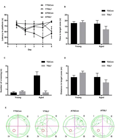
3.2. Behavioral Performance via MWM
suggested that 16-weeks’ Lf intervention might have no effect on the spatial learning and memory abilities of both young and middle-aged APP/PS1 mice.
Figure 2
3.3. Proteins Involved in Aβ metabolism and Phosphorylation of Tau Protein
p-Tau ser396 and p-Tau ser404 protein expression were observed from both hippocampus and cortex between TGCon and TGLf groups for both young and middle-aged mice (Figure 3A-D).
3.4. Proteins Involved in Neuro-inflammation and Synaptic Plasticity
between YTGCon and YTGLf group (Figure 4A&B). For the middle-aged APP/PS1 mice, a significant elevation in synaptophysin protein expression was observed in cortex from ATGLf group relative to ATGCon, while there was no significant difference for GFAP, Ibα1, BDNF and PSD95 protein expression in both hippocampus and cortex, as well as synaptophysin in hippocampus between ATGCon and ATGLf group (Figure 4C&D).
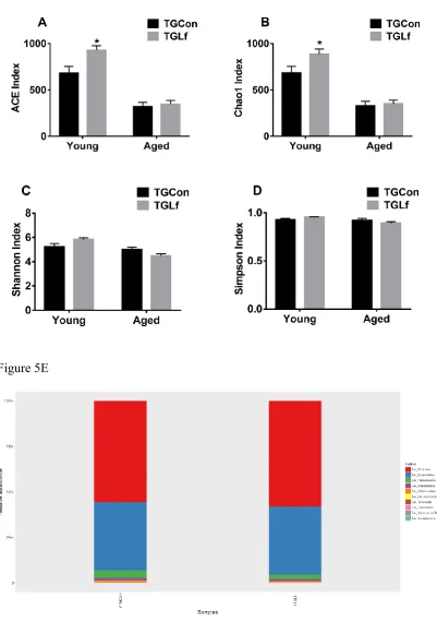
3.5. Compositions and Overall Structure of Gut Microbiota
group compared to YTGCon group (Figure 5A&B), while Shannon and Simpson index showed no significant difference between groups within the same age (Figure 5C&D). By the taxonomic analysis of OTU representative sequences, the distributions of the top 10 bacteria at phylum and genus level in young and middle-aged mice were shown in Figure 5E-H, respectively. For the young APP/PS1 mice, Firmicutes, Bacteroidetes, and Proteobacteria were the dominant bacteria at the phylum level
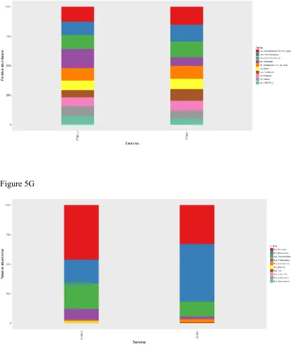
(Figure 5E), and Lachnospiraceae_NK4A136_group, Ruminiclostridium, and Ruminiclostridium_9 dominated the microbiota at the genus level (Figure 5F). For the
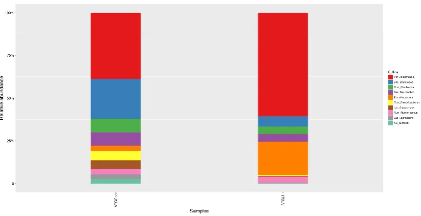
middle-aged APP/PS1 mice, the top 3 bacteria at the phylum level was Firmicutes, Bacteroidetes, and Verrucomicrobia (Figure 5G), at the genus level was Akkermansia,
Bacteroides and Oscillospira (Figure 5H). We further analyzed the difference of gut
microbiota at both phylum and genus level between each two groups within the same age by Mann-Whitney U test. As shown in table 1, for the young APP/PS1 mice, there was no significant difference for the top 10 bacteria between YTGCon and YTGLf group at the phylum level; while at the genus level, the relative abundance of Bacteroides and Alistipes of YTGLf group was lower than that of YTGCon group, and
the relative abundance of Oscillibacter in YTGLf group was higher than that of YTGCon group. For the middle-aged APP/PS1 mice, the relative abundance of the phylum Proteobacteria, as well as Oscillospira, Coprococcus and Ruminococcus at the genus level from ATGLf group was significantly reduced compared to ATGCon group.
composition (Figure 6A&B). Specifically, an evidence clustering was identified between the TGCon and TGLf group for both the young and middle-aged mice. The observation suggested that significant difference in gut microbial community structure existed between the YTGCon and YTGLf, as well as ATGCon and ATGLf. The two principal component scores accounted respectively for 27.61% and 18.22% of the total variations for the YTGCon and YTGLf, also 22.33% and 16.34% of the total variations for the ATGCon and ATGLf. We used LEfSe analysis to compare the statistical differences in microbial communities between TGCon and TGLf group for both the young and middle-aged mice. As shown in Figure 6C, there were eight bacterial biomarkers (i.e. Oscillibacter, Anaerotruncus, EF096579_g, EU454405_g, Mollicutes_RF39, EU474361_g, EU774448_g, and EF096976_g) significantly
Figure 5E
Figure 5G
Table 1 The relative abundance of the top 10 gut bacterial genera at the phylum and genus level (%) from both young and middle-aged
APP/PS1 mice
level YTGCon YTGLf level ATGCon ATGLf
phylum phylum
Firmicutes 55.43±4.00 57.74±4.18 Firmicutes 17.19±5.70 8.30±1.64
Bacteroidetes 37.62±4.77 37.37±4.17 Bacteroidetes 7.54±5.72 12.21±4.69
Proteobacteria 3.80±0.66 2.49±0.18 Verrucomicrobia 8.00±5.47 3.17±2.60
Actinobacteria 1.48±0.24 1.17±0.23 Proteobacteria 3.42±1.69 0.52±0.12*
Deferribacteres 0.54±0.05 0.44±0.04 Actinobacteria 0.41±0.16 0.68±0.29
Verrucomicrobia 0.36±0.09 0.19±0.05 [Thermi] 0.52±0.51 0.00±0.00
Tenericutes 0.19±0.08 0.21±0.06 TM7 0.05±0.03 0.19±0.12
Fusobacteria 0.17±0.10 0.05±0.03 Tenericutes 0.05±0.02 0.01±0.00
Cyanobacteria 0.05±0.04 0.01±0.01 Cyanobacteria 0.01±0.01 0.00±0.00
genus genus
Lachnospiraceae_NK4 A136_group
3.05±0.26 3.68±0.45
Akkermansia 8.00±5.47 3.17±2.60
Ruminiclostridium 2.69±0.18 3.48±0.61 Bacteroides 4.79±4.36 0.32±0.20
Ruminiclostridium_9 2.83±0.18 3.27±0.30 Oscillospira 1.64±0.56 0.22±0.07*
Bacteroides 3.88±0.75 1.71±0.29* Desulfovibrio 1.62±1.56 0.24±0.11
Rikenellaceae_RC9_gu t_group
2.49±0.48 2.64±0.32
Allobaculum 0.61±0.29 1.03±0.57
Blautia 1.96±0.21 2.13±0.20 Faecalibacterium 1.18±1.16 0.02±0.02
Oscillibacter 1.47±0.15 2.37±0.28* Coprococcus 1.03±0.77 0.03±0.01*
Roseburia 1.72±0.24 2.03±0.28 Ruminococcus 0.63±0.15 0.19±0.04*
4. Discussion
We demonstrated that 16 weeks of lactoferrin intervention in both young and middle-aged male APP/PS1 mice might have no effect on cognitive function, consistently, no alterations in key markers involved in Aβ, tau pathology, neuro-inflammation and synaptic plasticity were observed post Lf intervention. However, we demonstrated that Lf intervention could broadly affect gut microbiota profiles, and the effects might be different for young and middle-aged mice. To be specific, in the young APP/PS1 mice, Lf elevated α diversity index including ACE and Chao 1, and reduced the relative abundance of the genera Bacteroides and Alistipes and elevated Oscillibacter, in addition, Oscillibacter, Anaerotruncus,
EF096579_g, EU454405_g, Mollicutes_RF39, EU474361_g, EU774448_g, and
EF096976_g were specifically abundant post Lf intervention via LEfSe analysis. In
the middle-aged APP/PS1 mice, the relative abundance of the phylum Proteobacteria, as well as the genera Oscillospira, Coprococcus and Ruminococcus was significantly reduced post Lf intervention, additionally, S24_7, Bacteroidia, Bacteroidetes and Methylobacterium were specific via LEfSe analysis post Lf intervention.
intervention on cognition via different AD mouse models with specific emphasis on different forms of Lf.
Existing evidence suggest that Lf could modulate gut microbiota profiles in different species [16-18]. For example, in very low birth weight infants, two daily recombinant human lactoferrin intake from day 1-28 of life reduced Enterobacter and Klebsiella, while increased Citrobacter in feces [16]. Zhang et al. [18] reported that oral administration of Bovine Lactoferrin-Derived Lactoferricin (Lfcin) B could efficiently maintain gut microbiota homeostasis in enterohemorrhagic Escherichia coli (EHEC) O157:H7 mouse model. Our study is the very first to demonstrate that 16 weeks intervention of Lf might affect gut microbiota profiles differently in young and middle-aged APP/P1 mice.
(1) Effects of Lf on gut microbiota profiles in young APP/PS1 mice
nonalcoholic steatohepatitis patients [31]. Members of genus Bacteroides, i.e. Bacteroides fragil have been reported to excrete a serious of complex neurotoxins that
can boost inflammation including surface lipopolysaccharide (LPS) and toxic proteolytic peptides [32]. Elevated Alistipes has also been reported to be associated with improved gut microbiota composition after dietary intervention for HFD animals [33]. Additionally, Ma et al. [34] reported that genera Bacteroides and Alistipes were negatively associated with the maintenance of intestine redox in tea polyphenols treated HFD fed mice. The reduction of Bacteroides and Alistipes post Lf in young mice might also suggest these specific bacteria play function roles in the oxidative stress and anti-inflammatory response. The LEfSe analysis demonstrated that Oscillibacter, Anaerotruncus, EF096579_g, EU454405_g, Mollicutes_RF39,
EU474361_g, EU774448_g, and EF096976_g were specifically abundant in young
APP/PS1 mice post Lf intervention. The functional roles of these abundant bacteria post Lf intervention require further exploration.
(2) Effects of Lf on gut microbiota profiles in middle-aged APP/PS1 mice
genera Ruminococcus post Lf intervention suggest that Lf might improve gut microbiota profiles in middle-aged APP/PS1 mice, while the functional roles of reduction in Oscillospira and Coprococcus post Lf intervention require further exploration. The LEfSe analysis demonstrated that S24-7, Bacteroidia, Bacteroidetes and Methylobacterium were specifically abundant post Lf treatment from middle-aged APP/PS1 mice. Similar to those in young APP/PS1 mice, the functional roles of these abundant bacteria post Lf intervention in middle-aged APP/PS1 mice also require further exploration.
In summary, we demonstrated 16 weeks’ Lf intervention had no effect on cognitive function, and key AD related markers including Aβ, tau pathology, neuro-inflammation and synaptic plasticity from both young and middle-aged APP/PS1 mice; while Lf might exert diversified effects on gut microbiota profiles for APP/P1 mice with different ages. Our findings could indicate that dietary Lf might be beneficial for gut microbiota homeostasis although might have no effects on cognition.
Author Contributions: Conceptualization, Z.W., L.Q.Q. and J.Y.X.; Methodology &Investigation, H.Z., G.W., L.L. and W.D.; Writing-Original Draft Preparation, H.Z., G.W., J.Y.X. and Z.W.; Writing-Review & Editing, Z.W. and L.Q.Q.; Supervision, Z.W.; Funding Acquisition, Z.W. and L.Q.Q. All authors have read and agreed to the published version of the manuscript.
National Key R&D Program of China, and the Priority Academic Program Development of Jiangsu Higher Education Institutions (PAPD).
Cited References
1. Baker, E. N.; Baker, H. M., A structural framework for understanding the multifunctional character of lactoferrin. Biochimie 2009, 91, 3-10.
2. Legrand, D., Overview of Lactoferrin as a Natural Immune Modulator. J Pediatr
2016, 173 Suppl, S10-15.
3. Mulder, A. M.; Connellan, P. A.; Oliver, C. J.; Morris, C. A.; Stevenson, L. M., Bovine lactoferrin supplementation supports immune and antioxidant status in healthy human males. Nutr Res 2008, 28, 583-589.
4. Zhang, Y.; Lima, C. F.; Rodrigues, L. R., Anticancer effects of lactoferrin: underlying mechanisms and future trends in cancer therapy. Nutr Rev 2014, 72, 763-773.
5. Jacobsen, L. C.; Sorensen, O. E.; Cowland, J. B.; Borregaard, N.; Theilgaard-Monch, K., The secretory leukocyte protease inhibitor (SLPI) and the secondary granule protein lactoferrin are synthesized in myelocytes, colocalize in subcellular fractions of neutrophils, and are coreleased by activated neutrophils. J Leukoc Biol 2008, 83, 1155-1164.
6. Fillebeen, C.; Descamps, L.; Dehouck, M. P.; Fenart, L.; Benaissa, M.; Spik, G.; Cecchelli, R.; Pierce, A., Receptor-mediated transcytosis of lactoferrin through the blood-brain barrier. J Biol Chem 1999, 274, 7011-7017.
8. Guo, C.; Yang, Z. H.; Zhang, S.; Chai, R.; Xue, H.; Zhang, Y. H.; Li, J. Y.; Wang, Z. Y., Intranasal Lactoferrin Enhances alpha-Secretase-Dependent Amyloid Precursor Protein Processing via the ERK1/2-CREB and HIF-1alpha Pathways in an Alzheimer's Disease Mouse Model. Neuropsychopharmacology 2017, 42, 2504-2515. 9. Wang, L.; Sato, H.; Zhao, S.; Tooyama, I., Deposition of lactoferrin in fibrillar-type senile plaques in the brains of transgenic mouse models of Alzheimer's disease. Neurosci Lett 2010, 481, 164-167.
10. Kawamata, T.; Tooyama, I.; Yamada, T.; Walker, D. G.; McGeer, P. L., Lactotransferrin immunocytochemistry in Alzheimer and normal human brain. Am J Pathol 1993, 142, 1574-1585.
11. An, L.; Sato, H.; Konishi, Y.; Walker, D. G.; Beach, T. G.; Rogers, J.; Tooyama, I., Expression and localization of lactotransferrin messenger RNA in the cortex of Alzheimer's disease. Neurosci Lett 2009, 452, 277-280.
12. Alzheimer’s Disease International. World Alzheimer Report 2018 . London (GB): ADI; 2018. https://www.alz.co.uk/research/ WorldAlzheimerReport2018.pdf.
13. Jiang, C.; Li, G.; Huang, P.; Liu, Z.; Zhao, B., The Gut Microbiota and Alzheimer's Disease. J Alzheimers Dis 2017, 58, 1-15.
14. Zhao, Y.; Dua, P.; Lukiw, W. J., Microbial Sources of Amyloid and Relevance to Amyloidogenesis and Alzheimer's Disease (AD). J Alzheimers Dis Parkinsonism
2015, 5, 177.
disease. CNS Neurol Disord Drug Targets 2014, 13, 305-311.
16. Sherman, M. P.; Sherman, J.; Arcinue, R.; Niklas, V., Randomized Control Trial of Human Recombinant Lactoferrin: A Substudy Reveals Effects on the Fecal Microbiome of Very Low Birth Weight Infants. J Pediatr 2016, 173 Suppl, S37-42. 17. Hu, P.; Zhao, F.; Zhu, W.; Wang, J., Effects of early-life lactoferrin intervention on growth performance, small intestinal function and gut microbiota in suckling piglets. Food Funct 2019, 10, 5361-5373.
18. Haiwen, Z.; Rui, H.; Bingxi, Z.; Qingfeng, G.; Jifeng, Z.; Xuemei, W.; Beibei, W., Oral Administration of Bovine Lactoferrin-Derived Lactoferricin (Lfcin) B Could Attenuate Enterohemorrhagic Escherichia coli O157:H7 Induced Intestinal Disease through Improving Intestinal Barrier Function and Microbiota. J Agric Food Chem
2019, 67, 3932-3945.
19. Sun, H.; Liu, M.; Sun, T.; Chen, Y.; Lan, Z.; Lian, B.; Zhao, C.; Liu, Z.; Zhang, J., et al., Age-related changes in hippocampal AD pathology, actin remodeling proteins and spatial memory behavior of male APP/PS1 mice. Behav Brain Res 2019, 376, 112182.
20. Shen, L.; Liu, L.; Ji, H. F., Alzheimer's Disease Histological and Behavioral Manifestations in Transgenic Mice Correlate with Specific Gut Microbiome State. J Alzheimers Dis 2017, 56, 385-390.
Food Funct 2019, 10, 7299-7307.
22. Ling, C. J.; Min, Q. Q.; Yang, J. R.; Zhang, Z.; Yang, H. H.; Xu, J. Y.; Qin, L. Q., Lactoferrin Alleviates the Progression of Atherosclerosis in ApoE(-/-) Mice Fed with High-Fat/Cholesterol Diet Through Cholesterol Homeostasis. J Med Food 2019, 22, 1000-1008.
23. Takeuchi, T.; Matsunaga, K.; Sugiyama, A., Antidepressant-like effect of milk-derived lactoferrin in the repeated forced-swim stress mouse model. J Vet Med Sci 2017, 79, 1803-1806.
24. Chen, N.; Lei, T.; Xin, L.; Zhou, L.; Cheng, J.; Qin, L.; Han, S.; Wan, Z., Depot-specific effects of treadmill running and rutin on white adipose tissue function in diet-induced obese mice. J Physiol Biochem 2016, 72, 453-467.
25. Lv, M.; Yang, S.; Cai, L.; Qin, L. Q.; Li, B. Y.; Wan, Z., Effects of Quercetin Intervention on Cognition Function in APP/PS1 Mice was Affected by Vitamin D Status. Mol Nutr Food Res 2018, 62, e1800621.
26. Quast, C.; Pruesse, E.; Yilmaz, P.; Gerken, J.; Schweer, T.; Yarza, P.; Peplies, J.; Glockner, F. O., The SILVA ribosomal RNA gene database project: improved data processing and web-based tools. Nucleic Acids Res 2013, 41, D590-596.
27. Lozupone, C.; Lladser, M. E.; Knights, D.; Stombaugh, J.; Knight, R., UniFrac: an effective distance metric for microbial community comparison. ISME J 2011, 5, 169-172.
2011, 12, R60.
29. Carro, E.; Bartolome, F.; Bermejo-Pareja, F.; Villarejo-Galende, A.; Molina, J. A.; Ortiz, P.; Calero, M.; Rabano, A.; Cantero, J. L., et al., Early diagnosis of mild cognitive impairment and Alzheimer's disease based on salivary lactoferrin. Alzheimers Dement (Amst) 2017, 8, 131-138.
30. Liu, H.; Wu, H.; Zhu, N.; Xu, Z.; Wang, Y.; Qu, Y.; Wang, J., Lactoferrin protects against iron dysregulation, oxidative stress, and apoptosis in MPTP-induced Parkinson's disease in mice. J Neurochem 2019.
31. de Faria Ghetti, F.; Oliveira, D. G.; de Oliveira, J. M.; de Castro Ferreira, L.; Cesar, D. E.; Moreira, A. P. B., Influence of gut microbiota on the development and progression of nonalcoholic steatohepatitis. Eur J Nutr 2018, 57, 861-876.
32. Lukiw, W. J., Bacteroides fragilis Lipopolysaccharide and Inflammatory Signaling in Alzheimer's Disease. Front Microbiol 2016, 7, 1544.
33. Guo, W. L.; Pan, Y. Y.; Li, L.; Li, T. T.; Liu, B.; Lv, X. C., Ethanol extract of Ganoderma lucidum ameliorates lipid metabolic disorders and modulates the gut microbiota composition in high-fat diet fed rats. Food Funct 2018, 9, 3419-3431. 34. Ma, H.; Zhang, B.; Hu, Y.; Wang, J.; Liu, J.; Qin, R.; Lv, S.; Wang, S., Correlation Analysis of Intestinal Redox State with the Gut Microbiota Reveals the Positive Intervention of Tea Polyphenols on Hyperlipidemia in High Fat Diet Fed Mice. J Agric Food Chem 2019, 67, 7325-7335.
36. Konikoff, T.; Gophna, U., Oscillospira: a Central, Enigmatic Component of the Human Gut Microbiota. Trends Microbiol 2016, 24, 523-524.
37. Mancabelli, L.; Milani, C.; Lugli, G. A.; Turroni, F.; Mangifesta, M.; Viappiani, A.; Ticinesi, A.; Nouvenne, A.; Meschi, T., et al., Unveiling the gut microbiota composition and functionality associated with constipation through metagenomic analyses. Sci Rep 2017, 7, 9879.
38. Rajilic-Stojanovic, M.; Jonkers, D. M.; Salonen, A.; Hanevik, K.; Raes, J.; Jalanka, J.; de Vos, W. M.; Manichanh, C.; Golic, N., et al., Intestinal microbiota and diet in IBS: causes, consequences, or epiphenomena? Am J Gastroenterol 2015, 110, 278-287.
Figure legends
Figure 1 Glucose and Insulin Tolerance Test.
Glucose levels (A) at 15 min, 30 min, 45 min, 60 min, 90 min, 120 min and the total area under the curve (AUC) (B) during ITT. Glucose levels (C) at different timepoints and AUC (D).
Figure 2 Behavioral Performance via MWM
(A) Mean escape latency to hidden platform on five days of navigation test. During probe trails, time spent in the target quadrant (B), number of crossing the previous hidden platform (C), and swimming distance in the target quadrant (D) were recorded. (E) Representative motion tracking of the mice in the different groups. All values were presented as means ± SEM. * p<0.05 versus age-matched TGCon group.
Figure 3 Aβ aggregation and phosphorylation of Tau Associated Protein
Expression
In young APP/PS1 mice, protein expression of IDE, ADAM10, BACE1, Cathepsin B, P-Tau ser396 and P-Tau ser404 in hippocampus (A) and cortex (B) were detected by western blotting. In middle-aged APP/PS1 mice, the above proteins were shown in hippocampus (C) and cortex (D) via western blotting. All values were presented as mean + SEM. A.U. means arbitrary units. Representative blots were shown at right panel in A-D.
Figure 4 Neuro-inflammation Related Markers and Synaptic Plasticity
middle-aged APP/PS1 mice, the above proteins were shown in hippocampus (C) and cortex (D) via western blotting. All values were presented as mean + SEM. A.U. means arbitrary units. Representative blots were shown at right panel in A-D.
Figure 5 Alterations in Gut Microbiota Composition
The alpha diversity of ACE index (A), Chao1 index (B), Shannon index (C), Simpson index (D) displays the microbial diversity of each group. Relative abundance of top 10 microbes in young APP/PS1 mice at the rank of phylum (E) and genus (F), and in middle-age APP/PS1 mice at the rank of phylum (G) and genus (H) were shown.
Figure 6 PCoA based on the abundance of OTUs and LEfSe analysis