This is an author produced version of
Entropy-based EEG Time Interval Selection for
Improving Motor Imagery Classification
.
White Rose Research Online URL for this paper:
http://eprints.whiterose.ac.uk/100769/
Proceedings Paper:
Soleymanpour, R. and Arvaneh, M. (2017) Entropy-based EEG Time Interval Selection for
Improving Motor Imagery Classification. In: 2016 IEEE International Conference on
Systems, Man, and Cybernetics (SMC). 2016 IEEE International Conference on Systems,
Man, and Cybernetics (SMC 2016), 9 - 12th Oct 2016, Budapest, Hungary. IEEE . ISBN
978-1-5090-1897-0
https://doi.org/10.1109/SMC.2016.7844864
© 2016 IEEE. Personal use of this material is permitted. Permission from IEEE must be
obtained for all other users, including reprinting/ republishing this material for advertising or
promotional purposes, creating new collective works for resale or redistribution to servers
or lists, or reuse of any copyrighted components of this work in other works.
promoting access to
White Rose research papers
[email protected] http://eprints.whiterose.ac.uk/Entropy-based EEG
Time Interval Selection for
Improving Motor Imagery Classification
Rahim Soleymanpour
School of Electrical and Computer Engineering University of Science and Technology, Mazandaran
Behshahr, Iran [email protected]
Mahnaz Arvaneh
Dept. of Automatic Control and Systems Engineering University of Sheffield
Sheffield, United Kingdom [email protected]
Abstract: Classification of different motor imagery tasks using electroencephalogram (EEG) signals is challenging, since EEG presents individualized temporal and spatial characteristics that are contaminated by noise, artifacts and irrelevant mental activities. In most applications, the EEG time interval on which feature extraction algorithms operate is fixed for all subjects, whereas the start time and the duration of motor imagery-based brain activities can vary from subject to subject. To improve the classification accuracy, this paper proposes a novel entropy-based algorithm to accurately identify the time interval that motor imagery has been performed. The proposed algorithm searches through different time intervals across trials and finds the one with minimum irregularity. The hypothesis behind the proposed algorithm is that when motor imagery is performed, the activities of the neurons in the motor cortex tend to become more synchronized and less irregular. We evaluate our proposed algorithm using a publicly available motor imagery-based BCI dataset. The experimental results show that the proposed algorithm selects the EEG intervals leading to superior BCI performance compared to fixed EEG intervals that are commonly used for all subjects.
Keywords— brain-computer Interface, EEG time interval,
motor imagery, multi scale entropy
I. INTRODUCTION
Brain-computer interface (BCI) aims to improve the quality of life of people with severe disability by providing a new pathway for communication and control [1], [2]. This technology can replace, restore, improve, and supplement the natural central nervous system by analyzing and decoding brain signals and translating them to control commands [1], [2]. In BCI applications, electroencephalogram (EEG) is commonly used to measure electrical activities of brain due to its high temporal resolution and lower cost compared to other modalities such as functional magnetic resonance imaging (fMRI), functional near-infrared spectroscopy (fNIRS), etc.,
Several BCI systems operate based on EEG patterns generated by performing different motor imagery tasks. Performing motor imagery generally leads to short-term inhibition and suppression of the sensory rhythms across motor cortex, which are called event-related de-synchronization (ERD) and event-related de-synchronization (ERS) respectively [3], [4]. However, accurate detection of
performed motor imagery task is challenging due to noise and non-stationarity inherent in the recorded EEG signals. Consequently, advanced signal processing and machine learning algorithms are applied on short EEG time intervals to mitigate effects of noise and non-stationarity and subsequently improve the BCI performance [5]. As an example, common spatial pattern (CSP) is a commonly used algorithm in BCI that has a great influence on its performance [6]. The CSP algorithm is a spatial filter that maximizes the difference between variances of two classes of EEG signals. Nevertheless, the BCI performance greatly depends on the EEG frequency band and the time interval on which CSP is applied on [7], [8], [9].
There are several BCI research studies published on automatic selection of most discriminative EEG frequency bands before employing CSP [8]. However, relatively very few studies have focused on automatic selection of relevant EEG time intervals. Typically, the EEG time interval on which the CSP algorithm operates is unspecifically set to a fixed value for all subjects (e.g. 0.5 to 2.5 s after onset of cue) [10]. However, the start time and the duration of motor imagery-based brain activity can vary from subject to subject. Using a fixed EEG time interval for all subjects often leads to deteriorated BCI performance due to either excluding some relevant information or including irrelevant brain activities that cause great non-stationarity across trials. Ang et al. suggested to select a subject-specific time interval among a few options based on cross validation results on training data [8]. However, this is a very time consuming approach.
In this paper, we propose a new entropy-based algorithm to identify individualized time EEG time intervals for motor imagery BCIs. The hypothesis behind the proposed algorithm is that when the brain responses to a stimulus, in the corresponding brain region, activities of neurons become more regular and synchronized and less noisy in comparison with the neutral state [11]. Based on this hypothesis, our proposed algorithm identifies the best EEG time interval, which is more regular than other ones across trials, by using the entropy theory. Entropy metric is a well-known approach in measuring the irregularity of physiological signals [12]. Generally, entropy increases with the degree of disorder and is maximum for completely random systems. Sample entropy (SE) [13] and
permutation entropy (PE) [14] are two algorithms proposed to compute the degree of regularity in time series. Recently, authors in [15] have proposed a multiscale sample entropy (MSE) algorithm to improve the calculation of entropy for complex physiological signals. They showed the effectiveness of MSE in measuring the degree of irregularities using cardiac inter-beat signals.
In this paper, we propose a novel MSE-based EEG time interval selection algorithm. Our proposed algorithm measures the complexity and irregularity of the EEG signals, and identifies when the brain has started performing motor imagery and how long performing motor imagery has lasted. The effectiveness of the proposed algorithm is evaluated using the publicly available dataset 2a from BCI competition IV [16]. The performance of the proposed algorithm is also compared with the state-of-the-art algorithm where a fixed EEG time interval is used for all subjects.
II. METHODS
A. Multiscale Sample Enthropy (MSE)
MSE has been used as a measure of irregularities across multiple time scales. Costa et al. proposed an improved version of the MSE algorithm to show its effectiveness in measuring the degree of irregularities of cardiac interbeat time series [15]. In this paper, we adapted the MSE algorithm proposed in [15] to be applied on EEG signals. The following steps illustrate the proposed MSE algorithm for measuring the degree of irregulary across samples of an EEG signal recorded from a single channel.
1. Let
X
{
x
1,
x
2,..,
x
L}
denotes an EEG signal withL
samples recorded from a single channel. As suggested in [15, 17], the coarse-graining procedure is applied on the signalX
to construct the new signalY
{
y
b}
Cb1with the length of C . In fact, the coarse-graining procedure divides
X
to C non-overlapping epochs with a length of
each. Thereafter,y
bis calculated by averaging the EEG samples of theb
th epoch (see Fig. 1 as examples). This procedure can be summarized as:( ) ( 1) 1 1 1 , i i b b i L y x i C
(1) where is called the scale factor. The coarse-graining procedure not only reduces the computation time of the MSE algorithm by down-sampling the signal but also attenuates the high frequency noise.2. Given
Y
, in this step, the vectors of m consecutive data points are formed asY
,m( ) {
i
y
i,
y
i1,...,
y
i m 1}
, wherei
{1, 2,...,
c
m
1}
. m is referred to as the embedding dimension.3. In the third step, the distances between all the possible pairs of
Y
,m( )
i
andY
,m( )
j
are calculated, where,
{1, 2,...,
1}
i j
c
m
andi
j
. The lower distance between two vectors indicates more similarity and less irregularity between them. We used Chebyshev method to measure the distance between two vectors which is the greatest of their differences along any coordinate dimensions. Indeed, other distanse methods can be also used to do this. Next, the number of paired vectors that had distances less thanr
are calculated, wherer
is a small predefined value. We will explain in Section IV how we chose ther
value in this study. Subsequently, we calculate Bm( )r as the probebility of having,m ,m
[Y ( ), Y ( )] ,
d i j r i j for the given
Y
.4. In this step, , 1
( )
m
B r is calculated by repeting the second and the third steps with m+1.
5. Consequently, MSE is defined as:
, 1 , ( ) ( , m, , ) ln . ( ) m m B r MSE r X B r (2)
It should be added that a lower value of MSE implies more similarity in time series.
Fig. 1. The scheme illustrating how to construct the coarse-grained data from original data over two different scales, adapted from [17].
B. The proposed MSE-based interval selection algorithm
The hypothesis behind the proposed algorithm is that when the brain responses to an external stimulus, the activities of the neurons in the associated regions tend to become more synchronized. In other words, during a short time interval when the user is performing motor imagery, the brain signals generated from motor cortex become more regular, and irrelevant neural activities as well as muscle artifacts are minimized. Based on this hypothesis, in this paper, we investigate whether or not MSE can be used to identify when the brain has started performing motor imagery and how long performing motor imagery has lasted.
The proposed MSE-based EEG interval selection algorithm is applied on band-passed EEG data. Since the CSP algorithm changes the spatial structures of the EEG signals, we apply the
proposed method before the CSP algorithm to check the generated patterns of brain signals before any changes in their spatial structure.
In the first step of our proposed MSE-based interval selection algorithm, the EEG trials of the training set recorded from motor cortex (i.e. C3, C4, Cz) are divided to a number of overlapping intervals. Fig. 2 displays how an EEG trial is divided into multiple intervals. In this figure, T and T respectively indicate the length of the EEG interval and the sliding shift from the previous EEG interval. The procedure of dividing an EEG trial to multiple sub-windows can be repeated with different values of T and T. In this study, we set T to the fixed value of 0.5 s to reduce the search space and consecuently the computational time.
Fig. 2: Dividing an EEG trial to n+1 intervals with the length of T
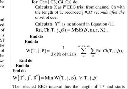
Algorithm 1 presents the proposed MSE-based EEG interval selection algorithm. As shown in Algorithm 1, the average MSE of each EEG interval is calculated across all the training trials and all the selected motor cortex channels (i.e. C3, C4, Cz) using
W T j ß
, ,
, where T is the length of the EEG interval started j T seconds after the onset of the cue. Thereafter, the EEG interval that resulted in the minimum average MSE is selected as the time interval with minimum irregularity during performing motor imagery. We believe that this interval is an effective EEG interval for discriminating between the two performed motor imagery tasks as in this time interval the irrelevant brain activities that cause irregularity and non-stationarity in the activities of motor cortex are minimum. We will validate this hypothesis in the result section by comparing the classification accuracy obtained using the MSE-based selected time intervals with the results of the state-of-the-art BCI algorithm that uses a fixed EEG time interval for all the subjects.Algorithm 1 MSE-based EEG interval selection
W(T,j,ß) is the average MSE of EEG intervals with the length T started j T seconds after the onset of cue,
m is embedding dimension (in this study m= 2), r is matching threshold (in this study r= 0.2* std),
T= sliding shift (in this study 0.5 sec), for
= 1: number of scale factors do for different lengths of T do for j= 0: n dofor i= 1: number of training trials do
for Ch= [ C3, C4, Cz] do
Calculate X as ith EEG trial from channel Ch with
the length of T, recorded j T seconds after the onset of cue,
Calculate
Y
as mentioned in Equation (1),
R(i,
Ch T j
, , , )
MSE
( , , , X)
m r
, End do End do
of trial 1 s R(i, , , , ), 1 of trials , , 3 i ch Ch T j W T j ß
End do End do End do
* * *
Min
,
,
,
,
,
, ,
W T
j
ß
W
T
j
ß
T
j
The selected EEG interval has the length of T* and starts
j* T sec after onset of stimulus.
III. EXPERIMENTS
A. Data description
In this paper, we used the publicly available dataset 2a from BCI Competition IV [16]. This dataset has been provided by the BCI research group at the University of Gras. It includes four different motor imagery tasks; left hand, right hand, both feet, and tongue. In this study, we use only left hand and right hand motor imagery tasks. The signals were recorded from nine subjects using 22 EEG channels with the sampling rate of 250 Hz. The EEG data for each subject comprised of a training and a testing set of which each set included 72 trials for each motor imagery task. The training and test sessions were conducted on different days. Each trial began with a preparation beep sound. After 2 s a visual cue instructed the participants to perform one of the four above-mentioned motor imagery tasks for 4 s followed by a few seconds rest.
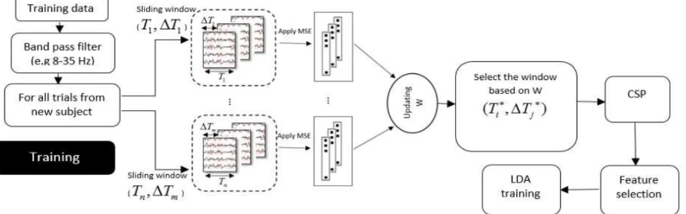
B. Data processing
Fig. 3 shows the proposed procedure applied for classification of motor imagery signals. As shown in Fig. 3, first, 8-35 Hz band pass filter was applied on the raw EEG signals since this frequency band included the range of frequencies that are mainly involved in performing motor imagery [18]. The filtering was performed using a Chebyshev filter. In the training phase, the next step was dividing the EEG signals into a number of time windows and selecting the EEG time window with the lowest average MSE as described in Section II.b. Thereafter, the spatially filtered signals were obtained using the first and the last three spatial filters of CSP. Finally, the variances of the spatially filtered signals were applied as the inputs of the LDA classifier. Note that, in this paper, we did not reject any trials or electrodes.
As mentioned, we assume that T is fixed to 0.5s in order to reduce the research space. The maximum value of time
window is chosen 4.5s, because of the timing of the paradigm of the dataset.
IV. RESULT AND DISCUSSION
A. Selecting the parameters of the proposed algorithm The parameters of the proposed MSE-based EEG interval selection algorithm should be carefully chosen as their values may have great influence on the performance. These parameters include the matching threshold (r), the embedding dimension (m), the length of the coarse-grained signal (C), and the scale factor (ß) [19]. In the following, we discuss how to define the values of the abovementioned parameters.
Matching threshold (r): Recent studies on MSE suggested to set the r value as either 0.15 or 0.2 of the standard deviation of the signal [12, 13]. Accordingly, we defined the r value as 0.2 of the standard deviation of the investigated EEG time interval as it achieved better classification results in our pilot experiment compared to the other suggested value.
Embedding dimension (m): the value of 2 was assigned for this parameter as suggested in [19].
Data length (C): The minimum data length for calculating the sample entropy equals to 10m, however, for a better estimation higher values such as 20m are recommended [20]. Since, the length of data is different using different scale factors, we propose using the following equation to identify the minimum length of the data under examination:
(
)
10 ,
m1,
, max,
sf
T
N
(3)where fs and T are respectively the sampling rate and the
duration of the EEG interval in second. m and ß indicate embedding dimension and scale factor, respectively.
Scale factor (ß): For different lengths of EEG intervals, the maximum value of ß is selected such that (3) is satisfied. For example, since in this study fs and m are 250Hz and 2
respectively, ß should be 4 or less for 2 s time windows. B. Performance Comparison
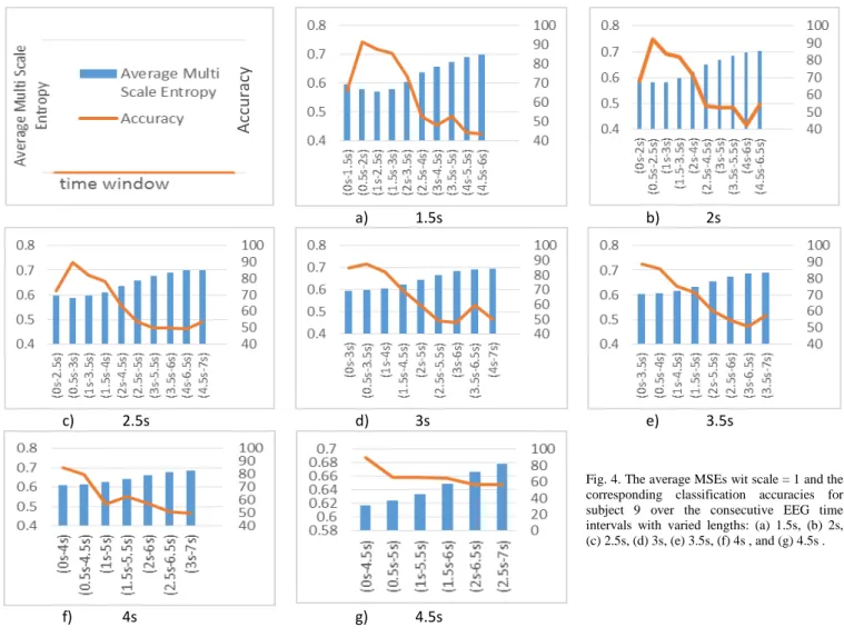
Fig. 4 presents the average MSEs (for ß=1) as well as classification results of different EEG time intervals for subject 9. In Fig. 4, the x-axes present different EEG time intervals, and the right and left y-axes indicate the classification accuracy and the average MSE, respectively. Besides, different sub-figures are corresponding to different lengths of EEG intervals.
As Fig. 4 shows, EEG time intervals with lower average MSEs resulted in higher classification accuracies. Indeed, this behavior can be observed in all the subfigures. In addition, the duration of an EEG interval has a great influence on the classification result. For example, when the time window is greater than 3.5s, the average MSE increases and the classification accuracy decreases, since the time window exceeds the duration of motor imagery and includes other irrelevant neural activities. On the other hand, when the EEG interval is too short, some parts of relevant information is missed resulting in a degraded classification accuracy.
Comparing the average MSE values across all the subfigures, we can see the window (1s-2.5s) in Fig 4.a has the lowest average MSE. Indeed, the highest obtained classification accuracy also belongs to this time interval. In summary, this figure clearly demonstrates the effectiveness of the proposed MSE-based EEG interval selection algorithm for subject 9.
As demonstrated in Table I, to evaluate the influence of different scale factors, the classification results for all nine subjects were obtained using EEG time intervals that were selected based on different scale factors. Table I shows that on average the value 3 for the scale factor yielded the highest average classification accuracy. However, the results of the scale factor 3 are not statistically better than the results obtained using the other scale factors (p>0.05). According (3), selecting large scales may make calculating MSE for short EEG intervals inaccurate. For example, the scale greater than 3 for the length 1.5s is not acceptable. On the other hand, the computation time of the MSE algorithm is remarkably faster when the scale is larger due to down sampling of the signal.
In our proposed algorithm, we calculated the MSE values for all the possible scale factors over a number of multiple EEG time intervals. Thereafter, the time interval with the lowest MSE is selected for feature extraction and classification. The results of our proposed algorithm based on considering all possible scale factors are presented in Table II.
Table II shows that the proposed algorithm outperformed the state of the art algorithms where fixed EEG intervals were used for all the subjects. Our proposed algorithm performed on
average 3.5%, 5.6%, and 6.1% better than using the fixed time window of (0.5s to 2.5s), (0s to 3s) and (1.5s to 3s) respectively. Interestingly, the paired t-test revealed that compared to the results of using the fixed EEG time intervals of (0 s to 3s) and (1.5s to 3s), the superior performance of our algorithm is statistically significant (i.e. p=0.002).
V. CONCLUSION
In this paper, we proposed an algorithm for automatic identification of the most relevant EEG time windows applied for motor imagery classification. We believe by initiation of a motor imagery task, synchronization of the relevant neuronal population decreases the irregularity of the corresponding brain signals. To this line, we proposed an entropy-based approach, to select the EEG time interval with minimum irregularity across trials. The classification accuracy in the BCI application has been increased by choosing this new dynamic window for our feature extraction step. The success of our proposed algorithm in improving BCI performance was presented using a publicly available motor imagery dataset. The experimental results suggest that the concept of entropy can be potentially
a) 1.5s b) 2s
c) 2.5s d) 3s e) 3.5s
Fig. 4. The average MSEs wit scale = 1 and the corresponding classification accuracies for subject 9 over the consecutive EEG time intervals with varied lengths: (a) 1.5s, (b) 2s, (c) 2.5s, (d) 3s, (e) 3.5s, (f) 4s , and (g) 4.5s . f) 4s g) 4.5s
A
cc
u
rac
y
used in many different EEG-based applications to extract more informative temporal features.
REFERENCES
[1] J. R. Wolpaw, , et al. “Brain–computer interfaces for communication and control,” Clin. Neurophysiol., vol. 113, no. 6, pp. 767–791, 2002. [2] E. A. Curran and M. J. Stokes, “Learning to control brain activity: a
review of the production and control of EEG components for driving brain–computer interface (BCI) systems,” Brain Cogn., vol. 51, no. 3, pp. 326–336, 2003.
[3] G. Pfurtscheller and C. Neuper, “Motor imagery activates primary sensorimotor area in humans,” Neurosci. Lett., vol. 239, no. 2, pp. 65– 68, 1997.
[4] G. Pfurtscheller and F. L. Da Silva, “Event-related EEG/MEG synchronization and desynchronization: basic principles,” Clin. Neurophysiol., vol. 110, no. 11, pp. 1842–1857, 1999.
[5] A. Bashashati, et al. “A survey of signal processing algorithms in brain– computer interfaces based on electrical brain signals,” J. Neural Eng., vol. 4, no. 2, p. R32, 2007.
[6] H. Ramoser, J. Muller-Gerking, and G. Pfurtscheller, “Optimal spatial filtering of single trial EEG during imagined hand movement,” IEEE Trans. On Rehabil. Eng. vol. 8, no. 4, pp. 441–446, 2000.
[7] G. Dornhege, et al. “Combined optimization of spatial and temporal filters for improving brain-computer interfacing,” IEEE Trans. On Biomed. Eng. vol. 53, no. 11, pp. 2274–2281, 2006.
[8] K. K. Ang, et al.“Filter bank common spatial pattern (FBCSP) in brain-computer interface,” IEEE International Joint Conference on Neural Networks, pp. 2390–2397,2008.
[9] H. Ghaheri and A. Ahmadyfard, “ Temporal windowing in CSP method for multi-class Motor Imagery Classification,” 20th Iranian Conference in Electrical Engineering( ICEE), pp. 1602–1607,2012. [10] M. Arvaneh, et al., “EEG data space adaptation to reduce intersession
nonstationarity in brain-computer interface,” Neural Comput., vol. 25, no. 8, pp. 2146–2171, 2013.
[11] M. M. Churchland, et al., “Stimulus onset quenches neural variability: a widespread cortical phenomenon,” Nat. Neurosci., vol. 13, no. 3, pp. 369–378, 2010.
[12] M. U. Ahmed and D. P. Mandic, “Multivariate multiscale entropy: A tool for complexity analysis of multichannel data,” Phys. Rev. E, vol. 84, no. 6, p. 061918, 2011.
[13] J. S. Richman and J. R. Moorman, “Physiological time-series analysis using approximate entropy and sample entropy,” Am. J. Physiol.-Heart Circ. Physiol., vol. 278, no. 6, pp. H2039–H2049, 2000. [14] C. Bandt and B. Pompe, “Permutation entropy: a natural complexity
measure for time series,” Phys. Rev. Lett., vol. 88, no. 17, p. 174102, 2002.
[15] M. Costa, A. L. Goldberger, and C.-K. Peng, “Multiscale entropy analysis of complex physiologic time series,” Phys. Rev. Lett., vol. 89, no. 6, p. 068102, 2002.
[16] M. Tangermann, et al. “Review of the BCI competition IV,” Front Neurosci, vol. 6, no. 55, p. 2, 2012.
[17] M. Costa, A. L. Goldberger, and C.-K. Peng, “Multiscale entropy analysis of biological signals,” Phys. Rev. E, vol. 71, no. 2, p. 021906, 2005.
[18] J. Müller-Gerking, G. Pfurtscheller, and H. Flyvbjerg, “Designing optimal spatial filters for single-trial EEG classification in a movement task,” Clin. Neurophysiol., vol. 110, no. 5, pp. 787–798, 1999.
[19] B. J. Gow, et al., “Multiscale entropy analysis of center-of-pressure dynamics in human postural control: methodological considerations,” Entropy, vol. 17, no. 12, pp. 7926–7947, 2015.
[20] M. Kirchner, et al, “Evaluation of the temporal structure of postural sway fluctuations based on a comprehensive set of analysis tools,” Phys. Stat. Mech. Its Appl., vol. 391, no. 20, pp. 4692–4703, 2012.
TABLE II.COMPARING CLASSIFICATION RESULTS OF THE PROPOSED ALGORITHM WITH THE RESULTS WHEN FIXED EEG INTERVALS ARE
APPLIED FOR ALL SUBJECTS
fixed time window proposed
MSE-base algorithm (0.5s-2.5s) (0 s-3s) (1.5s-3s) selected
time Acc. Scale
sub 1 91.5 86.8 82.1 (0.5s-4s) 95.3 5 sub 2 45.3 60.4 54.7 (0s-1.5s) 66 3 sub 3 97.2 93.4 92.5 (0.5.-3s) 98.2 4 sub 4 65.1 59.4 65.1 (1s-4s) 66 4 sub5 68.9 62.3 63.2 (0.5s-2.5s) 68.9 3 sub 6 57.5 62.3 65.1 (2s-4.5s) 69.8 3 sub 7 74.5 67 63.2 (0.5s-3.5s) 68.9 4 sub 8 95.3 95.3 92.5 (1.5-3.5s) 93.4 3 sub 9 92.5 82.1 85.8 (0.5s-2.5s) 92.5 5 Mean 76.4 74.3 73.8 79.9
TABLE I. CLASSIFICATION RESULTS AND THEIR CORRESPONDING SELECTED TIME INTERVALS OF THE PROPOSED ALGORITHM FOR DIFFERENT SCALE FACTORS
Scale=1 Scale=2 Scale=3 Scale=4 Scale=5
Subject selected
time Acc. selected time Acc.
selected time Acc. selected time Acc. Selected time Acc. sub 1 (0s-1.5s) 83 (0s-1.5s) 83 (0.5s-3s) 92.5 (0.5s-4s) 95.3 (0.5s-4s) 95.3 sub 2 (0s-1.5s) 66 (0s-1.5s) 66 (0s-1.5s) 66 (0s-2s) 57.5 (0s-2.5s) 52.8 sub 3 (0s-1.5s) 78.3 (0s-2s) 84 (0.5.-3s) 98.2 (0.5.-3s) 98.2 (0s-2.5s) 89.6 sub 4 (1s-4s) 66 (1.5s-4s) 64.2 (1s-4s) 66 (1s-4s) 66 (1s-4s) 66 sub5 (0.5s-2s) 68 (0.5s-2.5s) 68.9 (0.5s-2.5s) 68.9 (0.5s-3.5s) 59.4 (1.5s-4s) 63 sub 6 (2s-4.5s) 69.8 (2s-4.5s) 69.8 (2s-4.5s) 69.8 (2s-4.5s) 69.8 (2s-4.5s) 69.8 sub 7 (1.5s-3s) 63.2 (0.5s-3.5s) 68.9 (1s-3.5s) 61.3 (0.5s-3.5s) 68.9 (0.5s-2.5s) 74.5 sub 8 (1s-2.5s) 86.8 (1.5-3.5s) 93.4 (1.5-3.5s) 93.4 (1s-3.5s) 95.3 (1s-3.5s) 95.3 sub 9 (1s-2.5s) 87.7 (0.5s-3s) 89.6 (0.5s-2.5s) 92.5 (0.5s-2.5s) 92.5 (0.5s-2.5s) 92.5 Mean 74.3 76.4 78.7 78.10 77.6
![Fig. 1. The scheme illustrating how to construct the coarse-grained data from original data over two different scales, adapted from [17]](https://thumb-us.123doks.com/thumbv2/123dok_us/9000478.2797866/3.893.133.819.81.1127/scheme-illustrating-construct-coarse-grained-original-different-adapted.webp)