Modeling of Urinary Bladder Deformation Within Passive and Active
Regimes
I. Vlastelica1, D. Veljkovic1, V. Rankovic1, B. Stojanovic1, M. Rosic2, M. Kojic1,3
1 Center for Scientific Research of Serbian Academy of Sciences and Arts and University of
Kragujevac
Jovana Cvijica bb, Kragujevac, Serbia e-mail: [email protected]
2 Institute for Physiology, Faculty of Medicine, University of Kragujevac
Svetozara Markovica 69, Kragujevac, Serbia
3 Harvard School of Public Health, Harvard University
665 Huntington Ave., Boston, MA 02115, USA
Abstract
Finite element model for urinary bladder mechanical response during passive filling and muscle activation is presented in the paper. The nonlinear elastic model for the tissue is used according to the Fung potential for the strain energy. Filling of the bladder is simulated by the pressure increase with the passive state of muscles. When the filling phase is completed, muscles are activated with the tripled pressure increase. This modeling is in accordance with the real physiological processes. Data are used from experiments on the rabbit urinary bladder.
The computed distribution of stresses reveals non-uniform increase of stresses within muscle fibers during activation.
Key-words: urinary bladder, FE modeling, Fung’s tissue model, muscle activation
1. Introduction
Urinary bladder is an organ which functions by increasing its volume and shape during filling phase, followed by empting process when it returns to approximately initial shape. Between the filling and empting phases, a jump of the internal pressure occurs due to activation of muscles within the bladder wall tissue. The proper functioning of the urinary bladder depends on the activation which is related to the pressure increase and to the stress jump within the muscle fibers.
It is of interest from the physiological (medical) point of view to investigate the mechanical stress/strain states generated during urinary bladder filling, activation and empting phases. The aim of this report is to elucidate stress generation within the tissue by using one of the common biological models for the passive response and a nonlinear model for generation of active stresses within muscle fibers.
The measured data about the constitutive behavior of the urinary bladder tissue are used to determine material constants of the Fung 2D model by a fitting procedure.
2. Finite element model
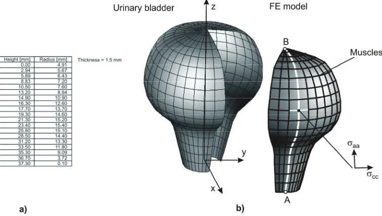
The urinary bladder is modeled by shell finite elements (Dvorkin and Bathe [2], Kojic et al. [3], [4]). The bladder is approximated by a body with axial symmetry and one quarter of the shell model is discretized, as shown in Fig. 1. The wall profile and thickness given in the figure corresponds to measurements in vitro of a rabbit urinary bladder. The muscle fibers are modeled by line elements in the axial and circumferential directions.
Fig. 1. Finite element model of a rabbit urinary bladder. a) Coordinates of the wall profile; b)
Finite element model. The wall is discretized by 4-node finite elements and muscles are represented by line elements in the axial and circumferential directions. One quarter of the bladder is modeled due to symmetry conditions. The stresses within the tissue are axial stress
aa
σ in the direction of the tangent to the wall profile, and circular (circumferential) stress σcc.
It is assumed that the wall tissue behaves as nonlinear elastic material with hardening typical for biological materials [3]. We use here a Fung’s potential for the strain energy function of the form (Fung et al. [5], Fung [6])
(
2 2)
1 1 2 2 4 1 2
exp 2 1
2
c
W = ⎡⎣ a E +a E + a E E − ⎤⎦ (1)
where c(dimension of stress),and , , (dimensionless)a a a1 2 4 are material constants; and E E1, 2 are the Green-Lagrange strains (in the principal strain directions). The principal Piola-Kirchhoff stresses are:
1 1
W S
E
∂ =
∂ , 2 2
W S
E
∂ =
The principal stresses Cauchy stresses σ1 and σ2 follow from the relation / 2
i i i
S =σ λ as
(
)
(
)
(
)
(
)
2 2 2
1 1 1 1 4 2 1 1 2 2 4 1 2
2 2 2
2 2 2 2 4 1 1 1 2 2 4 1 2
exp 2
exp 2
c a E a E a E a E a E E
c a E a E a E a E a E E
σ λ
σ λ
= + + +
= + + + (3)
where λ λ1, 2 are the stretches. The constitutive matrix Cij = ∂σi /∂ej is obtained by the proper differentiations of the above expressions for stress, where ej are small strains with the configuration at start of load step (or at last equilibrium iteration) as the reference configuration. In these differentiations we have to use the relations: dEi/∂ =λi λi and ∂ei /∂ =λi λi.
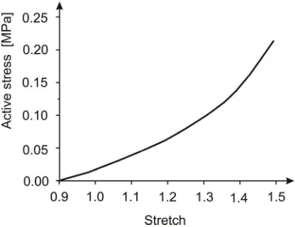
The muscle fibers are modeled by line finite elements. During passive state it is assumed that the fibers have the same characteristics as tissue, i.e. there is no additional stiffness of the wall. When the muscle net is activated, the active stresses are generated. The active stress within a muscle fiber is expressed as
( ) ( )
0a a
i fi t i
σ = σ λ (4)
where a
i
σ acts in the fiber direction ni, a
( )
if t is the activation function (function of time), and
( )
0 iσ λ is the reference stress which depends on the fiber stretch λi. We used here the reference stress dependence on stretch to correspond to a hardening stress-stretch relationship shown in Fig. 2.
Fig. 2. Activereference stress within muscle fiber in terms of the fiber stretch σ λ0
( )
.3. Results
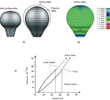
elastic model differs from that obtained from Fung’s model and has the character which does not agree with experiments. Also, the principal strains are in the axial and circumferential directions due to axial symmetry and the Fung’s model with the only normal strains is applicable.
Fig. 3. Urinary bladder deformation. a) Initial and deformed configuration before activation; b)
Field of effective strain, 2
3 ij ij
e= e e , at the end of filling (activation); c) Pressure-volume
relationship obtained using Fung’s nonlinear elastic 2D model. Data: linear elastic material model (E=0.05MPa,ν =0.49) and Fung’s model (equation (1):
1 2 4
0.003372, 0.6, 0.43, 0.49
C= a = a = a = ).
Material constants for the models are given in the figure caption of Fig. 3.
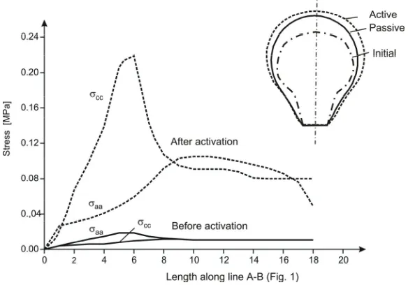
axial and circumferential fibers at end of filling and after activation is shown in Fig. 4. Before activation axial and circular stresses are relatively small and close to each other in the entire domain. The pressure is increased during activation for 300%, as observed experimentally for human urinary bladder. It can be seen that very large increase of stresses within fibers occurs due to the activation. The increase of circular stress is very high in the lower region because the radius of the bladder changes significantly in that region. On the other hand the axial stress increases gradually from the bottom to the top and reaches a plateau in the middle upper part.
Fig. 4. Distribution of axial (σaa) and circumferential (σcc) stresses along the line A-B shown in Fig. 1, before and after activation (Fung’s model).
4. Conclusions
From the presented results of the analysis it can be seen that useful information can be obtained about the stress-strain state within the urinary bladder wall during the filling phase and during activation. Stresses and strains during the filling phase are small and almost uniform. However, the stresses reach very high values when activation of muscle fibers occurs and the distribution is non-uniform, what can be important insight with respect to the bladder functioning.
Acknowledgments
References
[1] N. Zdravkovic, Solution of problem of connective tissue and muscle mechanics by finite element method, Ph. D. thesis (in Serbian), Faculty of Mechanical Engineering, University of Kragujevac, 2000.
[2] Dvorkin E. N., Bathe K. J., A continuum mechanics based four-node shell element for general nonlinear analysis, Eng. Comput., 1, 77-88, 1984.
[3] Kojic M, Slavkovic R, Zivkovic M, Grujovic N (1998). The Finite Element method-Linear Analysis. (in Serbian) Faculty of Mech. Engrg., Univesity of Kragujevac, Serbia.
[4] M. Kojic, N. Filipovic, B. Stojanovic, N. Kojic, Computer Modeling in Bioengineering – Theoretical Background, Examples and Software, J. Wiley and Sons, in press. [5] Fung Y C, Fronek K, Patitucci P, Pseudoelasticity of arteries and the choice of its
mathematical expression, Am. J. Psyhol. 237, H620-H631, 1979.