GSJ: Volume 6, Issue 11, November 2018, Online: ISSN 2320-9186
www.globalscientificjournal.com
Epidemiology of Acute Lymphoblastic Leukemia
with reference to prevalence and genetics
Muhammad Zubair, Shabana Khadim, Tayyaba Razzaq, Tahara Ashraf, Maham Riaz, Nafees Alam, Sabila Afzal , Syed shakeel Shah , Muhamamd Shahid
Abstract: Acute lymphoblastic leukemia is an internecine disarray of lymphoid cells that affects the both adolescent and being mature. Acute lymphoblastic leukemia also called acute lymphocyte leukemia is a kind of leukemia in which too many lymphocytes in bone marrow. Pakistan and western countries are combative with acute lymphoblastic leukemia in an alpine degree. This cancer participates 32% of all cancers. It is amassed bourgeois in children and it is more common in boys than in girls as the male to female ratio is 1.3 to 1. Acute lymphoblastic leukemia is caused by chromosomal aberrations like deletions, translocations, rearrangements, hyperdiploidy and hypodiploidy etc. Advances in our discerning of etiology of acute lymphoblastic leukemia suggest that refashion upstairs of this disease peculiarly target the genetic imperfection of acute lymphoblastic leukemia cells. In acute lymphoblastic leukemia, the lymphocytes are not adequate to conflict contagiousness anemia effortless hemorrhage. Acute lymphoblastic leukemia can compass to our central nervous system. Speculation remain meager in adolescent and being mature. In this review we provide a concise examination of pathobiology studies in acute lymphoblastic leukemia with a focus on treatments genetics and sub types. The possible risk factors include environmental factors, genetic and different transfusion agents. The disease associated with acute lymphoblastic leukemia has been also reviewed. We also reviewed diagnosis and different therapies for the treatment of acute lymphoblastic leukemia.
Keywords: Acute lymphoblastic leukemia, cancer of lymphocytes, childhood cancer, genetics of ALL, types of ALL, treatment of ALL
—————————— ——————————
Introduction:
Acute lymphoblastic leukemia also called acute lymphocyte leukemia is a type of leukemia in which there are many lymphocytes in the bone marrow and red blood cells. Lymphocytes are the type of A granulocytes and A granulocytes are the type of white blood cells. Lymphocytes helps make up the body’s defense system. Acute lymphoblastic leukemia is more common in children, it can also affect adults.
Acute lymphoblastic leukemia in the western countries has been well described. Acute lymphoblastic leukemiais most common cancer seen in children accounting in 25 %. Boys are affected more with a sex ratio 1.3:1.The age distribution in developed countries shows a marked value early peak between 2-5 years followed by small peak between 10-12 years. Reasons of these difference may because of less number of common Acute lymphoblastic Leukemiain early peak and more T cells in the second peak .(Yasmeen and Ashraf 2009)
Muhammad Zubair +92 3049521781
([email protected]) under graduate at university of Narowal, Punjab, Pakistan
Shabana Khadim ([email protected]) Tayyaba Razzaq ([email protected]) Tahara Ashraf ([email protected]) Maham Riaz ([email protected]) Nafees Alam
Corresponding author: Sabila Afzal
[email protected] Muhammad zubair
In acute lymphoblastic leukemia the lymphocytes are not able to fight well against the infections. The number of lymphocyte increase in blood and bone marrow. There is less room temperature for healthy red blood cells, white blood cells and platelets. This may cause many infections such as anemia and easy bleeding. Acute lymphoblastic leukemia may also spread to our central nervous system easily.(Sharma and Sarangdevot 2012)Leukemia first case was reported by HughesBenet in 1845 in an adult. Leukemia in child was first reported by Dr Henry Fuller in 1846. Leukemia disease remained common lethal until the discovery of effective chemotherapy. Over 50 years many new methods of diagnosis and treatment of this disease have evolved which lead to improve survival.(Yasmeen and Ashraf 2009.)
Genetics of the acute lymphoblastic Leukemia:
acute lymphoblastic leukemia genome. Beyond hyperdiploidy, ETV6-RUNX1 fusion, the other classical lesions, there is whole world somatic mutations, short deletions, translocations and epigenetic lesions that can riddle the coding and non coding acute lymphoblastic leukemia genome and the affected cells are collectively called as leukemic cells. Some lesions are targetable today with available agents like involving kinases in the newly recognized Ph-like leukemias. The deletion occur at the IKZF1 lkaros gene.
Moriyama also talk about inherited genetic variations in childhood acute lymphoblastic leukemia. Studies have shown that genetic variants are the major cause of interpatient variability in acute lymphoblastic leukemia. The relative risks of the acute lymphoblastic leukemiaare modest and may not justify. Notable exceptions are present such as high rate of the inherited TP53 variants (Li Fraumeni Syndrome) in rare low-hypo diploid acute lymphoblastic leukemia. The situation may be change in future by medication and therapy. Jabbour provide an overview of the development of the monoclonal antibodies in the acute lymphoblastic leukemia patients. Leukemia associated antigens such as CD19, CD20 and CD22 are expressed in large amount in the acute lymphoblastic leukemic patients and antibodies are also developed to target these antigens. Antibodies may also be used in different formats including the naked antibodies such as rituximab and ofatumumab, be specific antibodies such as bilatumumab or those attached to toxins such as inotuzumab ozagamycin. These agents can be used alone for causing this cancer. (Pui, Relling et al. 2004)
Karyotype of patients:
Cytogenetic is the application which may be used in the study of the acute lymphoblastic leukemia. The application of the cytogenetic for the clinical study of the acute lymphoblastic leukemia has been very useful. About one-third of the children representing the acute lymphoblastic leukemia have a high hyper-diploid (HeH) karyotype. HeH may be defined as the presence of the 51 to 66 chromosomes. These results were collected by the Medical Research Council (MRC) and Eastern Cooperative Oncology Group (ECOG). (Nordlund, Bäcklin et al. 2013)
Mutation in chromosome:
Chromosomal mutations play an important role in the acute lymphoblastic leukemia. Acute lymphoblastic leukemia in the young children is due to abnormality in the gene MLL on chromosome band 11q2.(Ross 1998)
Prevalence of acute lymphoblastic leukemia:
Acute lymphoblastic leukemia was diagnosed in less than15 year’s age of children from1994 to 2004 at the oncology unit of National Institute of Child Health (NICH) and Children Cancer Hospital, Karachi. According to this acute lymphoblastic leukemia was contributes about 32% (611/1890) of all cancers. Majority of the patients belongs to Karachi 59% and interior Sindh 27% while all others found in all over the country. 6.5 years is the medium age range for this cancer which is between 3 and 15 years mostly. NICH is a government 400 bedded hospital and Children Cancer Hospital, Karachi where those children are treated which can’t pay the fee or we can say that on the charity bases children are treated. There were 200-225 new cases of this cancer every year.
All children were under the age of 15 years.(Yasmeen and Ashraf 2009)
Acute lymphoblastic leukemia is more common in boys than in girls. The sex ratio was found male to female 1.3:1. However, the relative frequency of acute lymphoblastic leukemia is same overall the world also.If we consider age as the factor for acute lymphoblastic leukemia than there are two peaks in early developed countries according to the above two sectors. A large peak between 2 and 5 years and a small peak between 10 and 12 years with medium age of 4 years. In Pakistan the first peak is between 2 and 5 years and the second peak is 10 and 12 years but the median age is 6.5 years studied by the above sectors. However, they did not perform the immune phenol typing in their patients so not known exactly. The reason may be that more numbers of T cells are found in second peak.(Gurney, Davis et al. 1996)
Table 1: age distribution of acute lymphoblastic leukemia in Pakistan and Western countries
Peaks Age (years) Pakistan Western countries First high
peak
2-5 2-5 years 2-5 years
Second high peak
10-12 10-12 years 10-12 yeas
Average 4-7 years 6.5 years 4 years
(Gurney, Davis et al. 1996)
Pathobiology:
Acute lymphoblastic leukemia is caused due to the mutations (during translocation) in the a granulocytes, either in T lymphocytes or B lymphocytes. Due to these mutations, the stem cells of white blood cells (haemopoietic cells) are unable to differentiate into their respective destined cells to perform their normal body function. The goal of this pathology of acute lymphoblastic leukemia is to identify between which chromosomes the mutation had occurred, and to recommend about the treatment to cure the patients against this disease.(Wang and Dick 2005)
Causes of acute lymphoblastic leukemia:
The causes of acute lymphoblastic leukemia can be:
Chromosomal translocation:
By approaching the genotypic studies, we came to know that some genes (transcription factors) undergo mutation and they either boost up or slow down a reaction. Hence these genes are called oncogenic genes (mutated genes) that lead to cause Leukemia’s.(Armstrong and Look 2005)
Acute lymphoblastic leukemia by B-lymphocytes: Example: Fusion occurs between TEL-AML, generated by the t(12;21)(p13;q22) chromosomal translocation. So if these both genes combine they will lose their normal function of doing complete haemopoiesis and resulting product (protein) will not involve in the normal development of cells and causing the leukemia’s due to mutation in B precursor cells.(Pui, Pei et al. 2010)
T-cells have mutation in NOTCH1 gene. This is a gene which encodes for Trans membrane receptor that regulates normal T-cell development. NOTCH 1 mutation is caused by the γ-secretase, a multicomponent membrane-associated enzyme, is used for NOTCH1 signaling through mutant NOTCH receptors in T-cell acute lymphoblastic leukemia. For the activation of the NOTCH receptor, a substrate, γ-secretase should be placed on it. This complex which will then further go to the nucleus and regulates a number of responder genes, including the MYC oncogene and pre-Tα to cause irregular and uncontrollable growth of cells causing leukemia. And the cure is γ-secretase inhibitor to prevent the ICN (part of NOTCH1) to move into nucleus to active the MYC oncogene and causing cancer. (Weng, Ferrando et al. 2004)
Cooperating mutations:
One mutation can meet with another mutation to cause leukemia’s. For example:
Biallelic deletion or epigenetic silencing of the cyclin-dependent kinase inhibitor 2A gene (CDKN2). This CDKN2 encodes two factors, a) p16INK4A and 2) p14ARF. These two are the tumor suppressor molecules. And if this gene is inactivated, it will neutralize the TP53 and retinoblastoma pathways and then cause acute lymphoblastic leukemia. It can be in both B and T cells.(Pui, Pei et al. 2010)
Besides these genes, other genes of B ad T cells are studied which undergo mutations and causing leukemia’s. Some models were made (e.g., in mice) to aggregate more than two mutations to show its effects on causation of leukemia’s. Two models of zebrafish were studied.
Model of T-cell acute lymphoblastic leukemia, which has a MYC gene, and it will start many cancer causing cells by activating other genes.(Langenau, Feng et al. 2005)
Model of B-cell precursor leukemia which haveTEL-AML1 oncoprotein. (Sabaawy, Azuma et al. 2006)
By studying these cooperating mutations up to their genetics and molecular basis of disease (leukemia’s) antibodies against ALL are recommended and by giving different anti-leukemic drugs, patients can be cured and the differentiation of stem cells of WBCs can be possible then up to a normal rate almost.(Croce 2008)
Genetic Subtypes:
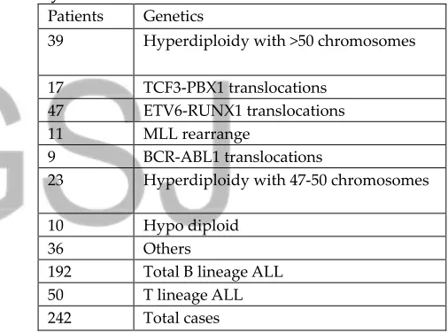
These subtypes are on the basis of genetics of acute lymphoblastic leukemia. The study of molecular genetics of the acute lymphoblastic leukemia also helps in the classification of this cancer.Charles studied the genetic alternations in the 242 patients and B lineage ALL was found in more patients. There are two main subtypes of acute lymphoblastic leukemia based on genetics of acute lymphoblastic leukemia. These are B lineage acutelymphoblastic leukemia andT lineage acute lymphoblastic leukemia
B lineage ALL:
For this scientist perform the genome analysis of pediatric acute lymphoblastic leukemia patients using some techniques such as high resolution, single-nucleotide polymorphism arrays and DNA genome sequencing. They showed that chromosomal aberrations including deletions, point mutation, chromosomal rearrangement are responsible for the B lineage ALL. Due to these reasons
disturbance in the B cells development occurs. There are 40% cases of B progenitor ALL. PAX5 gene is more responsible for the B lineage ALL. B lineage ALL includes translocations t(9;22)*BCR-ABL1+, t(1;19)*TCF3-PBN1+, t(12;21)*ETV6-RUNX1+, rearrangements of MLL, hyperdiploidy and hypodiploidy (table 1).(Greaves and Wiemels 2003)
T lineage ALL:
Very rare cases of the T cell acute lymphoblastic leukemia are due to chromosomal translocation. More than 50% of the T-ALL are responsible for this major subtype. It is very dangerous type of acute lymphoblastic leukemia found in children and adults. It is mainly due to acquired chromosomal translocation and may be due to other genetic and epigenetic abnormalities. Chromosomal translocations involve NOTCH1. NOTCH1 is a gene plays an important role for the encoding of receptors which are responsible for the development of normal T cells. Due to this mutation, abnormal T cells are formed which are responsible for this cancer.(Ferrando, Neuberg et al. 2002)
Table 2: B lineage ALL and T lineage ALL in 242 patients studied by Charles et al
Patients Genetics
39 Hyperdiploidy with >50 chromosomes
17 TCF3-PBX1 translocations 47 ETV6-RUNX1 translocations 11 MLL rearrange
9 BCR-ABL1 translocations
23 Hyperdiploidy with 47-50 chromosomes
10 Hypo diploid 36 Others
192 Total B lineage ALL 50 T lineage ALL 242 Total cases
(Ferrando, Neuberg et al. 2002)
Biologic characterization of leukemic cells
B-cells and T-cells:
Acute lymphoblastic leukemia caused by any primitive lymphoid cells ( B- cells or T- cells ,Mixed Phenotype acute leukemic cells and Natural Killer acute lymphoblastic leukemia cells ) at specific developmental stages. These cells have multi lineage potential. Acute myeloid lymphoblastic cells identified by Auer rods, monocyte -associated with esterase’s but all Acute lymphoblastic leukemia identified by immune-phenotyping because it have no morphological or cytochemical features.(Pui, Raimondi et al. 1991)
Identified in 3,000 – 4,000 patients, 2/3 are children in America each year.(Cortes and Kantarjian 1995)
lineage cells (approximately 25% in adults) CD5 and CD7 for immature T-cells antigens markers. ii) For myeloid cells CD13 or CD33.(Pui, Raimondi et al. 1991)
Further characteristics identified by normal B- lineage and T- lineage cells maturation. Therapeutic distinction occurs between mature B- cells (light chains of immunoglobulin present on surface) precursors and T- cells immuno-phenotype. For this antigens are used. In one fourth children and one third adults with acute lymphoblastic leukemia myeloid- associated antigens expresses. It’s only useful for immunological leukemia with minimal primitive leukemia not for therapeutic implications. In few patients both CD2 and CD7 ( lymphoid-associated molecules) identified(Look 1997)
Risk assessment:
Three risk categories are formed low, standard (average) and high risk. T-cell and B-cell acute lymphoblastic leukemia patients with poor prognosis in standard risk B- cells precursor acute lymphoblastic leukemia. In low risk, 1-9 years old children identified B- cell precursor Acute lymphoblastic leukemia(Nachman, Sather et al. 1993).
In this less than 50,000 per cubic millimeter leukocyte are present. Higher age groupspatients in high risk criteria. This explains on the base of genetic abnormalities. In less than one year old children 65-80 percent MLL gene rearrangements occur and have poor prognosis. In adults MLL gene rearrangements and BCR-ABL fusion causes poor prognosis at higher rate.(Smith, Arthur et al. 1996)
Other way to predict high risk assessment is clearance of B and T- cells. If clearance of B-lineage and T-lineage slow then patients have poor prognosis. PCR and immunological methods used in minimal residual disease when there are 10 billion leukemic cells in patients. Molecular or immunological remission ( less than 0.01 % of nucleated bone marrow leukemic cells).(Gaynon, Desai et al. 1997)
Blast cells immune-phenotype and genotype of BCR-ABL fusion in LC shows high risk leukemia. Haploid and hypoploid patients show low prognosis. Initial leukocyte count given standard risk leukemic patients. In standard response patients have (less than 100,000 per cubic millimeter) leukocyte counts.(Conter, Schrappe et al. 1997)
Subservient alarm:
In cerebrospinal fluid (CSF), leukemic cells presence indicates to prevent relapse of central nervous system (CNS) diseases. In 20-30 year old individuals, BCR-ABL fusion is high and low in 1-9 year old children. But at some level it is vice versa. Survival rate is higher in children than in adults. Treatment in younger above 25-30 year old adults easy as compared to 35-65 years old due to late responses to chemotherapy with low immunity. Patients with B-cell and T-B-cell ALL have large number of leukemic B-cells, hyper-uricemia, hyper-kalemia and hyper-phosphatemia. Secondary hypo-calcemia is common. Intervenous hydration, sodium bicarbonate to alkalizeurine, hyper-uricemia, AlOH in high concentration occurs in acute lymphoblastic leukemia patients.(Masson, Synold et al. 1996)
Mixed phenotype acute leukemic cells (MPALC):
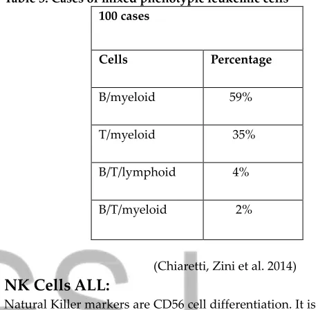
Mixed phenotype is rare 4% in ambiguous lineage co-exist with two separate blast cells T-or B-cells acute lymphoblastic leukemia either myeloid or monocytic blast cells In single blast cell leukemic population with co-expressing of B-antigens (CD19, CD20) or T-cell antigens (CD3) and also myeloid antigens.Same expression of monocyteantigen (MPO, CD64 and lysozyme). Absence of lineage-specific antigen (MPO, cCD3, cCD22) identified in undifferentiated leukemia.(Chiaretti, Zini et al. 2014) Table 3: Cases of mixed phenotypic leukemic cells
100 cases
Cells Percentage
B/myeloid 59%
T/myeloid 35%
B/T/lymphoid 4%
B/T/myeloid 2%
(Chiaretti, Zini et al. 2014)
NK Cells ALL:
Natural Killer markers are CD56 cell differentiation. It is very rare in only 3% cases present in early T-cell antigens (CD7 and CD2).(Pui, Ribeiro et al. 1991)
Diseases associated with acute lymphoblastic
leukemia:
There are other diseases also that are associated with the acute lymphoblastic leukemia. The patients which have this disease are also faced with the diseases like Bloom’s syndrome, Down’s syndrome, Hepatitis B, lymph node or pleural effusion, tumor lysis syndrome, fever, chest Hyperuricaemia, hemoglobin less or more than normal value and other factors/parameters associated with acute lymphoblastic leukemia are also found (table 3).(Margolin 1997)
Parameters and their fraction associated with acute
lymphoblastic leukemia:
According to the above two center NICH and Children Cancer Hospital Karachi following parameters was studied.
Table 4: parameters associated with acute lymphoblastic leukemia with their reported and diagnostic value
Parameters Reported value Diagnostic value
Consangunity 52% 60%
W.B.C Above than
50,000mm3 17%
Above than 50,000mm3 30%
Hepatomegaly 67% 67%
Splenomegaly 58% 58%
Lymphdenopathy 50% 75%
Hyperuricaemia 28% 13%
(Margolin 1997)
Risk factors:
In the united state acute lymphoblastic leukemia has been increased from 1975 to 2002. Until 1980 in United State acute lymphoblastic has been the cause of death. These are environmental, genetic and others. (Belson, Kingsley et al. 2007)
Diagnosis of acute lymphoblastic leukemia
Phenotype:
Immunophenotyping cytometry is importantly requirement to diagonals leukemia lymphoblast and cell lineage truly. Therapeutic importance of T and B cell and B-cell precursor phenotypes are important in the identification of T and B cell also helpful various kind of leukemia lymphoblast’s diagonalise. Polyploidy associated antigen repression.(Tissing, Den Boer et al. 2007)
Genotype:
RT-PCR Fluorescence in situ hybridization flow cytometry and other techniques are used to detect as well as to study the chromosomal analysis i.e. specific fusion transcript gain as loss of cellular DNA contents, or specific chromosomes with prognostic or therapeutic relevance helpful in detecting or diagonals is of lymphoblastic leukemia Gene-Expression profiling also helpful in the identification of major subtypes of acute lymphoblastic leukemia. It is more important technique that can replace many current diagonals tic techniques.(Pui, Campana et al. 2001)
Pharmacogenetic:
Pharmacogenetic studies have focused on single gene identification on the basis of their effect or influence on the pharmacokinetics and pharmacological effects of anticancer drugs. Global gene expression profiling studies help in the determination and treatment of acute lymphoblastic leukemia. Polymorphisms and activity of thiopurine methyltransferase also enhance treatment outcome in acute lymphoblastic leukemia.(Pui, Pei et al. 2010)
Treatment of acute lymphoblastic leukemia:
For the acute lymphoblastic leukemia, Short term intensive chemotherapy treatment includes:
1. Remission-induction phase
2. Intensification (or consolidation) phase 3. Continuation therapy
4. CNS-directed treatment
5. Allogeneic haemopoietic stem-cell transplantation
1.
Remission-induction phase:
The purpose of this treatment method is a struggle to develop the normal differentiated cells (effective haemopoiesis) in the patients by giving them drugs. Drugs are of three types glucocorticoid (prednisone or dexamethasone), vincristine, and one or both of these drugs (asparaginase, anthracycline). Mostly people almost 90% are treated with this complex of drugs but those with mature and severe leukemia’s (e.g. in adults) almost 70%, they are
provided with four or more than four drug complex. With the use of cyclophosphamide and asparaginase in excess, in addition to this complex, the survival rate and life span of patient can be increased. But these drugs can affect the CNS by penetrating into it, so the dexamethasone is much better than glucocorticoids (prednisone or prednisolone) for treatment of acute lymphoblastic leukemia because this is less toxic than the later ones and hence no harm to CNS. Leukemia recovery by the use of asparaginase is associated with how much quantity of this drug is used and the duration of this dosage providing to patient as compared to the type of asparaginase used, e.g one type in one patient may show its toxicity low as compared to in another patient (high). Due to excess use of drugs, the complexity becomes increased in patients especially in older children and adults. So, the asparaginase and corticoids are replaced by less toxic drugs. (Kantarjian, Giles et al. 2006)
2.
Consolidation (intensification) treatment:
This method is used to check the resistance of the Leukemia cells against the drugs. This method involves the use of high-dose methotrexate plus mercaptopurine, reinduction treatment or with the same agent that are given initially for 20-30 weeks. Actually it is a reinduction method in which doses of above drugs increased and given after the intervals of time to check our desire results (in recovering of acute lymphoblastic leukemia). The dose of methotrexate depends upon patient’s capability to take it and it is the best drug for T-cell or high-risk B-cell leukemia e.g., due to TEL-AML1 or E2A-PBX1 fusion, the patients are treated and cured by increasing the concentration of methotrexate.(Nachman, Sather et al. 1998)
3.
Continuation treatment:
Almost half of the children cases are treated by this continuation method. And it will lead to the duration of two or two and half years. This method also needs the mercaptopurine and methotrexat to patients on daily basis. Thioguanine is strongest than mercaptopurine in this method but above a certain range of this dose, it will cause toxicity that will disturb CNS or CSF. Thioguanine is given about 40 mg/m² daily or some little from this which will response anti leukemic and will help in the eradication of acute lymphoblastic leukemia. Mercaptopurine is not a standard drug for acute lymphoblastic leukemia but thioguanine is given in short-term courses during this treatment.(Barredo, Devidas et al. 2006)
4.
CNS-directed treatment:
5.
Allogeneic
haemopoietic
stem-cell
transplantation:
This method is beneficial for the patients of sub-types of lymphoblastic leukemia which results after the mismatching of receiving and donor individuals during transplantation. This method can tell about the presence of the acute lymphoblastic leukemia and to a extent can cure it by autologous transplantation ( transplantation in which stem cells are removed from a person called donor then again given to the same person from which they were excluded). (Vey, Thomas et al. 2006)
Conclusion:
Acute lymphoblastic leukemia also called acute lymphocyte leukemia is a type of leukemia in which there are many lymphocytes in the bone marrow and red blood cells. It is also called as cancer accounting for 32% of all cancers. It is most common in children with the highest peak between 2 and 5 years but may also effects adults. In child it accounts up to leukemia in child was first reported by Dr.Henry Fuller in 1846. This disease is fetal and can be recovered by chemotherapy.(Fuller 1846)
Current status and Future Perspectives:
Although applying the electrifying reformations to reach the goal in treatments of acute lymphoblastic leukemia but a meaningful interference has overcome in earlier aid to all the patients who are
affected with this disease. To embellish these problems definite areas are assuring to focus for future laboratory and clinical studies.A more important challenge for future is to understand the relationship between mutations or gene fusion i.e chromatin modifiers genes, pattern of histone proteins modifications and special DNA methylation changes in primary acute lymphoblastic leukemia cells into a new therapeutic approaches.
In future to increase the understanding about biology of acute lymphoblastic leukemia, to improve the approaches wit currently available antileukaemic agents, basically is a need for the identification of new chemotherapeutic agents active against lymphoid leukemia and design innovative clinical studies for research purposes. Molecular biological techniques e.g. assessment of immunoglobin and T cell receptor gene rearrangements are directly used to analysis of acute lymphoblastic leukemia. These approaches are co join with methodology include ‚in situ hybridization‛ give liberty to assimilation of leftover leukemia. To improve this analysis further high-tech clarification will be enforced.
Ascertainment of genetic basis of individual acute lymphoblastic leukemia patients will help to adopt an appropriate strategies to its treatments regardless their complex clinical profile. It includes Inhibitors of FLT-3 for Leukemia such as TEL-AML1 positive acute lymphoblastic leukemia.
Bibliography
Armstrong, S. A. and A. T. Look (2005). "Molecular genetics of acute lymphoblastic leukemia." Journal of clinical oncology 23(26): 6306-6315.
Barredo, J. C., et al. (2006). "Isolated CNS relapse of acute lymphoblastic leukemia treated with intensive systemic chemotherapy and delayed CNS radiation: a pediatric oncology group study." Journal of clinical oncology 24(19): 3142-3149.
Belson, M., et al. (2007). "Risk factors for acute leukemia in children: a review." Environmental health perspectives 115(1): 138.
Chiaretti, S., et al. (2014). "Diagnosis and subclassification of acute lymphoblastic leukemia." Mediterranean journal of hematology and infectious diseases 6(1).
Conter, V., et al. (1997). "Role of cranial radiotherapy for childhood T-cell acute lymphoblastic leukemia with high WBC count and good response to prednisone. Associazione Italiana Ematologia Oncologia Pediatrica and the Berlin-Frankfurt-Münster groups." Journal of clinical oncology 15(8): 2786-2791.
Cortes, J. E. and H. M. Kantarjian (1995). "Acute lymphoblastic leukemia a comprehensive review with emphasis on biology and therapy." Cancer 76(12): 2393-2417.
Croce, C. M. (2008). "Oncogenes and cancer." New England Journal of Medicine 358(5): 502-511.
Edward, H., et al. (2001). "The domain of strategic management: history and evolution." Handbook of strategy and management 31.
Ferrando, A. A., et al. (2002). "Gene expression signatures define novel oncogenic pathways in T cell acute lymphoblastic leukemia." Cancer cell 1(1): 75-87.
Fuller, H. (1846). "Particulars of a case in which enormous enlargement of the spleen and liver, together with dilation of all the blood vessels of the body were found co-incident with a peculiarly altered condition of the blood." Lancet 2: 43-44.
Gaynon, P. S., et al. (1997). "Early response to therapy and outcome in childhood acute lymphoblastic leukemia: a review." Cancer: Interdisciplinary International Journal of the American Cancer Society 80(9): 1717-1726.
Greaves, M. F. and J. Wiemels (2003). "Origins of chromosome translocations in childhood leukaemia." Nature Reviews Cancer 3(9): 639.
Gurney, J. G., et al. (1996). "Trends in cancer incidence among children in the US." Cancer: Interdisciplinary International Journal of the American Cancer Society 78(3): 532-541.
Kantarjian, H., et al. (2006). "Nilotinib in imatinib-resistant CML and Philadelphia chromosome–positive ALL." New England Journal of Medicine 354(24): 2542-2551.
Langenau, D. M., et al. (2005). "Cre/lox-regulated transgenic zebrafish model with conditional myc-induced T cell acute lymphoblastic leukemia." Proceedings of the National Academy of Sciences 102(17): 6068-6073.
Lee, E. J., et al. (2001). "Brief-Duration High-Intensity Chemotherapy for Patients With Small Noncleaved–Cell Lymphoma or FAB L3 Acute Lymphocytic Leukemia: Results of Cancer and Leukemia Group B Study 9251." Journal of clinical oncology 19(20): 4014-4022.
Look, A. T. (1997). "Oncogenic transcription factors in the human acute leukemias." Science 278(5340): 1059-1064.
Margolin, J. F. (1997). "Acute lymphoblastic leukemia-central nervous system relapse." Principles and practice of pediatric oncology: 440-441.
Masson, E., et al. (1996). "Allopurinol inhibits de novo purine synthesis in lymphoblasts of children with acute lymphoblastic leukemia." Leukemia 10(1): 56-60.
Nachman, J., et al. (1993). "Young adults 16‐21 years of age at diagnosis entered on childrens cancer group acute lymphoblastic leukemia and acute myeloblastic leukemia protocols. Results of treatment." Cancer 71(S10): 3377-3385.
Nachman, J. B., et al. (1998). "Augmented post-induction therapy for children with high-risk acute lymphoblastic leukemia and a slow response to initial therapy." New England Journal of Medicine 338(23): 1663-1671.
Nordlund, J., et al. (2013). "Genome-wide signatures of differential DNA methylation in pediatric acute lymphoblastic leukemia." Genome biology 14(9): r105.
Pui, C.-H., et al. (2001). "Childhood acute lymphoblastic leukaemia–current status and future perspectives." The lancet oncology 2(10): 597-607.
Pui, C.-H., et al. (2004). "Acute lymphoblastic leukemia." New England Journal of Medicine 350(15): 1535-1548.
Pui, C.-H., et al. (1991). "Acute myeloid leukemia in children treated with epipodophyllotoxins for acute lymphoblastic leukemia." New England Journal of Medicine 325(24): 1682-1687.
Pui, C., et al. (2010). "Long-term results of St Jude Total Therapy Studies 11, 12, 13A, 13B, and 14 for childhood acute lymphoblastic leukemia." Leukemia 24(2): 371.
Rinsky, R. A., et al. (1981). "Leukemia in benzene workers." American journal of industrial medicine 2(3): 217-245.
Ross, J. A. (1998). "Maternal diet and infant leukemia: a role for DNA topoisomerase II inhibitors?" International Journal of Cancer 78(S11): 26-28.
Sabaawy, H. E., et al. (2006). "TEL-AML1 transgenic zebrafish model of precursor B cell acute lymphoblastic leukemia." Proceedings of the National Academy of Sciences 103(41): 15166-15171.
Sharma, S. and K. Sarangdevot (2012). "Nanoemulsions for cosmetics." IJARPB 1(3): 408-415.
Shu, X.-O., et al. (1996). "Parental alcohol consumption, cigarette smoking, and risk of infant leukemia: a Childrens Cancer Group study." JNCI: Journal of the National Cancer Institute 88(1): 24-31.
Smith, M., et al. (1996). "Uniform approach to risk classification and treatment assignment for children with acute lymphoblastic leukemia." Journal of clinical oncology 14(1): 18-24.
Tissing, W. J., et al. (2007). "Genomewide identification of prednisolone-responsive genes in acute lymphoblastic leukemia cells." Blood 109(9): 3929-3935.
Vey, N., et al. (2006). "Allogeneic stem cell transplantation improves the outcome of adults with t (1; 19)/E2A-PBX1 and t (4; 11)/MLL-AF4 positive B-cell acute lymphoblastic leukemia: results of the prospective multicenter LALA-94 study." Leukemia 20(12): 2155.
Wang, J. C. and J. E. Dick (2005). "Cancer stem cells: lessons from leukemia." Trends in cell biology 15(9): 494-501.
Weng, A. P., et al. (2004). "Activating mutations of NOTCH1 in human T cell acute lymphoblastic leukemia." Science 306(5694): 269-271.
Yasmeen, N. and S. Ashraf (2009). "Childhood acute lymphoblastic leukaemia; epidemiology and clinicopathological features." JPMA 59(150).
Zahm, S. H. and M. H. Ward (1998). "Pesticides and childhood cancer." Environmental health perspectives 106(Suppl 3): 893.