Genome-Wide Interaction Analyses between
Genetic Variants and Alcohol Consumption
and Smoking for Risk of Colorectal Cancer
Jian Gong1☯
, Carolyn M. Hutter2☯
, Polly A. Newcomb1, Cornelia M. Ulrich1,3, Stephanie
A. Bien1, Peter T. Campbell4, John A. Baron5, Sonja I. Berndt6, Stephane Bezieau7, Hermann Brenner8,9, Graham Casey10, Andrew T. Chan11, Jenny Chang-Claude12,
Mengmeng Du1, David Duggan13, Jane C. Figueiredo10, Steven Gallinger14, Edward L. Giovannucci15, Robert W. Haile10, Tabitha A. Harrison1, Richard B. Hayes16,
Michael Hoffmeister8, John L. Hopper17, Thomas J. Hudson18, Jihyoun Jeon1, Mark A. Jenkins17, Jonathan Kocarnik1, Se´bastien Ku¨ ry7, Loic Le Marchand19, Yi Lin1,
Noralane M. Lindor20, Reiko Nishihara21, Shuji Ogino22, John D. Potter1,23,
Anja Rudolph12, Robert E. Schoen24, Petra Schrotz-King25, Daniela Seminara26, Martha
L. Slattery27, Stephen N. Thibodeau28, Mark Thornquist1, Reka Toth25, Robert Wallace29, Emily White1, Shuo Jiao1, Mathieu Lemire18, Li Hsu1, Ulrike Peters1*, CCFR and GECCO 1 Public Health Sciences Division, Fred Hutchinson Cancer Research Center, Seattle, Washington, United States of America, 2 Division of Genomic Medicine, National Human Genome Research Institute, Bethesda, Maryland, United States of America, 3 Huntsman Cancer Institute, University of Utah, Salt Lake City, Utah, United States of America, 4 Epidemiology Research Program, American Cancer Society, Atlanta, Georgia, United States of America, 5 Department of Medicine, School of Medicine, University of North Carolina, Chapel Hill, North Carolina, United States of America, 6 Division of Cancer Epidemiology and Genetics, National Cancer Institute, Bethesda, Maryland, United States of America, 7 CHU Nantes, Service de Ge´ne´tique Me´dicale, Nantes, France, 8 Division of Clinical Epidemiology and Aging Research, German Cancer Research Center (DKFZ), Heidelberg, Germany, 9 German Cancer Consortium (DKTK), Heidelberg, Germany, 10 Keck School of Medicine, University of Southern California, Los Angeles, California, United States of America, 11 Division of Gastroenterology, Massachusetts General Hospital and Harvard Medical School, Boston, Massachusetts, United States of America; Channing Division of Network Medicine, Brigham and Women’s Hospital and Harvard Medical School, Boston, Massachusetts, United States of America, 12 Division of Cancer Epidemiology, German Cancer Research Center, Heidelberg, Germany, 13 Translational Genomics Research Institute, Phoenix, Arizona, United States of America, 14 Department of Surgery, University Health Network Toronto General Hospital, Toronto, Ontario, Canada, 15 Channing Division of Network Medicine, Brigham and Women’s Hospital, Boston Massachusetts, United States of America, 16 Division of Epidemiology, New York University School of Medicine, New York, New York, United States of America, 17 Melbourne School of Population Health, The University of Melbourne, Melbourne, Australia, 18 Ontario Institute for Cancer Research, Toronto, Ontario, Canada, 19 Epidemiology Program, University of Hawaii Cancer Center, Honolulu, Hawaii, United States of America, 20 Department of Health Sciences Research, Mayo Clinic, Scottsdale, Arizona, United States of America, 21 Dana-Farber Cancer Institute, Harvard School of Public Health, Boston, Massachusetts, United States of America, 22 Harvard Medical School, Department of Epidemiology, Harvard School of Public Health, Boston, Massachusetts, United States of America, 23 Centre for Public Health Research, Massey University, Wellington, New Zealand, 24 Department of Medicine and Epidemiology, University of Pittsburgh Medical Center, Pittsburgh, Pennsylvania, United States of America, 25 Division of Preventive Oncology, National Center for Tumor Diseases and German Cancer Research Center, Heidelberg, Germany, 26 Division of Cancer Control and Population Sciences, National Cancer Institute, Bethesda, Maryland, United States of America, 27 Department of Internal Medicine, University of Utah Health Sciences Center, Salt Lake City, Utah, United States of America, 28 Departments of Laboratory Medicine and Pathology and Laboratory Genetics, Mayo Clinic, Rochester, Minnesota, United States of America, 29 Department of Epidemiology, The University of Iowa, Iowa City, Iowa, United States of America
☯These authors contributed equally to this work. *[email protected]
a11111
OPEN ACCESS
Citation: Gong J, Hutter CM, Newcomb PA, Ulrich
CM, Bien SA, Campbell PT, et al. (2016) Genome-Wide Interaction Analyses between Genetic Variants and Alcohol Consumption and Smoking for Risk of Colorectal Cancer. PLoS Genet 12(10): e1006296. doi:10.1371/journal.pgen.1006296
Editor: Scott M. Williams, Case Western Reserve
University School of Medicine, UNITED STATES
Received: November 20, 2015 Accepted: August 11, 2016 Published: October 10, 2016
Copyright: This is an open access article, free of all
copyright, and may be freely reproduced, distributed, transmitted, modified, built upon, or otherwise used by anyone for any lawful purpose. The work is made available under theCreative Commons CC0public domain dedication.
Data Availability Statement: All relevant data are
within the paper and its Supporting Information files.
Funding: GECCO is supported by National Cancer
Abstract
Genome-wide association studies (GWAS) have identified many genetic susceptibility loci for colorectal cancer (CRC). However, variants in these loci explain only a small proportion of familial aggregation, and there are likely additional variants that are associated with CRC susceptibility. Genome-wide studies of gene-environment interactions may identify variants that are not detected in GWAS of marginal gene effects. To study this, we conducted a genome-wide analysis for interaction between genetic variants and alcohol consumption and cigarette smoking using data from the Colon Cancer Family Registry (CCFR) and the Genetics and Epidemiology of Colorectal Cancer Consortium (GECCO). Interactions were tested using logistic regression. We identified interaction between CRC risk and alcohol consumption and variants in the 9q22.32/HIATL1 (Pinteraction= 1.76×10⁻⁸; permuted
p-value 3.51x10⁻⁸) region. Compared to non-/occasional drinking light to moderate alcohol consumption was associated with a lower risk of colorectal cancer among individuals with rs9409565 CT genotype (OR, 0.82 [95% CI, 0.74–0.91]; P = 2.1×10⁻⁴) and TT genotypes (OR,0.62 [95% CI, 0.51–0.75]; P = 1.3×10⁻⁶) but not associated among those with the CC genotype (p = 0.059). No genome-wide statistically significant interactions were observed for smoking. If replicated our suggestive finding of a genome-wide significant interaction between genetic variants and alcohol consumption might contribute to understanding colo-rectal cancer etiology and identifying subpopulations with differential susceptibility to the effect of alcohol on CRC risk.
Author Summary
Alcohol consumption and smoking are associated with CRC risk. We performed a genome-wide analysis for interaction between genetic variants and alcohol consumption and cigarette smoking to identify potential new genetic regions associated with CRC. About 8,000 CRC cases and 8,800 controls were included in alcohol-related analysis and over 11,000 cases and 11,000 controls were involved in smoking-related analysis. We iden-tified interaction between variants at 9q22.32/HIATL1and alcohol consumption in rela-tion to CRC risk (Pinteraction= 1.76×10⁻⁸). If replicated our suggested finding of the
interaction between genetic variants and alcohol consumption might contribute to under-standing colorectal cancer etiology and identifying subpopulations with differential sus-ceptible to the effect of alcohol on CRC risk.
Introduction
Colorectal cancer (CRC) is the third-most common cancer in men and the second most com-mon cancer in women worldwide [1]. Both environmental and genetic factors are involved in the development of CRC [2–7]. Since 2007, genome-wide association studies (GWAS) have identified about 50 loci associated with CRC risk[8–11]. However, only a small portion of the familial aggregation of CRC is explained by these identified genetic loci, and additional variants associated with CRC susceptibility are more likely to be identified through analyses of interac-tions between genes and environmental risk factors [12,13]. Single nucleotide polymorphisms (SNP) that impact only a subgroup of the population or have opposite effects in different Bretagne Ge´ne´tique and the Ligue Re´gionale
subgroups are likely to produce weak main effects that cannot be easily detected by marginal association testing of the SNPs. However, these variants may be identified by testing for inter-actions between SNP and environmental risk factors (genome-wide interaction analysis) [14, 15]. These findings may provide etiologic insight into CRC and identify potentially susceptible subpopulations [14,15].
There is compelling evidence from epidemiologic studies that alcohol consumption and cig-arette smoking are associated with risk of CRC [16–25]. Both alcohol consumption and ciga-rette smoking influence disease risk through pathways involving multiple gene products and regulatory elements, providing potential for biological interactions [26–28]. Accordingly, alco-hol consumption and smoking are important lifestyle factors to study interactions with genetic variants. In this study, we performed a genome-wide interaction analysis using the large data-sets from the Colon Cancer Family Registry (CCFR) and the Genetics and Epidemiology of Colorectal Cancer Consortium (GECCO) [3] to identify SNPs that modify the effects of alcohol and smoking on CRC risk.
Results
In this study, we included 14 studies from the Colon Cancer Family Registry (CCFR) and the Genetics and Epidemiology of Colorectal Cancer Consortium (GECCO) as described previ-ously [3,29,30] and in theS1 TextandS1andS2Tables. Basic characteristics of the partici-pants, stratified by study center, are described inS1andS2Tables, respectively. We were able to harmonize measures of alcohol consumption across 8,058 cases and 8,765 controls and mea-sures of smoking across up to 11,219 cases and 11,382 controls. As seen for other common dis-eases, such as cardiovascular disdis-eases, alcohol consumption shows a different effect with CRC risk depending on the level of alcohol consumed. Heavy alcohol intake (>2 standard drinks per day) has been shown to be associated with increased risk of CRC [16,17,31] while light-to-moderate drinking (<2 standard drinks per day) may have little effect [18,19] or reduce risk of CRC [16,20–22] compared to non-drinkers. Consistent with these previous publications [16– 22,31] we observed an inverse association with CRC risk for light-to-moderate drinkers (OR = 0.91, P = 0.006,Fig 1A) but a positive association for heavy drinkers (OR = 1.22, P = 0.0004,Fig 1B) compared with non-/occasional drinkers. Modeling alcohol using this cate-gorical approach fitted the association between alcohol intake and CRC risk better than the continuous variable based on the Akaike Information Criterion (AIC) which was 12.42 smaller for the model including the two categorical variables compared with the model including the continuous variable (AIC = 23123.72 for continuous alcohol and AIC = 23111.3 for categorical alcohol)[32]. Given the opposite effect of light/moderate alcohol drinking vs. heavy drinking, it is critical that analyses further investigating the impact of alcohol on CRC, such as interaction analysis do this separately for light/moderate and heavy drinking. Ever-smokers and pack-years of cigarette smoking were positively associated with CRC risk (OR = 1.18 for ever vs. never smokers, P = 8.9×10⁻⁹; OR = 1.11 per 10 pack-years increase, P = 7.1×10⁻¹³,Fig 2A and 2B). None of the smoking and alcohol variables showed evidence of heterogeneous associations across studies (Pheterogeneity>0.16).
Using conventional logistic regression including multiplicative interaction terms, we identi-fied genome-wide significant interactions (at P<5×10⁻⁸) between 11 SNPs at the 9q22.32/
HIATL1(Hippocampus Abundant Transcript-Like 1) locus and light-to-moderate drinking with no evidence of heterogeneity across studies (Pheterogeneity>0.5 for any of the 11 SNPs) (S3
Table,Fig 3). All 11 SNPs were common variants with minor allele frequency (MAF) between 0.31–0.34 and genotyped or imputed with high accuracy (imputation r2>0.98,S3 Table). The most significant SNP was rs9409565 with Pinteraction= 1.76×10⁻⁸; permuted p-value 3.51x10⁻⁸
Genome-wide association study of prostate cancer identifies a second risk locus at 8q24. Nat Genet 2007 May;39(5):645-9), Colon CGEMS pancreatic cancer scan (PanScan) (Amundadottir, L et al. Genome-wide association study identifies variants in the ABO locus associated with susceptibility to pancreatic cancer. Nat Genet. 2009 Sep;41(9):986-90, and Petersen, GM et al. A genome-wide association study identifies pancreatic cancer susceptibility loci on chromosomes 13q22.1, 1q32.1 and 5p15.33. Nat Genet. 2010 Mar;42 (3):224-8), and the Lung Cancer and Smoking study (Landi MT, et al. A genome-wide association study of lung cancer identifies a region of chromosome 5p15 associated with risk for adenocarcinoma. Am J Hum Genet. 2009 Nov;85 (5):679-91). The prostate and PanScan study datasets were accessed with appropriate approval through the dbGaP online resource (http://cgems. cancer.gov/data/) accession numbers phs000207. v1.p1 and phs000206.v3.p2, respectively, and the lung datasets were accessed from the dbGaP website (http://www.ncbi.nlm.nih.gov/gap) through accession number phs000093.v2.p2. Funding for the Lung Cancer and Smoking study was provided by National Institutes of Health (NIH), Genes, Environment and Health Initiative (GEI) Z01 CP 010200, NIH U01 HG004446, and NIH GEI U01 HG 004438. For the lung study, the GENEVA Coordinating Center provided assistance with genotype cleaning and general study coordination, and the Johns Hopkins University Center for Inherited Disease Research conducted genotyping. PMH is supported by National Institutes of Health (R01 CA076366 to P.A. Newcomb). VITAL is supported byNational Institutes of Health (K05 CA154337). WHI is funded by the National Heart, Lung, and Blood Institute, National Institutes of Health, U.S. Department of Health and Human Services through contracts HHSN268201100046C, HHSN268201100001C, HHSN268201100002C, HHSN268201100003C, HHSN268201100004C, and HHSN271201100004C. The funders had no role in study design, data collection and analysis, decision to publish, or preparation of the manuscript.
Competing Interests: The authors have declared
(Table 1,Fig 4C). The genetic variant was located in an intergenic region (28kb downstream of
HIATL1and 70kb downstream ofFBP2,Fig 3). All the other 10 genome-wide significant SNPs were in strong linkage disequilibrium (LD) with rs9409565 (LD r2>0.8,S3 Table,Fig 3) and some of them were located within the geneHIATL1. The observed interaction for rs9409565 was similar in men and women and by cancer site (colon vs rectum) (Fig 4A and 4B,S4 Table). We did not observe any genome-wide significant interaction between any SNP and heavy drinking. No inflation was observed in the genome-wide SNP × alcohol interaction analysis (the inflation factorλ= 0.99 and 1.00 for light-to-moderate drinkers and heavy drinkers, respectively). To evaluate the potential confounding[33] by other lifestyle and environmental risk factors of the interactions between rs9409565 and light-to-moderate alcohol consumption in relation to CRC risk, we adjusted for smoking status (ever vs. never) and BMI (two variables have the highest correlation r = 0.15 and 0.13 with alcohol consumption in our data), as well as exercise, fruit and vegetable consumption in the conventional case-control logistic regression model. Our results did not change (multivariate adjusted interaction p-value = 4.34x10⁻⁸).
When stratified by genotype rs9409565, light-to-moderate alcohol consumption (compared to non/occasional alcohol consumption) significantly decreased CRC risk in individuals with CT genotype (prevalence, 45% vs 49%; OR, 0.82 [95% CI, 0.74–0.91]; P = 2.1×10⁻⁴) and TT genotype (prevalence, 42% vs 52%; OR,0.62 [95% CI, 0.51–0.75]; P = 1.3×10⁻⁶) but not in those with CC genotype (P = 0.059) (Table 1,S5 Table). The association between alcohol intake and CRC was also not heterogeneous within each genotype strata (p-heterogeneity>0.73;S1 Fig).
We also estimated absolute risks of CRC based on Surveillance, Epidemiology, and End Results (SEER) age-adjusted incidence rates (Table 2). Compared with non/occasional Fig 1. The association between CRC and alcohol consumption (non-/occasional drinkers [reference group]; light-to-moderate drinkers [a]; and heavy drinkers[b]). Men and women were analyzed separately in each study and age and study site (if applicable) were adjusted in model. Non-/occasional drinkers: drinking<1 gram of alcohol per day; light-to-moderate drinkers: drinking 1–28 grams of alcohol per day ([a] alcoholc1-28g/d); and heavy drinkers: drinking>28 grams of alcohol per day ([b] alcoholc>28g/d). OR: odds ratio; N = total number of subjects; case = number of cases. Colon23: Hawaii Colorectal Cancer Studies 2 and 3; DACHS: Darmkrebs: Chancen der Verhu¨tung durch Screening; DALS: Diet, Activity and Lifestyle Study; HPFS: Health Professionals Follow-up Study; HPFS_AD: Health Professionals Follow-up Study for colorectal adenoma; MEC: Multiethnic Cohort Study; NHS: Nurses’ Health Study; NHS_AD: Nurses’ Health Study for colorectal adenoma; PHS: Physicians’ Health Study; PLCO: Prostate, Lung, Colorectal and Ovarian Cancer; Screening Trial; VITAL: VITamins And Lifestyle; WHI: Women’s Health Initiative. het.pval: p value of heterogeneity.
drinking, light-to-moderate drinking was associated with 14.0 fewer CRC cases per 100,000 individuals carrying the rs9409565-CT genotype per year; 35.5 fewer CRC cases per 100,000 individuals carrying the rs9409565-TT genotype per year.
Using the Cocktail method as a two-step method that may improve power we did not observe any genome-wide significant SNP×alcohol interactions. Further, we did not observe any genome-wide significant interactions for SNP×smoking (smoking history and pack-years of smoking) using logistic regression or the Cocktail method.
Gene expression analyses
The SNP rs9409565 showing a significant interaction with alcohol is located in an intergenic region betweenHIATL1andFBP2. As there is a recombination hotspot lying between rs9409565 andFPB2(Fig 3), we focused the gene expression analysis onHIATL1, which is expressed in normal colon and rectal tissue. [34,35] Furthermore, based on our gene expres-sion data for 35 colorectal cancer cases (S2 Text), the expresexpres-sion levels of theHIATL1gene was significantly higher in tumor tissues compared with adjacent normal tissues (paired student t test, P<7.2×10⁻⁵,S2 Fig). This finding is consistent with a previous study [36] which is included in the UCSC Cancer Genomics Browser[37–39] and show that human colon tumors (n = 100) significantly over-expressedHIATL1compared to normal colon tissues (n = 5) [36] (Fisher exact test: P = 0.03). Similarly, we were able to reproduce this observation in 50 inde-pendent paired colorectal adenocarcinoma and adjacent normal samples from The Cancer Genome Atlas (TCGA) (paired student t test, P = 0.02,S2 Fig). Furthermore, we observed that Fig 2. The association between CRC and smoking (ever vs. never smokers [a]; pack-years of smoking [b]). Never smokers were assigned the value 0 for pack-years of smoking. OR: odds ratio; OR for pack-years of smoking is based on per 20 pack-years increase. Age, sex (if applicable), and study site (if applicable) were adjusted in model. ASTERISK: The French Association STudy Evaluating RISK for sporadic colorectal cancer; CCFR: Colon Cancer Family Registry; Colon23: Hawaii Colorectal Cancer Studies 2 and 3.; DACHS: Darmkrebs: Chancen der Verhu¨tung durch Screening; DALS: Diet, Activity and Lifestyle Study; HPFS:Health Professionals Follow-up Study; HPFS_AD: Health Professionals Follow-up Study for colorectal adenoma; MEC: Multiethnic Cohort Study; NHS: Nurses’ Health Study; NHS_AD: Nurses’ Health Study for colorectal adenoma; OFCCR: Ontario Familial Colorectal Cancer Registry; PMH-CCFR: Postmenopausal Hormone study-Colon Cancer Family Registry; PLCO: Prostate, Lung, Colorectal and Ovarian Cancer Screening Trial; VITAL: VITamins And Lifestyle; WHI: Women’s Health Initiative. CCFR is a collaborating study with GECCO. smk_ever: ever smokers; smk_pkyr20: pack-years of smoking; het. pval: p value of heterogeneity.
Fig 3. Regional association plot for the interaction analyses between moderate alcohol drinking and SNPs at 9q22.32/HIATL1. The– log10 of p values (left y-axis) are plotted against the SNP genomic position based on NCBI build 37 (x-axis); the estimated recombination rate from 1000 Genomes Project European populations are on the right y-axis and plotted in blue. The most significant SNP was denoted with purple diamond. SNPs are colored to reflect correlation with the most significant SNP. Gene annotations are from the UCSC genome browser. Gene
FAM22F is also known as NUTM2F.
doi:10.1371/journal.pgen.1006296.g003
Table 1. Stratification analysesaby genotypes of rs9409565 for the association between alcohol consumption and CRC.
Genotype Non/occasional drinkers Light-to-moderate drinkers Pinterationb
Case (n) Control (n) OR Case (n) Control (n) OR (95% CI) P value
rs9409565 CC 1,365 1,593 1.0 1,638 1,717 1.11 (1.00–1.23) 5.9E-02
CT 1,495 1,574 1.0 1,646 2,002 0.82 (0.74–0.91) 2.1E-04
TT 425 387 1.0 434 590 0.62 (0.51–0.75) 1.3E-06
1.76E-08
a
: non/occasional drinkers as the reference group. Non-/occasional drinkers: drinking<1 gram of alcohol per day; light-to-moderate drinkers: drinking 1–28 grams of alcohol per day. Men and women were analyzed separately in each study and age, study site (if applicable), and population structure were adjusted in model.
b
: P value of interaction term between SNP and alcohol consumption, permuted p-value = 3.51x10⁻⁸(p value of heterogeneity = 0.96).
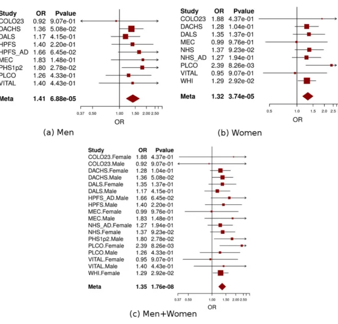
HIATL1showed significant differential expression across various levels of lifetime alcohol con-sumption in the colon tumor tissues (n = 28, ANOVA test P = 0.03,S3 Fig) and also had differ-ential gene expression across levels of alcohol consumption at reference time (the year before enrollment) in the normal colon tissues (n = 33) at P = 0.06 from ANOVA test (S4 Fig). In addition, for rs9409565 and rs9409567 (LD r2= 1.0 in CEU population), the two most signifi-cant SNPs at 9q22.32/HIATL1, are cis-acting quantitative trait loci (eQTL) forHIATL1 expres-sion in lymphoblastoid cell lines (P<7.0×10⁻⁶) and monocytes (P<5.8×10⁻¹²) [40,41], which is consistent with previously published eQTL results from GTEx, Genevar[42], Westra et al., and Lappalainen et al. showing that this these SNPs tag an eQTL locus in lymphoblastoid cells and related anatomical sources (including spleen, whole blood, esophagus muscularis, and Fig 4. Forest plot for meta-analysis of interaction analysis for rs9409565 and light-to-moderate drinking among men (a), women (b) and combined (c). Odds ratios (ORs) and 95% confidence intervals (95% CI) are presented for the multiplicative interaction between each additional copy of the count (or tested) allele (C) and light-to-moderate vs. non/occasional drinkers. The box sizes are proportional in size to the inverse of the variance for each study, and the lines visually depict the confidence interval. Results from the fixed-effects meta-analysis are shown as diamonds. The width of the diamond represents the confidence interval. P value of heterogeneity for (a), (b), and (c) is 0.93, 0.78, and 0.96, respectively.
sun-exposed skin) with p values ranging from 7x10-138to 4x10⁻⁶(S8 Table). In contrast, evalua-tion of eQTL in both normal (GTEx) and cancer colorectal tissue from TCGA for the
rs9409565 locus (r2>= 0.2 in Phase 3 1000 genomes EUR data) did not show any significant eQTL. The inability to detect an eQTL is likely because the enhancer tagged by the locus is active in some but not all cancer cell lines and the current reference cancer transcriptome data may not be large enough or molecularly representative of our study populationS5 Fig). Fur-thermore, we investigated whether any of the tagging SNPs are located in variant enhancer loci (VEL)reported by Akhtar-Zaidi et al.[43] using ChIP-seq (H3k27ac) enhancer signals. We observed that four of the variants (rs28406858, rs7042481, rs7858082, and rs9409510) in LD with rs9409565 (LD r20.6) were positioned within three gained cancer-specific VEL (S6 Fig).
Discussion
We identified a suggestive interaction between variants at 9q22.32/HIATL1and light-to-moder-ate alcohol consumption in relation to CRC risk. This is the first genome-wide significant GxE interaction reported for alcohol intake and risk of CRC and warrants replication in independent studies. Evidence for overlap between the discovered 9q22.32/HIATL1region with VEL as well as gene expression results support the relevance of the 9q22.32/HIATL1region for CRC risk.
Gene expression analyses indicated that a) SNPs identified in our study impactHIATL1
expression, b)HIATL1is involved in signaling pathways related to CRC and expression differs between normal and tumor CR tissue, and c)HIATL1expression in colon tissue differs by alco-hol consumption. The most significant variant rs9409565 is correlated with 142 variants (LD r20.5 in Phase 3 1000 Genomes European populations), which spanned across intronic regions and approximately 50kb downstream and 75kb upstream ofHIATL1. Nine of these variants (including rs9409550, rs4744345, rs9409546, rs9409778, and rs639276, all with inter-action P<5×10⁻⁸) fall within a transcriptionally active region in normal colon, rectal and duo-denal mucosa [44] as defined by epigenetic signals.[45] Furthermore, these variants fall in a region of enriched enhancer signal; although we note that currently available ChIP-seq data are not able to identify a putative transcription factor binding site at any of the tagged SNPs (S6 Fig). In support of our findings that HIATL1 expression is higher in tumor than adjacent nor-mal colorectal tissue, ChIP-seq (H3k27ac) enhancer signals suggest that this locus implicates a gained enhancer present in CR tumors that is absent in normal crypt cells (S6 Fig). In sum-mary, multiple data points suggest that the genetic variants we identified to interact with alco-hol on CRC risk are located in regulatory regions impacting the expression ofHIATL1and that
HIATL1expression varies by alcohol consumption.
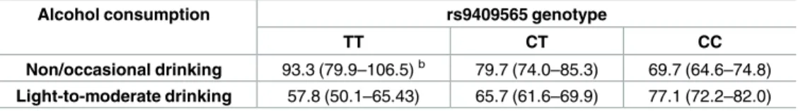
HIATL1is a member of the solute carrier (SLC) group of membrane transport, which enables the directed movement of substances (such as peptides, amino acids, proteins, metals, Table 2. Absolute riskaof CRC for alcohol consumption among individuals with different genotypes
of rs9409565.
Alcohol consumption rs9409565 genotype
TT CT CC
Non/occasional drinking 93.3 (79.9–106.5)b 79.7 (74.0–85.3) 69.7 (64.6–74.8) Light-to-moderate drinking 57.8 (50.1–65.43) 65.7 (61.6–69.9) 77.1 (72.2–82.0)
a
: Absolute risk calculation was based on Surveillance, Epidemiology, and End Results (SEER) age-adjusted CRC incidence rates between 1982–2011 among the White population of 74.5 per 100,000 men and women per year.
b
: the number of CRC cases per 100,000 individuals (95% CI).
and neurotransmitters) into or out of cells and plays an important role in a variety of cellular functions [46,47]. Although the detailed function ofHIATL1remains elusive, this gene was found to be expressed in a large range of animal species and it is highly evolutionarily con-served [48], suggesting an potentially important functional role. Transporter proteins are com-monly upregulated in many cancers [49,50] and take part in nutrient signaling to the mTOR pathway [51] which is an important signaling pathway in apoptosis and cancer [52–54]. Alco-hol may modify the effects ofHIATL1on CRC risk through its influence on the gene expres-sion ofHIATL1. Nonetheless, the precise mechanism(s) of the interaction between alcohol and
HIATL1on CRC risk remains unclear and further studies are needed.
Our Cocktail method for detecting G×E interactions did not identify the statistical interaction detected by the conventional logistic regression analysis because rs9409565 did not show strong statistical evidence for association with CRC risk in the marginal association analyses (P = 0.54, OR = 1.014) or with alcohol consumption (P = 0.22). Accordingly, this SNP was ranked low in step 1 of the Cocktail method, resulting in very stringent alpha-threshold for the interaction term in step 2. Although the conventional logistic regression analysis tends to be less powerful overall for genome-wide interaction analysis compared with the Cocktail method [14,55], it has greater power to detect an association if the marginal association of the SNP on disease or the correlation of the SNP with environmental factor are weak as it was the case for the observed interaction. In addition, no association between rs9409565 and alcohol consumption excluded the possibility that the observed interaction was due to the dependence between them [56]. We also explored the effect of rs9409565 and alcohol using other potentially more powerful single step approaches and observed a similar interaction effect in the Empirical Bayesian analysis[57] and a weaker interaction effect in the case-only analysis[58], which may be explained by the non-significant differential effect of alcohol on CRC in individual carrying the CC genotype (S6 Table).
consumption), additional adjustment for multiple comparisons may be needed. However, we note that the observed interaction at 9q22.32/HIATL1would remain borderline significant (alpha threshold = 5×10⁻⁸/2 = 2.5×10⁻⁸). The small numbers of heavy drinkers, particular in women, impeded the reliable estimation of interaction parameters and limited our power to identify significant interaction between SNP and heavy drinking. We focused gene expression analysis onHIATL1because rs9409565 is located in an intergenic region betweenHIATL1and
FBP2and further there is a recombination hotspot lying between rs9409565 and FPB2. If we expand gene expression analyses for all genes 500kb upstream or downstream 500kb of rs9409565 in the 35 pairs of colorectal tumor-normal tissue samples (S2 Text) we observed no significant result after false discovery rate (FDR) correction. The most significant results were forMIRLET7Fwhich has a p value of 0.001 for testing differential gene expression across vari-ous levels of lifetime alcohol consumption in normal tissues andPTPDC1which has a p value of 0.002 for testing differential gene expression across various levels of alcohol consumption at ref-erence time. Further studies are needed to confirm our findings.
Alcohol has a particularly detrimental effect on several cancers, possibly including CRC, in Asian subpopulations with genetic determined alcohol sensitivity[64–66]. However, as we have focused our analysis on European descent populations and did not observe significant differ-ences of the alcohol-CRC association between studies (phet = 0.16–0.76) we do not expect major underlying differences of the effect of alcohol in our study populations.
We did not perform stratification analyses by anatomical sites for our genome-wide GxE interaction analysis because the association of CRC with alcohol consumption (S7 Table) and smoking [23] did not vary according to anatomical site within the large bowel. Although we did observe potential interactions for alcohol consumption, we did not observe statistical evi-dence for genome-wide SNP x smoking interactions. This may be because smoking has a weaker association with CRC compared with alcohol intake [24,26,67], so we may have been underpowered even with more than 10,000 cases and 10,000 controls. We also may not have properly captured the most relevant smoking variables, such as duration of smoking or time since quitting smoking. The association between smoking and CRC risk are strongest for tumors that display certain molecular features such as microsatellite instability (MSI)-high and CpG island methylator phenotype (CIMP)-positive [68,69]. Because of the lack of MSI or CIMP data in several studies, we cannot perform stratification analysis by tumor characteristics for smoking-related analyses.
We note that it would be too early to make any recommendation on alcohol intake from our findings even after independent replication given that such recommendation need to be consid-ered in context of the effect of alcohol on all diseases. Furthermore, it will be important to inves-tigate the interactions between alcohol and genetic variants in larger studies to comprehensively evaluate the full impact of genetic variation on the effect of alcohol on colorectal cancer risk.
In summary, we identified a tentative novel interaction for CRC risk between alcohol intake and variants at 9q22.32/HIATL1. Further replication and functional studies are required to confirm our findings and understand the biologic implications of the interaction. This, in turn, could provide further insight into CRC etiology and may identify potentially susceptible subpopulations.
Materials and Methods
Ethics statement
Utah Institutional Review Board (DALS); Partners Human Research Committee (NHS and PHS); Harvard School of Public Health Institutional Review Board (HPFS); Fred Hutchinson Cancer Research Center Institutional Review Board (VITAL, overall study); Ethics Committee of the Medical Faculty of the University of Heidelberg (DKFZ); NCI Special Studies Institu-tional Review Board (PLCO)]. For each participating study, participants or the next of kin in the case of deceased participants, provided either written informed consent to participate (Colo23, DACHS, DALS, MEC, PHS, PLCO, VITAL, WHI) or they provided implied written consent by the return of the mailed questionnaires (NHS, HPFS). Additional consent to review medical records was obtained through signed written consent.
Study population
We included 14 study centers from the CCFR and GECCO as described in theS1 TextandS1 andS2Tables. All colorectal cancer cases were defined as colorectal adenocarcinoma and con-firmed by medical records, pathologic reports, or death certificates. We included advanced colorectal adenoma, a well-defined colorectal cancer precursor [70,71], from two studies (S1 Text). Advanced adenoma was defined as an adenoma 1 cm or larger in diameter and/or with tubulovillous, villous, or high-grade dysplasia/carcinoma-in-situ histology. Colorectal ade-noma cases were confirmed by medical records, histopathology, or pathologic reports. Controls for adenoma cases had a clean sigmoidoscopic or colonoscopic examination. All participants provided informed consent and studies were approved by their respective Institutional Review Boards.
Genotyping, quality assurance/quality control and imputation
Average sample and SNP call rates, and concordance rates for blinded duplicates have been previously published [3]. In brief, genotyped SNPs were excluded based on call rate (<98%), lack of Hardy-Weinberg Equilibrium in controls (HWE, p<1 x 10⁻⁴), and low minor allele frequency (MAF<0.05). We imputed the autosomal SNPs of all studies to the Northern Euro-peans from Utah (CEU population) in HapMap II. SNPs were restricted based on per-study minor allele count>5 and imputation accuracy (R2>0.3). After imputation and quality-con-trol (QC) exclusion, approximately 2.7M SNPs were used in analysis.
All analyses were restricted to individuals of European ancestry, defined as samples cluster-ing with the Utah residents with Northern and Western European ancestry from the CEPH collection population in principal component analysis [72], including the HapMap II popula-tions as reference.
approximately equal to 14 grams of alcohol). We coded these categories using indicator vari-ables for the genome-wide interaction analysis. Smoking history was defined as never- and ever-smoking; pack-years of smoking was calculated by multiplying the average number of packs of cigarettes smoked per day by smoking duration (years). Smoking history (ever vs. never smoking) and pack-years (treated as a continuous variable) of smoking were used in genome-wide interaction analysis, separately.
Statistical analysis
Statistical analyses of all data were conducted centrally at the GECCO coordinating center on individual-level data to ensure a consistent analytical approach. Unless otherwise indicated, we adjusted for age at the reference time, sex (when appropriate), center (when appropriate), and the first three principal components from EIGENSTRAT to account for potential population substructure. The alcohol and smoking variables were coded as described above. Each directly genotyped SNP was coded as 0, 1, or 2 copies of the variant allele. For imputed SNPs, we used the expected number of copies of the variant allele (the “dosage”), which has been shown to give unbiased test statistics [74]. Genotypes were treated as continuous variables (i.e. log-addi-tive effects). Each study was analyzed separately using logistic regression models and study-specific results were combined using fixed-effects meta-analysis methods to obtain summary odds ratios (ORs) and 95% confidence intervals (CIs) across studies. We calculated the hetero-geneity p-values using Woolf ’s test [75]. Quantile-quantile (Q-Q) plots were assessed to deter-mine whether the distribution of the p-values was consistent with the null distribution (except for the extreme tail). Subjects with missing data for SNPs or environmental factors were excluded from the relevant analyses. Considering the potential male-female difference in alco-hol metabolism[76,77] and the different levels of alcohol consumption between sexes, we con-ducted the genome-wide interaction analysis for alcohol separately for men and women and used fixed effects meta-analysis to combine their results. All analyses were conducted using the R software (Version 3.0.1).
Two statistical methods that leverage SNPs and environmental factors interaction (G×E interaction) were used to detect potential disease associated loci. First, we used conventional case-control logistic regression analysis including G×E interaction term(s). As the alcohol con-sumption variable has three categories there are two interaction terms in the statistical models. Based on an increasing number of publications [78–83] providing a detailed discussion on the appropriate genome-wide significance threshold, which all arrive at similar values in the range of 5 x 10⁻⁷to 5 x 10⁻⁸for European populations, we decided to use an alpha level of 5 x 10⁻⁸as the genome-wide significance threshold, assuming about 1 million independent tests across the genome (0.05/1,000,000 = 5 x 10⁻⁸). For significant results we used permutation approach to determine the empirical p-value. We defined the number of permutation needed as 1/p-value (i.e., for a p-value of 5 x 10−81/5E-08 = 20,000,000). We permutated the case-control status 1/ p-value times and calculated the p values for the interaction from each meta-analyses to calcu-late the permuted p-value.
less stringent alpha-level cut-offs for interaction [86,87]. The second step of the Cocktail method is the testing step. We used either case-control (CC) or case-only (CO) logistic regres-sion to calculate a p-value for the interaction. If the G was assigned based on its low marginal association P value in the screening tests, we used CO test; if it was ranked because of a low cor-relation screening p-value, we used CC tests. We compared the test step p-value to the alpha-level cutoff for each SNP in a given group.
We calculated absolute risks for each genotype of the SNP showing significant G×E interac-tion. Briefly, based upon the Surveillance, Epidemiology, and End Results (SEER) age-adjusted colorectal cancer incidence rate (denoted by “I”) between 1982–2011 among the White popula-tion of 42.9 per 100,000 men and women per year, we estimated the reference incidence rate of colorectal cancer (denoted by “I_{reference}”) using the following formula: I_{reference} = I/(P (AA, non-E) + OR{Aa, non-E}×P(Aa, non-E) + OR{aa, non-E}×P(aa, non-E) + OR{AA, E}×P (AA, E) + OR{Aa, E}×P(Aa, E)) + OR{aa, E}×P(aa, E)), where P(genotype, E (or non-E)) is the prevalence of light-to-moderate drinking (or non/occasional drinking) in each corresponding genotype category among controls (non-cases). Based on this reference incidence rate of colo-rectal cancer (i.e., I_{reference}), we further calculated absolute colocolo-rectal cancer incidence rates within each subgroup defined by genotype of the SNP according to a light-to-moderate drinking or non/occasional drinking by multiplying the I_{reference} with each corresponding OR. Bootstrap methods were used to calculate the 95% CI of absolute risk estimates [88].
Expression analyses
We used different types of gene expression data to examine putative expression of genes identi-fied in our genome-wide interaction analysis, and to determine biological plausibility that the variants identified might impact CRC risk. First, we searched the Genotype-Tissue Expression project (GTEx) portal (http://www.broadinstitute.org/gtex/searchGenes)[34] and the Human Protein Atlas (http://www.proteinatlas.org)[35] to establish whether the implicated genes and corresponding proteins are expressed in human colon/rectal tissues. Second, we used several eQTL databases including the Browser at University of Chicago (http://eqtl.uchicago.edu/ Home.html),the Genevar (GENe Expression VARiation) at the Wellcome Trust Sanger Insti-tute (http://www.sanger.ac.uk/resources/software/genevar) [42], HaploReg (http://www. broadinstitute.org/mammals/haploreg/haploreg.php) (PMID:22064851), and the GTEx Portal Version 4(http://gtexportal.org/home/) (PMID: 26484569) to investigate whether any of the implicated SNPs may impact the expression of the nearby genes. A cis-eQTL analysis was also performed in TCGA COAD data in 356 Caucasian samples that have demographic and clinical data for 15,008 genes (S1 Text). Third, we analyzed expression data for the implicated genes from 35 pairs of colorectal tumor-normal tissue samples included in the ColoCare Cohort (S2 Text) as well as expression data from the Cancer Genome Atlas (TCGA;http://cancergenome. nih.gov) in 50 pairs of colorectal adenocarcinoma-normal tissue samples. We searched the UCSC Cancer Genomics Browser (https://genome-cancer.ucsc.edu) [37–39] to examine whether the implicated genes showed evidence of differential expression in colorectal tumor tissue and normal tissue. Last, we used the publically available data in the Gene Expression Omnibus site (http://www.ncbi.nlm.nih.gov/geo/) [89,90] and the gene expression data from normal colon (n = 33) and tumor (n = 28) tissue in the ColoCare Cohort (S2 Text) to investi-gate whether the expression of implicated genes are correlated with alcohol/smoking history.
Bioinformatics analysis
S1 Text, we queried multiple bioinformatics databases using the UCSC genome browser (http://genome.ucsc.edu), HaploReg (http://www.broadinstitute.org/mammals/haploreg/ haploreg.php), and literature review of published enhancer signatures of colon cancer.
Supporting Information
S1 Text. Description of study populations included in the Colon Cancer Family Registry (CCFR) and the Genetics and Epidemiology of Colorectal Cancer Consortium (GECCO); functional annotation of identified loci.
(DOCX)
S2 Text. Description of ColoCare study. (DOCX)
S1 Table. Descriptive characteristics for each study included in genome-wide interaction analysis for alcohol consumption.
(DOCX)
S2 Table. Descriptive characteristics for each study included in genome-wide interaction analysis for smoking.
(DOCX)
S3 Table. Significant findings for genome-wide interaction analyses with alcohol consump-tion.
(DOCX)
S4 Table. Stratification analyses by alcohol consumption for the association of CRC with rs9409565 in men and women and by cancer rite.
(DOCX)
S5 Table. Interaction between rs9409565 and alcohol consumption for CRC risk based on one reference group and stratified by genotype (last two rows) and by alcohol consumption (last column).
(DOCX)
S6 Table. Interactions between rs9409565 and alcohol consumption for CRC risk using Empirical Bayesian (EB) interaction analysis, case-control (CC) logistic regression and case-only (CO) interaction analysis.
(DOCX)
S7 Table. The association between CRC and alcohol consumption stratified by cancer site. (DOCX)
S8 Table. Expression Quantitative Trait Locus tagged by rs9409565 for genes in 1Mb. (DOCX)
S1 Fig. Association between CRC risk and light/moderate drinker vs non/occasional drinker, stratified by genotype across studies (the interaction estimates and p-values are slightly different from those shown inTable 1because the Forest plots are based on three separate stratified analyses while results inTable 1are derived from a single joint effect analysis)
(DOCX)
cases in TCGA (c,d). The 4 probes forHIATL1all showed thatHIATL1expression was signifi-cantly higher in tumor tissue than in normal tissue (Paired t test, P = 4.4×10⁻⁹to 7.2×10⁻⁵); the results from two probes that uniquely matchHIATL1transcript were shown in a (P = 7.2×10⁻⁵) & b (P = 5.1×10⁻⁷); These results were replicated in the colorectal tumor-normal-matched sam-ples from TCGA (c,d) (P = 0.025). In figures a, b, and c each line represent a colorectal cancer case connecting the values of gene expression in adjacent normal tissue to tumor tissue from that same case. In figure d the log2 transformed mean expression with 95% confidence interval is shown with a line connecting values of gene expression in tumor and adjacent normal tissue. (DOCX)
S3 Fig. The analysis of variance (ANOVA) to test differences in the expression ofHIATL1 between different levels of lifetime alcohol consumption in 28 colon tumor tissues (a: P value = 0.03; b: P value = 0.30). The lifetime alcohol consumption was categorized into four groups ([0, 4.7), [4.7,12.5), [12.5, 25.3), &>= 25.3 grams of alcohol/day). Y axis: normalized and log2 transformed values of gene expression; X axis: the lifetime alcohol consumption (grams of alcohol/day). Each dot in the figure represented a single sample.
(DOCX)
S4 Fig. The analysis of variance (ANOVA) to test differences in the expression ofHIATL1 between different levels of alcohol consumption at reference time (the year before enroll-ment) in 33 normal colon tissues (a: P value = 0.06; b: P value = 0.07). The alcohol consump-tion at reference time was categorized into four groups ([0, 4.7), [4.7, 12.5), [12.5, 25.3), &>= 25.3 grams of alcohol/day). Y axis: normalized and log2 transformed values of gene expression; X axis: the lifetime alcohol consumption (grams of alcohol/day). Each dot in the figure repre-sented a single sample. (a) and (b) reprerepre-sented the results from two probes uniquely matching
HIATL1transcript. (DOCX)
S5 Fig. The associations between rs9409567 andHIATL1in eQTL (expression quantitative trait loci) study (Stranger BE, et al. (2012) Patterns of cis regulatory variation in diverse human populations. PLoS Genetics.) among the Utah residents with Northern and Western European ancestry (CEU, n = 109) from Genevar (GENe Expression VARiation) in the Wellcome Trust Sanger Institute. Individual genotypes are plotted on a strip chart, where observed and permuted P values are labeled. r: Spearman's rho; P: observed P value; Pemp: p value of 10,000 permutations.
(DOCX)
S6 Fig. Functional annotation of rs9409567 and correlated SNPs in chromosome 9.
rs9409565 (shown as green bar) is correlated with 142 variants (r20.5 in 1000 Genomes Phase 3 European populations). The tagged variants span across intronic regions and approximately 50kb downstream and 75kb upstream ofHIATL1. Eigtheen of these variants fall within a tran-scriptionally active region in colorectal tissue, and four of these variants (rs7042481,rs7858085, rs9409510, rs28406858) are positioned within three variant enhancer loci (VEL, shown as orange bars).
(DOCX)
S7 Fig. Functional annotation of rs28406858 in the rs9409567 locus. Rs28406858 is shown as the orange bar and highlighted in blue. The variant is positioned in both a variant enhancer locus in the first intron ofHIATL1and a protein binding site for ELF1. Bioinformatic annota-tion suggests this variant is a strong candidate for funcannota-tional follow-up.
Acknowledgments
We thank Dr. Wei Sun for providing eQTL results on TCGA data. We thank Dr. Peter Scacheri for his guidance on the variant enhancer loci analysis.
ASTERISK: We are very grateful to Dr. Bruno Buecher without whom this project would not have existed. We also thank all those who agreed to participate in this study, including the patients and the healthy control persons, as well as all the physicians, technicians and students.
DACHS: We thank all participants and cooperating clinicians, and Ute Handte-Daub, Renate Hettler-Jensen, Utz Benscheid, Muhabbet Celik and Ursula Eilber for excellent techni-cal assistance.
GECCO: The authors would like to thank all those at the GECCO Coordinating Center for helping bring together the data and people that made this project possible. The authors also acknowledge COMPASS (Comprehensive Center for the Advancement of Scientific Strategies) at the Fred Hutchinson Cancer Research Center for their work harmonizing the GECCO epi-demiological data set. The authors acknowledge Dave Duggan and team members at TGEN (Translational Genomics Research Institute), the Broad Institute, and the Génome Québec Innovation Center for genotyping DNA samples of cases and controls, and for scientific input for GECCO.
HPFS, NHS and PHS: We would like to acknowledge Patrice Soule and Hardeep Ranu of the Dana Farber Harvard Cancer Center High-Throughput Polymorphism Core who assisted in the genotyping for NHS, HPFS, and PHS under the supervision of Dr. Immaculata Devivo and Dr. David Hunter, Qin (Carolyn) Guo and Lixue Zhu who assisted in programming for NHS and HPFS, and Haiyan Zhang who assisted in programming for the PHS. We would like to thank the participants and staff of the Nurses' Health Study and the Health Professionals Follow-Up Study, for their valuable contributions as well as the following state cancer registries for their help: AL, AZ, AR, CA, CO, CT, DE, FL, GA, ID, IL, IN, IA, KY, LA, ME, MD, MA, MI, NE, NH, NJ, NY, NC, ND, OH, OK, OR, PA, RI, SC, TN, TX, VA, WA, WY. The authors assume full responsibility for analyses and interpretation of these data.
PLCO: The authors thank Drs. Christine Berg and Philip Prorok, Division of Cancer Pre-vention, National Cancer Institute, the Screening Center investigators and staff or the Prostate, Lung, Colorectal, and Ovarian (PLCO) Cancer Screening Trial, Mr. Tom Riley and staff, Infor-mation Management Services, Inc., Ms. Barbara O’Brien and staff, Westat, Inc., and Drs. Bill Kopp, Wen Shao, and staff, SAIC-Frederick. Most importantly, we acknowledge the study par-ticipants for their contributions to making this study possible. The statements contained herein are solely those of the authors and do not represent or imply concurrence or endorsement by NCI.
PMH: The authors would like to thank the study participants and staff of the Hormones and Colon Cancer study.
WHI: The authors thank the WHI investigators and staff for their dedication, and the study participants for making the program possible. A full listing of WHI investigators can be found at:http://www.whi.org/researchers/Documents%20%20Write%20a%20Paper/WHI%
20Investigator%20Short%20List.pdf
Author Contributions
Conceptualization: JG CMH SAB LH UP.
Formal analysis: JG CMH SAB YL RT SJ LH.
Funding acquisition: UP.
Investigation: JG CMH PAN CMU SAB PTC JAB SIB SB HB GC ATC JCC MD DD JCF SG ELG RWH TAH RBH MH JLH TJH JJ MAJ JK SK LLM YL NML RN SO JDP AR RES PSK DS MLS SNT MT RT RW EW SJ ML LH UP.
Methodology: JG CMH PAN CMU SAB YL TAH EW SJ ML LH UP.
Project administration: TAH UP.
Resources: JG CMH PAN CMU SAB PTC JAB SIB SB HB GC ATC JCC MD DD JCF SG ELG RWH TAH RBH MH JLH TJH JJ MAJ JK SK LLM YL NML RN SO JDP AR RES PSK DS MLS SNT MT RT RW EW SJ ML LH UP.
Supervision: LH UP.
Validation: JG CMH PAN CMU SAB PTC JAB SIB SB HB GC ATC JCC MD DD JCF SG ELG RWH TAH RBH MH JLH TJH JJ MAJ JK SK LLM YL NML RN SO JDP AR RES PSK DS MLS SNT MT RT RW EW SJ ML LH UP.
Visualization: JG CMH SAB YL LH UP.
Writing – original draft: JG CMH SAB YL LH UP.
Writing – review & editing: JG CMH PAN CMU SAB PTC JAB SIB SB HB GC ATC JCC MD DD JCF SG ELG RWH TAH RBH MH JLH TJH JJ MAJ JK SK LLM YL NML RN SO JDP AR RES PSK DS MLS SNT MT RT RW EW SJ ML LH UP.
References
1. Ferlay J, S.H., Bray F, Forman D, Mathers C and Parkin DM, GLOBOCAN 2008 v1.2. Cancer Inci-dence and Mortality Worldwide: IARC CancerBase No. 10 [Internet], 2008(Lyon, France: International Agency for Research on Cancer; 2010. Available from:http://globocan.iarc.fr).
2. Peters U, et al., Meta-analysis of new genome-wide association studies of colorectal cancer risk. Hum Genet, 2012. 131(2): p. 217–34. doi:10.1007/s00439-011-1055-0PMID:21761138
3. Peters U, et al., Identification of Genetic Susceptibility Loci for Colorectal Tumors in a Genome-Wide Meta-analysis. Gastroenterology, 2013. 144(4): p. 799–807 e24. doi:10.1053/j.gastro.2012.12.020
PMID:23266556
4. Lichtenstein P, et al., Environmental and heritable factors in the causation of cancer—analyses of cohorts of twins from Sweden, Denmark, and Finland. N Engl J Med, 2000. 343(2): p. 78–85. doi:10. 1056/NEJM200007133430201PMID:10891514
5. Tenesa A and Dunlop MG, New insights into the aetiology of colorectal cancer from genome-wide association studies. Nat Rev Genet, 2009. 10(6): p. 353–8. doi:10.1038/nrg2574PMID:19434079
6. Cunningham D, et al., Colorectal cancer. Lancet, 2010. 375(9719): p. 1030–47. doi: 10.1016/S0140-6736(10)60353-4PMID:20304247
7. Brenner H, Kloor M, and Pox CP, Colorectal cancer. Lancet, 2014. 383(9927): p. 1490–502. doi:10. 1016/S0140-6736(13)61649-9PMID:24225001
8. Peters U, Bien S, and Zubair N, Genetic architecture of colorectal cancer. Gut, 2015. doi:10.1136/ gutjnl-2013-306705PMID:26187503
9. Al-Tassan NA, et al., Erratum: A new GWAS and meta-analysis with 1000Genomes imputation identi-fies novel risk variants for colorectal cancer. Sci Rep, 2015. 5: p. 12372. doi:10.1038/srep12372
PMID:26237130
10. Lemire M, et al., A genome-wide association study for colorectal cancer identifies a risk locus in 14q23.1. Hum Genet, 2015. 134(11–12): p. 1249–1262. doi:10.1007/s00439-015-1598-6PMID:
26404086
12. Thomas D, Gene—environment-wide association studies: emerging approaches. Nat Rev Genet, 2010. 11(4): p. 259–72. doi:10.1038/nrg2764PMID:20212493
13. van Ijzendoorn MH, et al., Gene-by-environment experiments: a new approach to finding the missing heritability. Nat Rev Genet, 2011. 12(12): p. 881; author reply 881. doi:10.1038/nrg2764-c1PMID:
22094952
14. Gauderman WJ, et al., Finding novel genes by testing G x E interactions in a genome-wide association study. Genet Epidemiol, 2013. 37(6): p. 603–13. doi:10.1002/gepi.21748PMID:23873611
15. Hutter CM, et al., Gene-environment interactions in cancer epidemiology: a National Cancer Institute Think Tank report. Genet Epidemiol, 2013. 37(7): p. 643–57. doi:10.1002/gepi.21756PMID:
24123198
16. Cho E, et al., Alcohol intake and colorectal cancer: a pooled analysis of 8 cohort studies. Ann Intern Med, 2004. 140(8): p. 603–13. doi:10.7326/0003-4819-140-8-200404200-00007PMID:15096331
17. Fedirko V, et al., Alcohol drinking and colorectal cancer risk: an overall and dose-response meta-analy-sis of published studies. Ann Oncol, 2011. 22(9): p. 1958–72. doi:10.1093/annonc/mdq653PMID:
21307158
18. Wei EK, et al., Comparison of risk factors for colon and rectal cancer. Int J Cancer, 2004. 108(3): p. 433–42. doi:10.1002/ijc.11540PMID:14648711
19. Longnecker MP, et al., A meta-analysis of alcoholic beverage consumption in relation to risk of colorec-tal cancer. Cancer Causes Control, 1990. 1(1): p. 59–68. doi:10.1007/BF00053184PMID:2151680
20. Fekjaer HO, Alcohol-a universal preventive agent? A critical analysis. Addiction, 2013. 108(12): p. 2051–7. doi:10.1111/add.12104PMID:23297738
21. Bergmann MM, et al., The association of pattern of lifetime alcohol use and cause of death in the Euro-pean Prospective Investigation into Cancer and Nutrition (EPIC) study. International Journal of Epide-miology, 2013. 42(6): p. 1772–1790. doi:10.1093/ije/dyt154PMID:24415611
22. Kontou N, et al., Alcohol consumption and colorectal cancer in a Mediterranean population: a case-control study. Dis Colon Rectum, 2012. 55(6): p. 703–10. doi:10.1097/DCR.0b013e31824e612a
PMID:22595851
23. Gong J, et al., A pooled analysis of smoking and colorectal cancer: timing of exposure and interactions with environmental factors. Cancer Epidemiol Biomarkers Prev, 2012. 21(11): p. 1974–85. doi:10. 1158/1055-9965.EPI-12-0692PMID:23001243
24. Botteri E, et al., Smoking and colorectal cancer: a meta-analysis. JAMA, 2008. 300(23): p. 2765–78. doi:10.1001/jama.2008.839PMID:19088354
25. Liang PS, Chen TY, and Giovannucci E, Cigarette smoking and colorectal cancer incidence and mor-tality: systematic review and meta-analysis. Int J Cancer, 2009. 124(10): p. 2406–15. doi:10.1002/ijc. 24191PMID:19142968
26. Varela-Rey M, et al., Alcohol, DNA methylation, and cancer. Alcohol Res, 2013. 35(1): p. 25–35. PMID:24313162
27. Oyesanmi O, et al., Alcohol consumption and cancer risk: understanding possible causal mechanisms for breast and colorectal cancers. Evid Rep Technol Assess (Full Rep), 2010(197: ): p. 1–151. PMID:
23126574
28. Cleary SP, et al., Cigarette smoking, genetic variants in carcinogen-metabolizing enzymes, and colo-rectal cancer risk. Am J Epidemiol, 2010. 172(9): p. 1000–14. doi:10.1093/aje/kwq245PMID:
20937634
29. Hutter CM, et al., Characterization of gene-environment interactions for colorectal cancer susceptibility loci. Cancer Res, 2012. 72(8): p. 2036–44. doi:10.1158/0008-5472.CAN-11-4067PMID:22367214
30. Newcomb PA, et al., Colon Cancer Family Registry: an international resource for studies of the genetic epidemiology of colon cancer. Cancer Epidemiol Biomarkers Prev, 2007. 16(11): p. 2331–43. doi:10. 1158/1055-9965.EPI-07-0648PMID:17982118
31. Research, W.C.R.F.A.I.f.C., Continuous Update Project Report. Food, Nutrition, Physical Activity, and the Prevention of Colorectal Cancer. 2011, Washington, DC: AICR.
32. Hilbe JM, Negative Binomial Regression. 2nd ed. 2011: Cambridge University Press. doi:10.1017/ CBO9780511811852
33. Vanderweele TJ, Ko YA, and Mukherjee B, Environmental confounding in gene-environment interac-tion studies. Am J Epidemiol, 2013. 178(1): p. 144–52. doi:10.1093/aje/kws439PMID:23821317
34. Consortium GT, The Genotype-Tissue Expression (GTEx) project. Nat Genet, 2013. 45(6): p. 580–5. doi:10.1038/ng.2653PMID:23715323
36. Kaiser S, et al., Transcriptional recapitulation and subversion of embryonic colon development by mouse colon tumor models and human colon cancer. Genome Biology, 2007. 8(7). doi: 10.1186/gb-2007-8-7-r131PMID:17615082
37. Goldman M, et al., The UCSC Cancer Genomics Browser: update 2013. Nucleic Acids Res, 2013. 41 (Database issue): p. D949–54. doi:10.1093/nar/gks1008PMID:23109555
38. Sanborn JZ, et al., The UCSC Cancer Genomics Browser: update 2011. Nucleic Acids Res, 2011. 39 (Database issue): p. D951–9. doi:10.1093/nar/gkq1113PMID:21059681
39. Zhu J, et al., The UCSC Cancer Genomics Browser. Nat Methods, 2009. 6(4): p. 239–40. doi:10. 1038/nmeth0409-239PMID:19333237
40. Zeller T, et al., Genetics and beyond—the transcriptome of human monocytes and disease susceptibil-ity. PLoS One, 2010. 5(5): p. e10693. doi:10.1371/journal.pone.0010693PMID:20502693
41. Veyrieras JB, et al., High-Resolution Mapping of Expression-QTLs Yields Insight into Human Gene Regulation. Plos Genetics, 2008. 4(10). doi:10.1371/journal.pgen.1000214PMID:18846210
42. Yang TP, et al., Genevar: a database and Java application for the analysis and visualization of SNP-gene associations in eQTL studies. Bioinformatics, 2010. 26(19): p. 2474–6. doi:10.1093/ bioinformatics/btq452PMID:20702402
43. Akhtar-Zaidi B, et al., Epigenomic enhancer profiling defines a signature of colon cancer. Science, 2012. 336(6082): p. 736–739. doi:10.1126/science.1217277PMID:22499810
44. Chadwick LH, The NIH Roadmap Epigenomics Program data resource. Epigenomics, 2012. 4(3): p. 317–324. doi:10.2217/epi.12.18PMID:22690667
45. Hoffman MM, et al., Unsupervised pattern discovery in human chromatin structure through genomic segmentation. Nat Methods, 2012. 9(5): p. 473–U88. doi:10.1038/nmeth.1937PMID:22426492
46. Schlessinger A, et al., Comparison of human solute carriers. Protein Science, 2010. 19(3): p. 412– 428. doi:10.1002/pro.320PMID:20052679
47. Hoglund PJ, et al., The Solute Carrier Families Have a Remarkably Long Evolutionary History with the Majority of the Human Families Present before Divergence of Bilaterian Species. Molecular Biology and Evolution, 2011. 28(4): p. 1531–1541. doi:10.1093/molbev/msq350PMID:21186191
48. Sreedharan S, et al., Long evolutionary conservation and considerable tissue specificity of several atypical solute carrier transporters. Gene, 2011. 478(1–2): p. 11–18. doi:10.1016/j.gene.2010.10.011
PMID:21044875
49. Nakanishi T and Tamai I, Solute Carrier Transporters as Targets for Drug Delivery and Pharmacologi-cal Intervention for Chemotherapy. Journal of PharmaceutiPharmacologi-cal Sciences, 2011. 100(9): p. 3731–3750. doi:10.1002/jps.22576PMID:21630275
50. Okudaira H, et al., Putative Transport Mechanism and Intracellular Fate of Trans-1-Amino-3-F-18-Fluorocyclobutanecarboxylic Acid in Human Prostate Cancer. Journal of Nuclear Medicine, 2011. 52 (5): p. 822–829. doi:10.2967/jnumed.110.086074PMID:21536930
51. Fan XT, et al., Impact of system L amino acid transporter 1 (LAT1) on proliferation of human ovarian cancer cells: A possible target for combination therapy with anti-proliferative aminopeptidase inhibitors. Biochemical Pharmacology, 2010. 80(6): p. 811–818. doi:10.1016/j.bcp.2010.05.021PMID:
20510678
52. Laplante M and Sabatini DM, mTOR signaling at a glance. Journal of Cell Science, 2009. 122(20): p. 3589–3594. doi:10.1242/jcs.051011PMID:19812304
53. Hoeffer CA and Klann E, mTOR signaling: At the crossroads of plasticity, memory and disease. Trends in Neurosciences, 2010. 33(2): p. 67–75. doi:10.1016/j.tins.2009.11.003PMID:19963289
54. Zoncu R, Efeyan A, and Sabatini DM, mTOR: from growth signal integration to cancer, diabetes and ageing. Nature Reviews Molecular Cell Biology, 2011. 12(1): p. 21–35. doi:10.1038/nrm3025PMID:
21157483
55. Hsu L, et al., Powerful cocktail methods for detecting genome-wide gene-environment interaction. Genet Epidemiol, 2012. 36(3): p. 183–94. doi:10.1002/gepi.21610PMID:22714933
56. Dudbridge F and Fletcher O, Gene-environment dependence creates spurious gene-environment interaction. Am J Hum Genet, 2014. 95(3): p. 301–7. doi:10.1016/j.ajhg.2014.07.014PMID:
25152454
57. Mukherjee B and Chatterjee N, Exploiting gene-environment independence for analysis of case-con-trol studies: an empirical Bayes-type shrinkage estimator to trade-off between bias and efficiency. bio-metrics, 2008. 64(3): p. 685–694. doi:10.1111/j.1541-0420.2007.00953.xPMID:18162111
59. Schumacher FR, et al., Genome-wide association study of colorectal cancer identifies six new suscep-tibility loci. Nat Commun, 2015. 6: p. 7138. doi:10.1038/ncomms8138PMID:26151821
60. Smith PG and Day NE, The design of case-control studies: the influence of confounding and interac-tion effects. Int. J Epidemiol, 1984. 13(3): p. 356–365. doi:10.1093/ije/13.3.356PMID:6386716
61. Skol AD, et al., Joint analysis is more efficient than replication-based analysis for two-stage genome-wide association studies. Nat Genet, 2006. 38(2): p. 209–213. doi:10.1038/ng1706PMID:16415888
62. Garcia-Closas M, Thompson WD, and Robins JM, Differential misclassification and the assessment of gene-environment interactions in case-control studies. Am J Epidemiol, 1998. 147(5): p. 426–433. doi:10.1093/oxfordjournals.aje.a009467PMID:9525528
63. Society AC, Cancer Facts and Figures 2014. 2014: Altanta, GA.
64. Eriksson CJ, Genetic-epidemiological evidence for the role of acetaldehyde in cancers related to alco-hol drinking. Adv Exp Med Biol, 2015. 815: p. 41–58. doi:10.1007/978-3-319-09614-8_3PMID:
25427900
65. Guo XF, et al., Meta-analysis of the ADH1B and ALDH2 polymorphisms and the risk of colorectal can-cer in East Asians. Intern Med, 2013. 52(24): p. 2693–9. doi:10.2169/internalmedicine.52.1202
PMID:24334570
66. Chen B, et al., A critical analysis of the relationship between aldehyde dehydrogenases-2 Glu487Lys polymorphism and colorectal cancer susceptibility. Pathol Oncol Res, 2015. 21(3): p. 727–33. doi:10. 1007/s12253-014-9881-8PMID:25573590
67. Houlston RS and Cogent, COGENT (COlorectal cancer GENeTics) revisited. Mutagenesis, 2012. 27 (2): p. 143–151. doi:10.1093/mutage/ger059PMID:22294761
68. Ogino S, et al., Molecular pathological epidemiology of epigenetics: emerging integrative science to analyze environment, host, and disease. Mod Pathol, 2013. 26(4): p. 465–84. doi:10.1038/ modpathol.2012.214PMID:23307060
69. Ogino S, et al., Molecular pathological epidemiology of colorectal neoplasia: an emerging transdisci-plinary and interdiscitransdisci-plinary field. Gut, 2011. 60(3): p. 397–411. doi:10.1136/gut.2010.217182PMID:
21036793
70. Brenner H, et al., Risk of progression of advanced adenomas to colorectal cancer by age and sex: esti-mates based on 840,149 screening colonoscopies. Gut, 2007. 56(11): p. 1585–1589. doi:10.1136/ gut.2007.122739PMID:17591622
71. Kinzler KW and Vogelstein B, Lessons from hereditary colorectal cancer. Cell, 1996. 87(2): p. 159–70. doi:10.1016/S0092-8674(00)81333-1PMID:8861899
72. Price AL, et al., Principal components analysis corrects for stratification in genome-wide association studies. Nat Genet, 2006. 38(8): p. 904–9. doi:10.1038/ng1847PMID:16862161
73. Alavanja MC, Brownson RC, and Benichou J, Estimating the effect of dietary fat on the risk of lung can-cer in nonsmoking women. Lung Cancan-cer, 1996. 14 Suppl 1: p. S63–S74. doi:10.1016/S0169-5002 (96)90211-1PMID:8785668
74. Jiao S, et al., The Use of Imputed Values in the Meta-Analysis of Genome-Wide Association Studies. Genet Epidemiol, 2011. 35(7): p. 597–605. doi:10.1002/gepi.20608PMID:21769935
75. Woolf B, On estimating the relation between blood group and disease. Ann Hum Genet, 1955. 19(4): p. 251–3. doi:10.1111/j.1469-1809.1955.tb01348.xPMID:14388528
76. Lieber CS, in Gender differences in alcohol metabolism and susceptibility. In Wilsnack RW, Wilsnack SC (eds). Gender and alcohol. New Brunswick, NJ: Rutgers Center of Alcohol Studies.
77. Frezza M, et al., High blood alcohol levels in women. The role of decreased gastric alcohol dehydroge-nase activity and first-pass metabolism. N Engl J Med, 1990. 322(2): p. 95–9. doi:10.1056/
NEJM199001113220205PMID:2248624
78. International HapMap, C., A haplotype map of the human genome. Nature, 2005. 437(7063): p. 1299– 320. doi:10.1038/nature04226PMID:16255080
79. Wellcome Trust Case Control, C., Genome-wide association study of 14,000 cases of seven common diseases and 3,000 shared controls. Nature, 2007. 447(7145): p. 661–78. doi:10.1038/nature05911
PMID:17554300
80. Risch N and Merikangas K, The future of genetic studies of complex human diseases. Science, 1996. 273(5281): p. 1516–1517. doi:10.1126/science.273.5281.1516PMID:8801636
81. Hoggart CJ, et al., Genome-wide significance for dense SNP and resequencing data. Genet Epide-miol, 2008. 32(2): p. 179–85. doi:10.1002/gepi.20292PMID:18200594
83. Dudbridge F and Gusnanto A, Estimation of significance thresholds for genomewide association scans. Genet Epidemiol, 2008. 32(3): p. 227–234. doi:10.1002/gepi.20297PMID:18300295
84. Kooperberg C and LeBlanc M, Increasing the power of identifying gene x gene interactions in genome-wide association studies. Genet Epidemiol, 2008. 32(3): p. 255–263. doi:10.1002/gepi.20300PMID:
18200600
85. Murcray CE, Lewinger JP, and Gauderman WJ, Gene-environment interaction in genome-wide asso-ciation studies. Am J Epidemiol, 2009. 169(2): p. 219–26. doi:10.1093/aje/kwn353PMID:19022827
86. Roeder K and Wasserman L, Genome-Wide Significance Levels and Weighted Hypothesis Testing. Stat Sci, 2009. 24(4): p. 398–413. doi:10.1214/09-STS289PMID:20711421
87. Ionita-Laza I, et al., Genomewide weighted hypothesis testing in family-based association studies, with an application to a 100K scan. Am J Hum Genet, 2007. 81(3): p. 607–14. doi:10.1086/519748PMID:
17701906
88. Efron B, 1977 Rietz Lecture—Bootstrap Methods—Another Look at the Jackknife. Annals of Statistics, 1979. 7(1): p. 1–26.
89. Edgar R, Domrachev M, and Lash AE, Gene Expression Omnibus: NCBI gene expression and hybrid-ization array data repository. Nucleic Acids Res, 2002. 30(1): p. 207–10. doi:10.1093/nar/30.1.207
PMID:11752295
![Fig 1. The association between CRC and alcohol consumption (non-/occasional drinkers [reference group]; light-to-moderate drinkers [a]; and heavy drinkers[b])](https://thumb-us.123doks.com/thumbv2/123dok_us/8273306.2191155/4.918.153.863.113.442/association-consumption-occasional-drinkers-reference-moderate-drinkers-drinkers.webp)
![Fig 2. The association between CRC and smoking (ever vs. never smokers [a]; pack-years of smoking [b])](https://thumb-us.123doks.com/thumbv2/123dok_us/8273306.2191155/5.918.133.860.113.460/fig-association-crc-smoking-smokers-pack-years-smoking.webp)